

Abstract
The cervical spine is a rare site where tophaceous gout has been identified. There are currently 15 described cases in the literature of gouty involvement of the cervical spine with only three cases involving only the atlanto-axial region. We add the fourth of such cases and only the second to be managed operatively.
Keywords: Gout, Cervical arthropathy, Atlanto-axial subluxation, Crystal arthropathy
Introduction
The first description of gout was described in 2600 BC when the Egyptians noted gouty arthritis of the big toe [1]. Gout is a disease which can be characterised by elevated uric acid levels as a result of defective purine metabolism and elevated acidity within the blood. It can also result from reduced elimination from the body in cases of renal failure. It can also result from high cell turnover in tumours. As a result of this, acute attacks of arthritis can occur when monosodium urate crystals become deposited on the articular cartilage of joints, tendons and any surrounding soft tissues. This can then become a chronic process whereby large masses of crystals are deposited causing gouty tophi [2].
The commonest sites of involvement for gouty arthritis include the first metatarsophalangeal joint of the big toe, ankle, knee, finger joints and elbow joint [3].
The cervical spine is a rare site where tophaceous gout has been identified. There are currently 15 described cases in the literature of gouty involvement of the cervical spine with only three cases involving only the atlanto-axial region [3–17] (Table 1) [17]. We add the fourth of such cases and only the second to be managed operatively [3]. This is, however, the first case not to be decompressed, and only received stabilisation.
Table 1.
All reported cases of cervical spine gouty tophi with the symptoms, treatment and follow up
| References | Level of involvement | Symptoms, management and outcome |
|---|---|---|
| Fraser et al. [17] | C2 | Cervical neck pain. Managed with colchicine and allopurinol. Symptoms resolved |
| Wazir et al. [3] | C1–2 | Cervical neck pain and progressive quadraparesis. Managed with decompression and fusion. Neurological improvement post operatively |
| Cabot et al. [5] | C4–7 | Cervical neck pain following a fall. Upper limb weakness and altered sensation. Managed with colchicine and allopurinol. 6-month follow-up demonstrated resolution of pain and weakness |
| Diaz et al. [6] | C4–5 | 1-week history of quadraparesis. Anterior decompression/fusion. Moderate clinical improvement form immobility to walking |
| Yen et al. [16] | C3–6 | 2-week history with progressive quadraparesis. Anterior microdiscectomies/fusion. Moderate clinical improvement from immobility to walking with a frame |
| Duprez et al. [7] | C3–5 | 1-year history of progressive quadraparesis. Poor compliance with gout medication. Anterior corpectomies, foraminotomies and fusion. Mild clinical improvement with partial neurological improvement post surgery |
| Sabharwal and Gibson [12] | C5–7 | Severe neck pain form C5–7 but no neurology with poor compliance to allopurinol and colchicine. Admitted with supervised administration of allopurinol and naproxen. Pain resolved |
| Van de Laar et al. [14] | Occiput-C3 | Spastic gain and reduced pain and vibration sensation from C2. Suspicion of malignancy lead to the diagnosis of gout. Anterior removal of tophi, pharmacological. Treated with Colchicine and allopurinol full resolution of symptoms |
| Alarcon and Reveille [4] | C6–7 | Presented with neck pain. Managed with colchicine and allopurinol with resolution of pain |
| Jacobs et al. [10] | C1–2, C5–6, C6–7 | Presented with a spastic quadraparesis. Managed in a Philadelphia collar because of a subsequent odontoid peg fracture. Regained normal strength with colchicine and allopurinol |
| Miller and Percy [11] | C5–6, C6–7 | Presented with neck pain. Managed with allopurinol. Symptoms resolved |
| Sequeira et al. [13] | C3–4 | 5-week history spastic quadraparesis. Biopsy and laminectomy. Improvement noted in the immediate post op period but then lost to follow-up discharged to chronic care hospital |
| Magid et al. [9] | C1–2, T2, T8–9 | C6 radiculopathy, back pain and T6 sensory and motor level 3-week history of becoming immobile. Decompression and Pharmacological. Pain has gone and able to mobilise with assistance treated with allopurinol and colchicine |
| Vinstein and Cockerill [15] | C3–4 | Presented with cervical neck pain and managed with allopurinol. Recovery of symptoms |
| Kersley et al. [8] | C1–2 | Severe neck and throat pain and head instability. Cervical collar, pharmacological. No improvement despite treatment with ACTH with death 3 years after presentation |
Case
A 57-year-old man with a 17-year progressive history of chronic gout, which affected his joints requiring hip arthroplasty, had become wheel chair bound due to the pain associated with gouty arthropathy. He could not tolerate allopurinol due to a hypersensitivity reaction causing a skin rash, fever and renal dysfunction. He was therefore being managed on Probenicide, a drug that increases uric acid excretion in the urine. His renal function was carefully monitored. Despite medical management for gout, he presented following a 1-year history of worsening neck pain, tingling in his arms and legs, when he sneezed or coughed, and described that he felt that his body could “no longer support his head”. On examination, he had 4/5 MRC power to his upper limb/lower limbs and brisk reflexes throughout. Throughout this time he remained on treatment with Probenicide.
An MRI scan was done, which had shown a large erosion and deposit on the odontoid peg causing thecal compression of the spinal cord. There was reported encroachment at the cervicomedullary junction (Fig. 1).
Fig. 1.
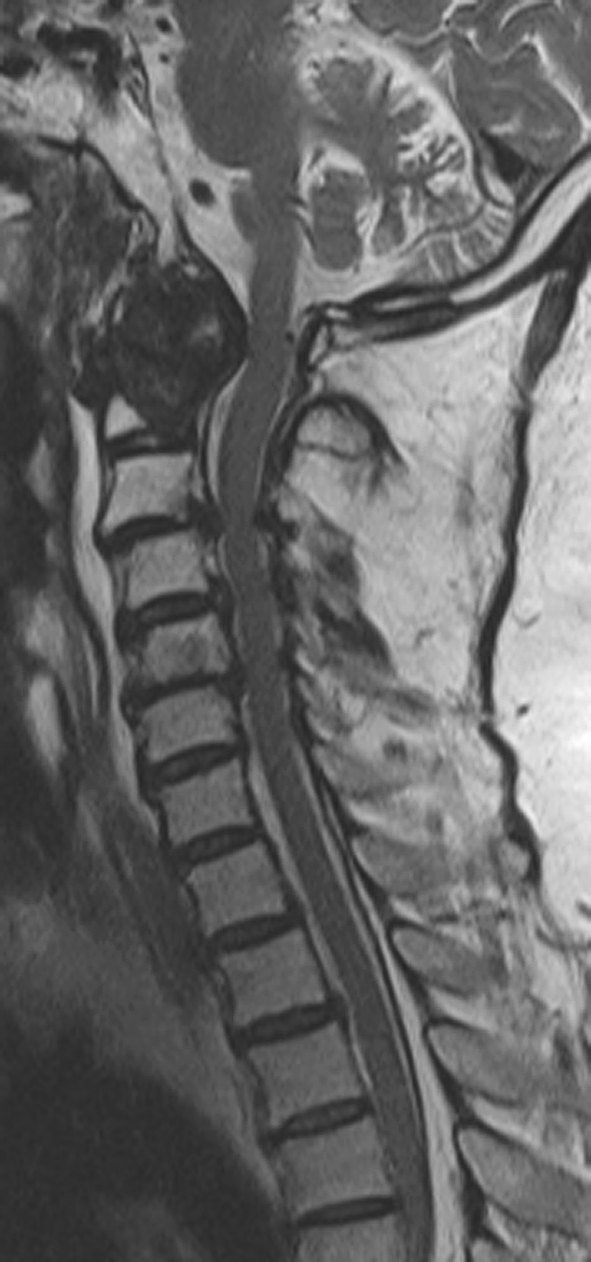
Sagittal T2-weighted MRI scan demonstrating thecal compression at the cervicomedullary region
He then went on to have a CT scan, which was reported as showing the odontoid peg appearing hypodense with amorphous intra-lesional calcification destructive in nature. There was extension to the left side to involve the lateral mass and posterior arch of C1, which looked moth-eaten. There was destruction of the anterior arch of C1 on the right side, with soft tissue breaching the retropharyngeal fat plane. The peg had become displaced anteriorly away from the inferior body of C2. The soft tissue was compressing the cervicomedullary junction (Fig. 2a, b) He was fitted in a halo brace to prevent any destabilisation.
Fig. 2.

a Sagittal CT scan demonstrating amorphous intra-lesional calcification at the C1–2 level. b Axial CT scan demonstrating amorphous intra-lesional calcification around the odontoid peg with destruction of C1
Possible differential diagnosis raised at this stage included rheumatoid pannus, gout, metastasis, myeloma, chondroma, chondrosarcoma and infection. He subsequently had a transoral biopsy that confirmed a diagnosis of gout with urate crystals that were negatively bi-refringent under polarised light.
It was decided that he would be a good candidate for a C0–C6 occipito-cervical fusion, which was up to level-C6 to prevent a stress riser from occurring causing further instability through fracture. A decompression was not performed because it was felt that the destructive nature of the gouty tophi, which appeared calcified was encroaching the dura and so inadvertent dural tear due to adhesions was a strong possibility. Therefore, the risks did not outweigh the benefits as a gouty decompression was likely to be critical should this have been done. He was therefore stabilised and managed medically with febuxostat (Fig. 3a, b) He is to date at 1-year follow-up doing very well with no further neck pain. His neurology has partially resolved in that he no longer has neck pain. He does however still have 4+/5 MRC power to all four limbs with some resolution from the initial presentation.
Fig. 3.

a Anterior posterior radiograph following occipito-cervical fusion (C0–C6). b Lateral radiograph following occipito-cervical fusion (C0–C6)
Conclusion
This case and others that have gone before it demonstrate that chronic neck pain and a history of gout should entertain a differential diagnosis of gout. Atlanto-axial subluxation should no longer be synonymous with only rheumatoid arthritis but also tophaceous gout. Once suspicion is raised about atlanto-axial instability management needs to come from temporary stabilisation (halo brace), definitive diagnosis (transoral biopsy) and then definitive treatment (occipito-cervical fusion). Decompression needs to be judged on an individual case by case basis and the authors suggest that a balance of risks versus benefits should be borne in mind when this decision is made. If neurological symptoms are mild to moderate then a stabilisation with medical management would seem appropriate. Calcification and encroachment of the dura could lead to a difficult decompression involving adhesions.
Conflict of interest
None of the authors has any potential conflict of interest.
References
- 1.Pillinger MH, Rosenthal P, Abeles AM. Hyperuricemia and gout: new insights into pathogenesis and treatment. Bull NYU Hosp Jt Dis. 2007;65(3):215–221. [PubMed] [Google Scholar]
- 2.James WD, Berger TG (2006) Andrews’ diseases of the skin: clinical dermatology. Saunders, Philadelphia/Elsevier, Amsterdam, p 546
- 3.Wazir NN, Moorthy V, Amalourde A, Lim HH. Tophaceous gout causing atlanto-axial subluxation mimicking rheumatoid arthritis: a case report. J Orthopaed Surg. 2005;13(2):203–206. doi: 10.1177/230949900501300220. [DOI] [PubMed] [Google Scholar]
- 4.Alarcon GS, Reveille JD. Gouty arthritis of the axial skeleton including the sacroiliac joints. Arch Intern Med. 1987;147:2018–2019. doi: 10.1001/archinte.147.11.2018. [DOI] [PubMed] [Google Scholar]
- 5.Cabot J, Mosel L, Kong A, Hayward M. Tophaceous gout in the cervical spine. Skelet Radiol. 2005;34:803–806. doi: 10.1007/s00256-005-0920-0. [DOI] [PubMed] [Google Scholar]
- 6.Diaz A, Porhiel V, Sabatier P, Taha S, Ragrgui O, Comoy J. Tophaceous gout of the cervical spine causing cord compression. Case report and review of the literature. Neurochirurgie. 2003;49:600–604. [PubMed] [Google Scholar]
- 7.Duprez TP, Malghem J, Berg BC, Noel HM, Munting EA, Maldague BE. Gout in the cervical spine: MR pattern mimicking diskovertebral infection. Am J Neuroradiol. 1996;17:151–153. [PMC free article] [PubMed] [Google Scholar]
- 8.Kersley GD, Mandel L, Jeffrey MR. Gout: an unusual case with softening and subluxation of the first cervical vertebra and splenomegaly. Ann Rheum Dis. 1950;9:282–304. doi: 10.1136/ard.9.4.282. [DOI] [PMC free article] [PubMed] [Google Scholar]
- 9.Magid SK, Gray GE, Anand A. Spinal cord compression by tophi in a patient with chronic polyarthritis: case report and literature review. Arthritis Rheum. 1982;24:1431–1434. doi: 10.1002/art.1780241117. [DOI] [PubMed] [Google Scholar]
- 10.Jacobs SR, Edeiken J, Rubin B, DeHoratius RJ. Medically reversible quadraparesis in tophaceous gout. Arch Phys Med Rehabil. 1985;66:188–190. [PubMed] [Google Scholar]
- 11.Miller JD, Percy JS. Tophaceous gout in the cervical spine. J Rheumatol. 1984;11:862–865. [PubMed] [Google Scholar]
- 12.Sabharwal S, Gibson T. Cervical gout. Br J Rheumatol. 1988;27:413–414. doi: 10.1093/rheumatology/27.5.413. [DOI] [PubMed] [Google Scholar]
- 13.Sequeira W, Bouffard A, Salgia K, Skosey J. Quadriparesis in tophaceous gout. Arthritis Rheum. 1981;24:1428–1430. doi: 10.1002/art.1780241116. [DOI] [PubMed] [Google Scholar]
- 14.Laar MA, Soesbergeb RM, Matricali B. Tophaceous gout of the cervical spine without peripheral tophi. Arthritis Rheum. 1987;30:237–238. doi: 10.1002/art.1780300224. [DOI] [PubMed] [Google Scholar]
- 15.Vinstein AL, Cockerill EM. Involvement of the spine in gout. A case report. Radiology. 1972;103:311–331. doi: 10.1148/103.2.311. [DOI] [PubMed] [Google Scholar]
- 16.Yen HL, Cheng CH, Lin JW. Cervical myelopathy due to gouty tophi in the intervertebral disc space. Acta Neurochir. 2002;144:205–207. doi: 10.1007/s007010200026. [DOI] [PubMed] [Google Scholar]
- 17.Fraser JF, Anand VK, Schwartz TH. Endoscopic biopsy sampling of tophaceous gout of the odontoid process. J Neurosurg Spine. 2007;7:61–64. doi: 10.3171/SPI-07/07/061. [DOI] [PubMed] [Google Scholar]