Abstract
A case of a 34-year-old female with unilateral cleft of atlas posterior arch associated with os odontoideum is reported. The patient had experienced neck pain for 6 months. Five days earlier to admission the pain aggravated as a result of mild head trauma from behind. Imaging examinations revealed C1–2 subluxation as well as the deformity. After 3 days of skull traction, a sound C1–2 reduction was achieved. Post atlantoaxial fusion using bilateral transarticular screws combined with C1 laminar hook on the intact side and autogenous bone graft was performed. On the sixth month of postoperative follow-up, CT revealed solid fusion was achieved. No related complications were detected within 3 years of follow-up. The clinical manifestations and imaging findings were presented. The incidence and etiopathogenesis of hypoplastic posterior arch of the atlas were concisely introduced. Techniques of post atlantoaxial fusion under circumstances of unilateral C1 posterior elements defects were discussed. The authors believe bilateral transarticular screws combined with C1 laminar hook on the intact side and autogenous bone graft can be applicable to atlantoaxial fusion on the premise of preoperative C1–2 reduction and C1 posterior arch remaining >1/2 of its full length.
Keywords: Atlantoaxial fusion, Os odontoideum, Unilateral cleft of atlas posterior arch
Introduction
Currarino et al. [1] classified the anomalous atlas (C1) posterior arch into five types on the basis of CT and radiographs. Type A: failure of the posterior midline fusion of the two hemiarches; Type B: unilateral cleft; Type C: bilateral clefts; Type D: total absence of the posterior arch with a persistent posterior tubercle; Type E: total absence of the posterior arch with missing posterior tubercle. Incidence of hypoplastic posterior arch of the atlas varied with the types. Type A defects occur in about 3–4% [2–4] of the population and represent 97% of all posterior defects. Type B through E defects have been reported to occur in 0.69% of the population [2–4]. Unilateral cleft of posterior arch of the C1 (belonging to Type B) associated with os odontoideum was extremely rare. To the best of our knowledge, there are four reported cases [5–8] of hypoplastic atlas associated with os odontoideum. In three cases, the atlas defects [5–7] were bipartite atlas (anterior-posterior arch median clefts), and the other case [8] was posterior median clefts. So this case report may be the first to describe a unilateral cleft of posterior arch of the C1 associated with os odontoideum. Anyway, the defects of C1 post arch bring challenges to planning atlantoaxial fusion when needed. In this article, a case of unilateral cleft of atlas posterior arch associated with os odontoideum was described. Techniques of atlantoaxial fusion for this condition were discussed.
Case report
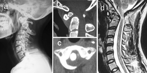
The patient was a 34-year-old female. She had experienced a neck pain for 6 months. In 5 days previous to admission the pain aggravated as a result of mild head trauma from behind. There were no complaints of sense or motion abnormity or gait irregularity. She denied any family history including Turner’s syndrome, Down’s syndrome or gonadal dysgenesis. Physical examinations reveal tenderness at the dorsal area of upper cervical spine and no neurological defect was found. Radiographs showed nonunion of odontoid process and atlantoaxial (C1–2) subluxation. CT confirmed the findings on radiographs and revealed a defect on the left side of posterior arch of the atlas. MRI showed the spinal cord was compressed due to the subluxation (Fig. 1).
Fig. 1.
Preoperative lateral radiography (a), CT (b, c), and MRI (d) of the patient
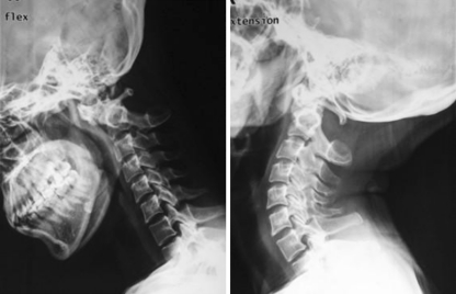
The patient took flexion–extension radiographs, which showed, on extension view, the C1–2 position was to some extent restored but was not satisfactory (Fig. 2). So Crutchfield skull traction was applied. After 3 days of traction, satisfactory C1–2 reduction was achieved and post atlantoaxial fusion was performed. After considerable analysis, we adopted a technique of bilateral transarticular screws combined with C1 laminar hook on the intact side and autogenous bone graft.
Fig. 2.
Lateral radiography before traction, flexion–extension views
After anesthesia, the patient was dorsicumbent in a wheel stretcher, and was protected with a tailor-made plaster bed. Then the patient was turned over together with the plaster bed to the operation table. C1–2 reduction was routinely checked under C-arm fluoroscopy just before operation. The surgery was carried out with a posterior approach and exposure of occiput to axis (C2). Bilateral transarticular screws were inserted successfully and a C1 laminar hook was mounted on the intact side of the posterior arch. A longitudinal rod was used to connect the screw and hook. To accomplish bone fusion, areas of bone graft were decorticated. A structural bone graft obtained from the posterior iliac crest was placed between the C1 posterior arch and the C2 spinous process. It was secured by proper pressure of instruments on the intact side. Cancellous bone fragments were then grafted between the laminae of C1–2 on the intact side.
The patient was free of the neck pain from the 5th day after operation and got solid bone union in 6 months. No related complications were detected within 3 years of follow-up.
Discussion
Etiopathogenesis of hypoplastic posterior arch of the atlas remains uncertain. Major speculation was a developmental failure of chondrogenesis [3, 9, 10]. Besides, the disorder may be related to inheritance [1, 9] and myelopathy associated with it may be more liable in orientals [3]. As described in the introduction, unilateral cleft of posterior arch of the atlas associated with os odontoideum was extremely rare.
In most cases, the hypoplastic posterior arch of the atlas is asymptomatic and is generally detected as an incidental finding needing no surgical treatments. Procedures may be indicated in the following cases: (1) myelopathy due to compression of surrounding soft tissue or nomadic posterior tubercle or hypoplastic spinal canal stenosis [3, 11, 12]. (2) Atlantoaxial instability [7, 8, 13, 14] due to injuries or other associating deformities. (3) Type C (absence of the bilateral posterior arches) and type D (absence of the bilateral posterior arches with isolated posterior tubercle) indicating high latent risks of acute or cumulative spinal cord injuries by the posterior tubercle or surrounding soft tissue especially in the course of the neck extension tend to be treated surgically [10, 15, 16]. In cases of (1) and (3), laminectomy or removal of the compressing factors can be sufficient. If there is C1–2 instability, internal fixation and fusion is indicated. Although occipital-cervical fusion can be considered, it should be the second choice for its extensive limitation of rotary motion. It is especially undesirable in case of C1–2 reduction.
Atlantoaxial fusion for unilateral cleft of posterior arch of the atlas is somewhat challenging highlighting matters of bone graft fixation and bone graft bed. Planning of the procedure should be based on precise evaluation of the extent and location of the posterior defect, conditions of the remains of C1 posterior elements and other parameters of the C1 and C2 on imaging. There are a few case of C1–2 fusion for defective arch of C1 reported in the previous literature [7, 8, 14] (Table 1). In general, screws fixation is mechanically superior saving from extensive external fixation. On the other hand, wirings played an important role before emergence of screw techniques or in case that screw techniques were limited.
Table 1.
Operations of C1–2 fusion for defective arch of C1
| No. | Author | Year | Diagnosis | Procedure |
|---|---|---|---|---|
| 1 | Callahan [8] | 1983 | Median cleft of posterior arch; os odontoideum | Bone grafts were placed bilaterally between C1 and 2 laminae. Wires were used respectively to bunch the grafts |
| 2 | Duong; Chadduck [14] | 1994 | Hypoplastic posterior arch; C1–2 subluxation | One graft was put into the posterior arch to patch the defect and the other two were used to connect the bilateral C1–2 laminae Wiring was used to secure all the junctions |
| 3 | Osti et al. [7] | 2006 | Bipartite atlas; os odontoideum | Bilateral transarticular screws fixation combined with sublaminar tension band wiring and autologous bone grafting |
In this case, although the symptoms were not severe and there was no neurological deficit, we recommended a surgical procedure because besides os odontoideum, definite atlantoaxial instability with spinal cord compression was revealed on the imaging appearances indicating a high potential risk for the development of myelopathy. It should be noted that the defect of the posterior arch itself is no indication for surgery and the aim of the operation is not to patch up the defect but to fix and fuse the C1–2 joint.
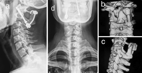
This patient got a sound C1–2 reduction through preoperative traction and CT revealed the posterior arch of atlas remained >1/2 of its full length. We used bilateral transarticular screws combined with C1 laminar hook on the intact side and autogenous bone graft (Fig. 3). Bilateral transarticular screws lay a solid foundation for the internal fixation [17–22] and the hook system serves as a measure to fix the bone graft through pressing. An additional hook system on the intact side, instead of wiring, was adopted which had advantages [23–25] of (1) more stability as a supplement to bilateral transarticular screws in restricting the flexion–extension, (2) more safety without passing wires though the arch, and (3) higher efficiency in securing the structural bone graft with a longer torque pressing the posterior arch as compared with C1 lateral mass screw. As limitations, the technique was based on premise that C1–2 reduction and sufficient remains of posterior arch. It was not applicable in case of the posterior arch remained <1/2 or unsatisfactory C1–2 reduction. It also required a structural bone graft because cancellous bone fragments alone can not support the posterior arch with a hook against the extension of cervical spine.
Fig. 3.
X-rays (a, d) taken on the 3rd day after operation and CT (b, c) taken in the 6th month after operation
Theoretically, in this case, C1 lateral mass and the C2 pedicle screws and rods with cancellous bone graft may be another good option. C1 lateral masses and C2 pedicles were intact in this case meeting anatomical qualifications of the technique. Reduction of C1 onto C2 can be accomplished by manipulation of the implants. Another advantage of this technique was that it minimized the dependence on the integrity of posterior arch of C1 [26, 27]. But this technique inserting 4 screws was technically demanding in preserving venous plexus and C2 nerve roots. When, although not usually needed, structural bone graft was used, additional wiring was often imperative resulting in more manipulation and risk of spine cord injuries.
We finally chose bilateral transarticular screws combined with C1 laminar hook on the intact side and autogenous bone graft on the premise of preoperative C1–2 reduction and C1 posterior arch remaining >1/2 of its full length. In the 6th month of postoperative follow-up, CT revealed solid bone fusion. No related complications were detected within 3 years of follow-up.
Conclusions
A case of unilateral cleft of atlas posterior arch associated with os odontoideum was reported. The authors offered an option for atlantoaxial fusion under circumstances of unilateral C1 posterior elements defects. Bilateral transarticular screws combined with C1 laminar hook on the intact side and autogenous bone graft can be applicable to atlantoaxial fusion on the premise that preoperative C1–2 reduction and C1 posterior arch remains >1/2 of its full length.
Acknowledgments
Supported by Science and Technology Commission of Shanghai Municipality (No. 08411952400).
Conflict of interest None of the authors has any potential conflict of interest.
References
- 1.Currarino G, Rollins N, Diehl JT. Congenital defects of the posterior arch of the atlas: a report of seven cases including an affected mother and son. AJNR Am J Neuroradiol. 1994;15:249–254. [PMC free article] [PubMed] [Google Scholar]
- 2.Devi BI, Shenoy SN, Panigrahi MK, Chandramouli BA, Das BS, Jayakumar PN. Anomaly of arch of atlas—a rare cause of symptomatic canal stenosis in children. Pediatr Neurosurg. 1997;26:214–217. doi: 10.1159/000121194. [DOI] [PubMed] [Google Scholar]
- 3.Phan N, Marras C, Midha R, Rowed D. Cervical myelopathy caused by hypoplasia of the atlas: two case reports and review of the literature. Neurosurgery. 1998;43:629–633. doi: 10.1097/00006123-199809000-00140. [DOI] [PubMed] [Google Scholar]
- 4.Friedman MB, Jacobs LH. Case report: computed tomography of congenital clefts of the atlas. Comput Radiol. 1985;9:185–187. doi: 10.1016/0730-4862(85)90164-7. [DOI] [PubMed] [Google Scholar]
- 5.Atasoy C, Fitoz S, Karan B, Erden I, Akyar S. A rare cause of cervical spinal stenosis: posterior arch hypoplasia in a bipartite atlas. Neuroradiology. 2002;44:253–255. doi: 10.1007/s00234-001-0740-4. [DOI] [PubMed] [Google Scholar]
- 6.Garg A, Gaikwad SB, Gupta V, Mishra NK, Kale SS, Singh J. Bipartite atlas with os odontoideum: case report. Spine (Phila Pa 1976) 2004;29:E35–E38. doi: 10.1097/01.BRS.0000106487.89648.88. [DOI] [PubMed] [Google Scholar]
- 7.Osti M, Philipp H, Meusburger B, Benedetto KP. Os odontoideum with bipartite atlas and segmental instability: a case report. Eur Spine J. 2006;15(Suppl 5):564–567. doi: 10.1007/s00586-005-0017-4. [DOI] [PMC free article] [PubMed] [Google Scholar]
- 8.Callahan RA, Lockwood R, Green B. Modified Brooks fusion for an os odontoideum associated with an incomplete posterior arch of the atlas. A case report. Spine Phila Pa 1976. 1983;8:107–108. doi: 10.1097/00007632-198301000-00017. [DOI] [PubMed] [Google Scholar]
- 9.Motateanu M, Gudinchet F, Sarraj H, Schnyder P. Case report 665. Congenital absence of posterior arch of atlas. Skeletal Radiol. 1991;20:231–232. doi: 10.1007/BF00241677. [DOI] [PubMed] [Google Scholar]
- 10.Sagiuchi T, Tachibana S, Sato K, Shimizu S, Kobayashi I, Oka H, Fujii K, Kan S. Lhermitte sign during yawning associated with congenital partial aplasia of the posterior arch of the atlas. AJNR Am J Neuroradiol. 2006;27:258–260. [PMC free article] [PubMed] [Google Scholar]
- 11.Connor SE, Chandler C, Robinson S, Jarosz JM. Congenital midline cleft of the posterior arch of atlas: a rare cause of symptomatic cervical canal stenosis. Eur Radiol. 2001;11:1766–1769. doi: 10.1007/s003300000755. [DOI] [PubMed] [Google Scholar]
- 12.Urasaki E, Yasukouchi H, Yokota A. Atlas hypoplasia manifesting as myelopathy in a child–case report. Neurol Med Chir (Tokyo) 2001;41:160–162. doi: 10.2176/nmc.41.160. [DOI] [PubMed] [Google Scholar]
- 13.Klimo P, Jr, Blumenthal DT, Couldwell WT. Congenital partial aplasia of the posterior arch of the atlas causing myelopathy: case report and review of the literature. Spine (Phila Pa 1976) 2003;28:E224–E228. doi: 10.1097/01.BRS.0000065492.85852.A9. [DOI] [PubMed] [Google Scholar]
- 14.Duong DH, Chadduck WM. Reconstruction of the hypoplastic posterior arch of the atlas with calvarial bone grafts for posterior atlantoaxial fusion: technical report. Neurosurgery. 1994;35:1168–1170. doi: 10.1227/00006123-199412000-00025. [DOI] [PubMed] [Google Scholar]
- 15.Richardson EG, Boone SC, Reid RL. Intermittent quadriparesis associated with a congenital anomaly of the posterior arch of the atlas. Case report. J Bone Joint Surg Am. 1975;57:853–854. [PubMed] [Google Scholar]
- 16.Sharma A, Gaikwad SB, Deol PS, Mishra NK, Kale SS. Partial aplasia of the posterior arch of the atlas with an isolated posterior arch remnant: findings in three cases. AJNR Am J Neuroradiol. 2000;21:1167–1171. [PMC free article] [PubMed] [Google Scholar]
- 17.Grob D, Crisco JJ, 3rd, Panjabi MM, Wang P, Dvorak J. Biomechanical evaluation of four different posterior atlantoaxial fixation techniques. Spine. 1992;17:480–490. doi: 10.1097/00007632-199205000-00003. [DOI] [PubMed] [Google Scholar]
- 18.Haid RW, Jr, Subach BR, McLaughlin MR, Rodts GE, Jr, Wahlig JB., Jr C1–C2 transarticular screw fixation for atlantoaxial instability: a 6-year experience. Neurosurgery. 2001;49:65–68. doi: 10.1097/00006123-200107000-00010. [DOI] [PubMed] [Google Scholar]
- 19.Fountas KN, Smisson HF, 3rd, Robinson JS., Jr C1–C2 transarticular screw fixation for atlantoaxial instability: a 6-year experience. Neurosurgery. 2002;50:672–673. [PubMed] [Google Scholar]
- 20.Low HL, Redfern RM. C1–C2 transarticular screw fixation for atlantoaxial instability: a 6-year experience, and C1–C2 transarticular screw fixation–technical aspects. Neurosurgery. 2002;50:1165–1166. doi: 10.1097/00006123-200205000-00047. [DOI] [PubMed] [Google Scholar]
- 21.Henriques T, Cunningham BW, Olerud C, Shimamoto N, Lee GA, Larsson S, McAfee PA. Biomechanical comparison of five different atlantoaxial posterior fixation techniques. Spine. 2000;25:2877–2883. doi: 10.1097/00007632-200011150-00007. [DOI] [PubMed] [Google Scholar]
- 22.Gluf WM, Schmidt MH, Apfelbaum RI. Atlantoaxial transarticular screw fixation: a review of surgical indications, fusion rate, complications, and lessons learned in 191 adult patients. J Neurosurg Spine. 2005;2:155–163. doi: 10.3171/spi.2005.2.2.0155. [DOI] [PubMed] [Google Scholar]
- 23.Guo X, Ni B, Wang M, Wang J, Li S, Zhou F. Bilateral atlas laminar hook combined with transarticular screw fixation for an unstable bursting atlantal fracture. Arch Orthop Trauma Surg. 2009;129:1203–1209. doi: 10.1007/s00402-008-0706-7. [DOI] [PubMed] [Google Scholar]
- 24.Ni B, Zhu Z, Zhou F, Guo Q, Yang J, Liu J, Wang F (2010) Bilateral C1 laminar hooks combined with C2 pedicle screws fixation for treatment of C1–C2 instability not suitable for placement of transarticular screws. Eur Spine J. doi:10.1007/s00586-010-1365-2 [DOI] [PMC free article] [PubMed]
- 25.Guo X, Ni B, Zhao W, Wang M, Zhou F, Li S, Ren Z. Biomechanical assessment of bilateral C1 laminar hook and C1–2 transarticular screws and bone graft for atlantoaxial instability. J Spinal Disord Tech. 2009;22:578–585. doi: 10.1097/BSD.0b013e31818da3fe. [DOI] [PubMed] [Google Scholar]
- 26.Harms J, Melcher RP. Posterior C1–C2 fusion with polyaxial screw and rod fixation. Spine (Phila Pa 1976) 2001;26:2467–2471. doi: 10.1097/00007632-200111150-00014. [DOI] [PubMed] [Google Scholar]
- 27.Goel A, Desai KI, Muzumdar DP. Atlantoaxial fixation using plate and screw method: a report of 160 treated patients. Neurosurgery. 2002;51:1351–1356. [PubMed] [Google Scholar]


