


Abstract
A case of caudal regression syndrome in which rehabilitation was obtained by lumbopelvic distraction and stabilization with external fixation Orthofix® is presented. The objective of the study is to describe the benefit of spine external fixator in caudal regression syndrome. Caudal regression syndrome is a rare and sporadic neural defect of distal spinal segments affecting the development of the spinal cord. It is characterized by vertebropelvic instability and essentially manifests as neurological deficit in the lower limbs and absence of bladder and bowel control. Pluridisciplinary management of this affection is complex. The clinical presentation and treatment of caudal regression syndromes are reviewed. A case of a boy for whom physical rehabilitation was obtained with external fixation Orthofix® is presented. Lumbopelvic stabilization was performed with autograft and allograft bone. A spine distraction was performed with external fixation with pedicular screws in L1 and L2 and in the pelvis bone. An elongation of 19 mm was obtained in 35 days. The external fixator was removed after 4 months and a spine cast was applied during 8 months. This treatment resulted in lumbopelvic nonunion of very low mobility. Lumbopelvic stability and the disappearance of pain were achieved. Sitting position was stable without hand support. At 14 years of follow-up, his condition is stable with possible deambulation at home with crutches. Lumbopelvic arthrodesis in caudal regression syndrome is difficult to obtain. The use of spine external fixator, however, allows trunk elongation and can achieve a stable sitting and upright position.
Keywords: Lumbosacral agenesia, Caudal regression, Lumbopelvic arthrodesis, External fixator
Introduction
Caudal regression syndrome is a rare and sporadic neural defect of distal spinal segments affecting the development of the spinal cord. Intelligence is preserved. The exact etiology is elusive, though maternal diabetes, genetic factors and hypoperfusion might be implicated [1, 2]. It is characterized by vertebropelvic instability, and manifests as neurological deficit in the lower limbs and absence of bladder and bowel control [3–5]. Management of this affection is complex and must be pluridisciplinary. Treatment of vertebral instability by lumbopelvic arthrodesis is particularly difficult. We presently report a case of caudal regression syndrome in which increased functional ability was obtained by lumbopelvic distraction and stabilization with external fixation Orthofix®.
Case report
A 1-year-old boy with incomplete paraplegia, no bladder and bowel control, and lower limbs deformity was addressed in 1992 for further management. He was the offspring of a non-consanguineous marriage. His mother was not a diabetic. He did not have other significant medical history. Antenatal ultrasound had not been done. He had small buttocks, a flat perineum, and an equinovarus deformity. He had no sensory deficit in the lower limbs and was able to move them. No other malformation or impairment was detected on clinical examination. Radiographies showed total sacral and partial lumbar spinal agenesis below the third lumbar vertebral body (L3 hemivertebrae) of type 1 of Bollini [3] and a bilateral dislocation of the hip (Fig. 1a, b). On RMI and 3D CT-scan, the malformation was visualized with the spinal agenesis below L3, an interruption of the dural sac and a contact between the two iliac bones (Figs. 2, 3). The hips were stabilized with a bilateral Salter’s operation.
Fig. 1.

a AP X-ray of the spine. Agenesis of the spine under L3 with bilateral hips dislocation. b Lateral X-ray of the spine
Fig. 2.

MRI showing spine agenesis under L3 with abnormalities of the myelon
Fig. 3.

3D CT-scan showing agenesis of the spine under L3 and contact between the two iliac bones
At the age of 3 years, the sitting position was instable with a spinopelvic kyphosis. The kyphotic bearing was responsible for bowel obstruction with constipation and pain due to a conflict between iliac crests and ribs. A lumbopelvic fusion was performed with autograft (2 tibial sticks) and allograft bone (1 femoral head). To increase trunk height and distance between the ribs and iliac crests, a spine distraction was performed with external fixation Orthofix® with pedicular screws in L1 and L2 and in the pelvis bone (Fig. 4, 5). With progressive distraction, an elongation of 19 mm was obtained in 35 days. Distraction was then stopped due to neurogenic pains in the lower limbs. The external fixator was removed after 4 months and a spine plaster cast was applied during 2 months, followed by a plastic corset for 6 months. This treatment resulted in lumbopelvic non-union of very low mobility. Lumbopelvic stability and the disappearance of pain were nevertheless achieved. Sitting position was stable without hand support. At 14 years of follow-up, his condition is stable with possible deambulation at home with crutches and stable sitting position (Figs. 6, 7).
Fig. 4.
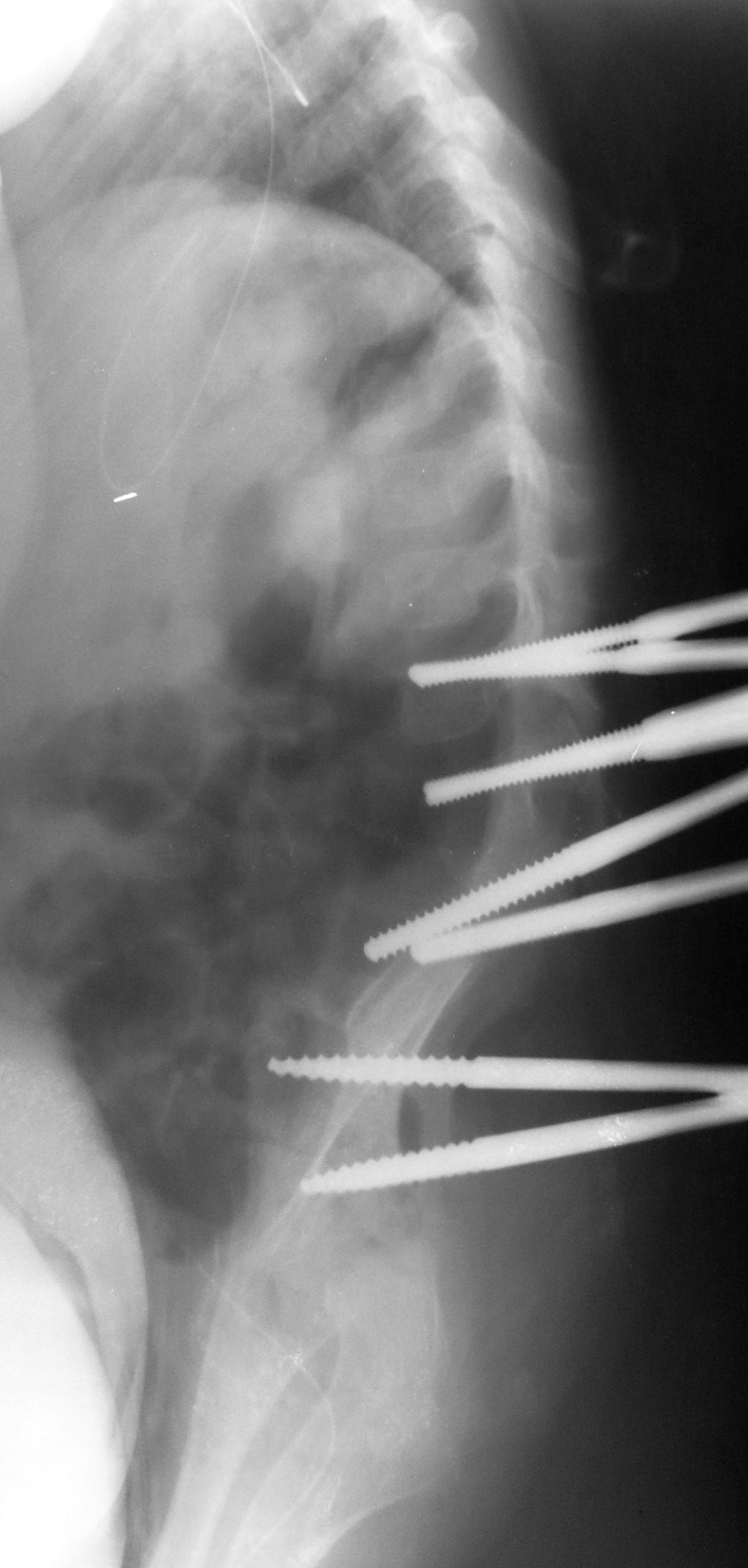
Lateral X-ray with the pedicular screws in LI and L2 and in the iliac bone
Fig. 5.

External fixator Orthofix®
Fig. 6.

Lateral X-ray of the spine in sitting position at 14 years of follow-up
Fig. 7.
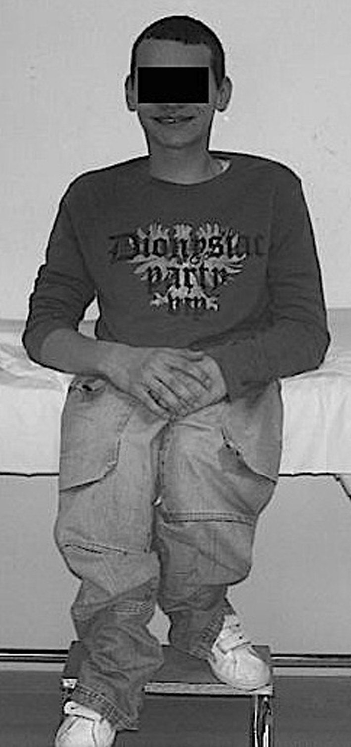
Sitting position is stable
Discussion
Caudal regression syndrome represents a continuum of congenital malformations ranging from agenesis of lumbosacral spine to the most severe cases of sirenomelia with fusion of lower extremities or limb duplication with major visceral abnormalities [6, 7]. Duhamel coined the term of caudal regression syndrome in 1964 to describe this entity [8]. A number of associated anomalies (musculoskeletal, gastrointestinal, genitourinary systems and miscellaneous) have been described, attesting to its heterogeneous nature [2]. These malformations are a consequence of abnormal development of the structures derived from the caudal mesoderm of embryo [2]. A neural tube defect, occurring before 28 days of gestation, has been described in particular [2]. The etiology is unknown. Genetic predisposition, vascular hypoperfusion and maternal diabetes have been suggested as possible causative factors [1]. Chan et al. and Goto et al. have implicated maternal metabolic derangements of diabetes as the essential cause for multiple malformations, among which is the caudal regression syndrome [9, 10]. A possible drug-related etiology for extreme caudal agenesis in a human fetus has also been suggested [11].
In caudal regression syndrome, which often involves several systems, each system and its pathology must be treated on their own merits. The pathologies that need special attention are orthopedic deformities and bladder and bowel continence, along with preservation of renal function. The treatment is therefore difficult and must be performed by a multidisciplinary team involving pediatrician, pediatric surgeon, orthopedic surgeon, physiotherapist and social worker [2].
In the case that we report herein, the patient had an incomplete paraplegia with an unstable sitting position, a kyphotic bearing that induced pain and impairment of intestinal transit. The vertebropelvic kyphosis results from the instability of the pelvis under the spine and is responsible for trunk shortening and unstable sitting position. Trunk shortening and abdominal collapse create an increase in intra-abdominal pressure that hampers intestinal transit [4]. Trunk shortening also induces pain due to a conflict between ribs and iliac crest. This is why we chose to treat both causes, trunk shortening and kyphosis, by using an external fixator allowing distraction and kyphosis correction.
As mentioned by Dumont [4], the pelvic and lower leg hypotrophy may make it difficult to obtain sufficient bone autograft and surface for graft union. The use of external fixator in spine surgery is uncommon, particularly in children. Dubousset [12] used Ilizarov fixator in several particular cases. Reyes-Sanchez et al. [13] used external fixator Orthofix® to achieve a correction by dynamic distraction, compression and derotation of the severe scoliotic curve. Pedicular fixation is solid and allows distraction, which enables to correct the trunk shortening and the abdominal collapse. Cutaneous problems that may arise on osteosynthesis material do not exist with this technique. The child’s positioning is, however, delicate due to the retraction of the lower limbs. Lumbopelvic arthrodesis is difficult to obtain whichever type of osteosynthesis is chosen [4, 5]. Arthrodesis must nevertheless be undertaken to eliminate pelvic instability and to achieve the sitting and upright position.
Conclusion
Caudal regression syndrome is becoming increasingly rare, as antenatal diagnosis often leads to medical interruption of pregnancy. Orthopedic treatment is difficult, comprising multiple surgical procedures on the lower limbs and spine. The use of spine external fixator allows trunk elongation and correction of kyphosis, and thereby reduces pain and transit impairment and can achieve a stable sitting and upright position. Lumbopelvic arthrodesis is, however, always difficult to obtain.
Conflict of interest statement
None of the authors has any potential conflict of interest.
References
- 1.Adra A, Cordero D, Mejides A, Yasin S, Salman F, O’Sullivan MJ. Caudal regression syndrome: etiopathogenesis, prenatal diagnosis, and perinatal management. Obstet Gynecol Surv. 1994;49:508–516. doi: 10.1097/00006254-199407000-00028. [DOI] [PubMed] [Google Scholar]
- 2.Singh SK, Singh RD, Sharma A. Caudal regression syndrome—case report and review of literature. Pediatr Surg Int. 2005;21:578–581. doi: 10.1007/s00383-005-1451-4. [DOI] [PubMed] [Google Scholar]
- 3.Bollini G (1989) Agénésies sacrées et lombo-sacrées. Chirurgie et orthopédie du rachis de l’enfant. Sauramps Medical, Paris, pp 349–356
- 4.Dumont CE, Damsin JP, Forin V, Carlioz H. Lumbosacral agenesis. Three cases of reconstruction using Cotrel-Dubousset or L-rod instrumentation. Spine. 1993;18:1229–1235. doi: 10.1097/00007632-199307000-00018. [DOI] [PubMed] [Google Scholar]
- 5.Rieger MA, Hall JE, Dalury DF. Spinal fusion in a patient with lumbosacral agenesis. Spine. 1990;12:1382–1384. doi: 10.1097/00007632-199012000-00032. [DOI] [PubMed] [Google Scholar]
- 6.Bastiani-Griffet F, Bollini G, Griffet J, Berard J, Bouyala JM. La pygomélie. Chir Ped. 1990;31:333–336. [PubMed] [Google Scholar]
- 7.Griffet J, Bastiani-Griffet F, Jund S, Moreigne M, Zabjek K. Duplication of the leg–renal agenesis: congenital malformation syndrome. J Pediatr Orthop. 2000;9:306–308. doi: 10.1097/01202412-200010000-00015. [DOI] [PubMed] [Google Scholar]
- 8.Duhamel B. From the mermaid to anal imperforation: the syndrome of caudal regression. Arch Dis Child. 1964;36:152–155. doi: 10.1136/adc.36.186.152. [DOI] [PMC free article] [PubMed] [Google Scholar]
- 9.Chan BWH, Chan KS, Koide T, Yeung SM, Leung MBW, Copp AJ, Loeken MR, Shiroishi T, Shum ASW. Maternal diabetes increases the risk of caudal regression caused by retinoic acid. Diabetes. 2002;51:2811–2816. doi: 10.2337/diabetes.51.9.2811. [DOI] [PubMed] [Google Scholar]
- 10.Goto MP, Goldman AS. Diabetic embryopathy. Curr Opin Pediatr. 1994;6:486–491. doi: 10.1097/00008480-199408000-00023. [DOI] [PubMed] [Google Scholar]
- 11.Rojansky N, Fasouliotis SJ, Ariel I, Nadjari M. Extreme caudal agenesis. Possible drug-related etiology? J Reprod Med. 2002;47:241–245. [PubMed] [Google Scholar]
- 12.Dubousset J (1991) La technique d’Ilizarov. Procédé de sauvetage en chirurgie du rachis. Rev Chir Orthop :Suppl I77:144
- 13.Reyes-Sanchez A, Rosales LM, Miramontes V. External fixation for dynamic correction of severe scoliosis. Spine J. 2005;5:418–426. doi: 10.1016/j.spinee.2004.11.013. [DOI] [PubMed] [Google Scholar]