Abstract
A rotator cuff tear causes shoulder pain and limits movement of the shoulder joint. A chronic degenerative change or impingement is the reason for a rotator cuff tear. Diagnosis is made based on medical history and, physical and radiological examinations. Other causes of shoulder pain include calcific tendinitis, degenerative arthropathy, joint dislocation, fracture, and primary or metastatic neoplasm. However, metastatic cancer in the shoulder joint is difficult to diagnosis. We experienced a case in which a 46-year-old female patient complained of left shoulder pain and limited joint mobility, and these symptoms were due to metastatic breast cancer in the shoulder.
Keywords: neoplasm metastasis, rotator cuff, shoulder
Shoulder pain occurs in 6.6 to 25 people out of every 1000, and is the third most frequent musculoskeletal disease after low back pain and knee pain [1-3]. The shoulder joint is the joint with the largest range of motion in the body, and the cause of shoulder pain is not just limited to the shoulder joint but can also be caused by lesions in the surrounding area. Common causes of shoulder pain are rotator cuff pathology, adhesive capsulitis, calcific tendinitis, degenerative joint disease, dislocation, fracture, acute trauma, and tumors [4].
The authors have recently experienced a case in which a 46-year-old female patient who complained of left shoulder pain and limitation of motion (LOM). After taking a medical history and giving a physical examination, rotator cuff pathology with accompanied rotator cuff tear was suspected but no abnormalities were seen in the ultrasonography. Through magnetic resonance imaging (MRI) and bone scan, metastatic breast cancer believed to have been completely removed by a previous surgery was observed in the humeral head. Herein, we report a case of metastatic breast cancer in the humeral head of the shoulder joint in a 46-year-old woman along with a literature review.
CASE REPORT
The 46-year-old female patient visited our department complaining of left shoulder pain that started after frequent use of the arm 1 month prior to her visit. The pain was of a dull nature with a visual analogue scale (VAS) of 35/100 mm and when the left arm was moved, the pain intensified with a VAS of 100/100 mm. She also complained of LOM in the left shoulder joint and disruption in sleep from the pain.
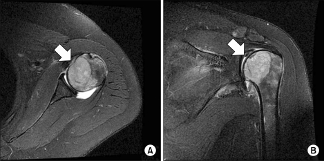
The patient had been diagnosed with right breast cancer 6 years prior to her visit and had a modified radical mastectomy done. She had been regularly visiting a surgical clinic until recently, was taking tamoxifen every day, had a whole body bone scan and ultrasonography done in the breast area once a year, and PET-CT (Positron Emission Tomography-Computed Tomography) taken once every two years to observe progress, but there were no traces of recurrence or metastasis. In the physical examination, there were no irregularities in the inspection, but there was tenderness in the palpated left greater tuberosity. The LOM in her left shoulder joint during active movement was as follows: 150° flexion and 50° extension from the sagittal plane; 110° abduction, 50° adduction from the coronal plane; 40° external rotation. Regarding the internal rotation to the back, her left hand could reach her 1st lumbar vertebra. Meanwhile, regarding passive movement, the patient complained of pain between 90 to 120° in abduction, but there was no LOM in any direction. The patient was positive for the Neer test, positive for the empty can test, and positive for the drop arm test; therefore, rotator cuff pathology with accompanied supraspinatus tendon rupture was suspected. Hence, a laboratory examination and simple x-ray was done but there were no abnormalities, and the left shoulder area was examined using an ultrasonography by our department, but the anticipated rotator cuff pathology could not be confirmed. An ultrasound-guided left suprascapular nerve block was done to treat the pain in the shoulder area, but the alleviation of the pain was insignificant. Thus, an ultrasonography was requested to be done by the Department of Radiology, but there were no abnormalities found in the rotator cuff. The patient complained of continuous pain and LOM so an MRI and bone scan were done. In the MRI examination, a cancer lesion could be observed in the humeral head (Fig. 1), and in the bone scan, abnormalities suspected from a metastatic tumor were found in the humeral head and 10th thoracic vertebra. The patient was transferred to the Department of Internal Medicine and was diagnosed with metastatic breast cancer in the humeral head and 10th thoracic vertebra and received chemotherapy and radiotherapy. Currently, six months later, there are no observations of metastasis into other areas and the cancer lesion in the humeral head has not changed.
Fig. 1.
Magnetic resonance imaging. T2-weighted axial image (A) and coronal image (B) shows osteolytic lesion involving left humeral head and metaphysis.
DISCUSSION
The rotator cuff is comprised of the supraspinatus, infraspinatus, subscapularis, and teres minor. These secure the shoulder joint so it can move and allows for the normal functioning of the shoulder. However, the rotator cuff is easily worn down and susceptible to degeneration so it is the weakest part of the shoulder joint. Therefore, rotator cuff tears frequently occur, and this weakens the shoulder joint and causes pain [5]. A rotator cuff tear occurs from the mixed effects of various mechanisms such as mechanical impact, degenerative changes, circulatory disorders, and joint abrasion. Miller and Dlabach [6] contend that the supraspinatus is most susceptible to tear in the rotator cuff. The tearing of the supraspinatus tendon occurs from abrasion by being caught between the humeral head and the acromion or acromioclavicular joint rather than mechanical impact [7,8].
For diagnosis of rotator cuff tear, history taking and physical examination are important. The patient finds it hard to lift up their arm and it is characteristic that active movement is limited but passive movement is free in the physical examination. In addition, when lowering the arm, drop arm sign appears where the patient does not have the strength or the arm is dropped due to pain. The findings of a physical examination are different for the tears of each tendon. In a supraspinatus tendon tear, painful arc sign is detected between 60 and 120° in abduction. When the arm is raised to a certain level, the final lift can be performed easily. In addition, when both arms are spread about 40° and the elbow joints are extended to where the thumbs are pointing down as if spilling water out of a cup, pain is generated when resistance is applied to the upper area (empty can test). In an infraspinatus tendon tear, when the arm is passively rotated to the maximum range of the external rotation, the patient is not able to hold this position actively and the arm is dropped from body or internally rotated (lag sign). In a subscapularis tendon tear, when the patient's arm is behind the back, there is a lack of muscle strength making it difficult to be able to stretch out from the back while overcoming resistance (lift off test) [6].
The radiological diagnostic method for rotator cuff tears uses ultrasonography or MRI examination. Rafii et al. [9] reported that the sensitivity of rotator cuff tears in MRI was 95% in the case of full thickness tears and 84% in the case of partial tears, while Quinn et al. [10] reported that the sensitivity of MRI examinations regarding rotator cuff tears was 84% and specificity was 97%. Burk et al. [11] compared observations from MRI and arthrography with direct observations during surgery in 16 cases and reported that MRI and arthrography had 92% sensitivity and 100% specificity. There are also reports on the usefulness of ultrasonography in rotator cuff tears, in which Brandt et al. [12] reported that when compared with observations during arthroscopic surgery in 38 cases, ultrasonography had 57% sensitivity and 76% specificity. As can be seen, ultrasonography is known to be less accurate compared to MRI in diagnosing rotator cuff tears.
Ultrasonography has many merits in that real time testing and treatment is possible; there is no risk of exposure to radiation so it can be used safely in pregnant women and children, and it is cheap and non-invasive. However, there are limitations to ultrasonography such as seeing deep structures like bowels because it cannot penetrate air and cannot pass through bone or metal so there are limits to evaluating bone marrow or the safety of orthopaedic hardware.
The most common malignant neoplasm in women is breast cancer, taking up 32% of the malignant neoplasms. With the development of modern diagnosis techniques and treatment modalities, the mortality rate is decreasing, but it is still the most common cause of death in women in their forties [13]. Coleman and Rubens [14] assert that full recovery from breast cancer is possible if surgically removed in the initial stage, and that bone metastasis occurs in 69% of all breast cancer cases. According to Martin and Moseley [15], breast cancer easily spreads to the spine, rib, pelvis, and proximal long bone, and the rate of metastasis reaches 70%. Thomas et al. [16] reported that cancer cells that spread to bone marrow accelerates osteolysis and osteoclast reactions and can cause pathological fractures or pain.
In shoulder pain caused by metastatic cancer, there are reported cases of lung cancer that has spread to the humerus [4], and kidney cancer that has spread to the humerus and scapula [17]. Computed tomography, MRI, and bone scan are used to diagnose cancer that has spread to the bone, and the irregular osteolytic lesions that appear in these images are the most common observations for skeletal metastasis [18].
The patient in our study had received surgery in the past for breast cancer but had shown no trace of recurrence or metastasis in progress observations through regular visits. Since she complained of pain in the left shoulder, had limited active joint movement but free passive joint movement, and was positive for drop arm sign and empty can test, rotator cuff pathology with a supraspinatus tendon tear was suspected; however, there were no images indicating tearing in the ultrasonography images. Thus, MRI and bone scan were done, and as a result, breast cancer that had spread to the bone was found. Recently, the use of ultrasonography in pain clinics is increasing. However, it is necessary for healthcare practitioners to realize that ultrasonography cannot penetrate bone and that additional examinations such as MRI may be needed when there is discord with the clinical symptoms. In addition, cancerous lesions or skeletal metastasis must be considered when musculoskeletal pain occurs in patients thought to have fully recovered from cancer.
References
- 1.Badley EM, Tennant A. Changing profile of joint disorders with age: findings from a postal survey of the population of Calderdale, West Yorkshire, United Kingdom. Ann Rheum Dis. 1992;51:366–371. doi: 10.1136/ard.51.3.366. [DOI] [PMC free article] [PubMed] [Google Scholar]
- 2.van der Windt DA, Koes BW, de Jong BA, Bouter LM. Shoulder disorders in general practice: incidence, patient characteristics, and management. Ann Rheum Dis. 1995;54:959–964. doi: 10.1136/ard.54.12.959. [DOI] [PMC free article] [PubMed] [Google Scholar]
- 3.Bjelle A. Epidemiology of shoulder problems. Baillieres Clin Rheumatol. 1989;3:437–451. doi: 10.1016/s0950-3579(89)80003-2. [DOI] [PubMed] [Google Scholar]
- 4.Jung YH, Woo SH, Jeon SG, Lee WY, Lim YH, Yoo BH. Right shoulder pain due to metastatic lung cancer: a case report. Korean J Pain. 2008;21:164–167. [Google Scholar]
- 5.Stevenson JH, Trojian T. Evaluation of shoulder pain. J Fam Pract. 2002;51:605–611. [PubMed] [Google Scholar]
- 6.Miller RH, Dlabach JA. Shoulder and elbow injuries. In: Canale ST, Beatty JH, editors. Campbell's operative orthopaedics. 11th ed. Philadelphia: Mosby Elsevier; 2007. pp. 2609–2631. [Google Scholar]
- 7.Penny JN, Welsh RP. Shoulder impingement syndromes in athletes and their surgical management. Am J Sports Med. 1981;9:11–15. doi: 10.1177/036354658100900102. [DOI] [PubMed] [Google Scholar]
- 8.Hawkins RJ, Kennedy JC. Impingement syndrome in athletes. Am J Sports Med. 1980;8:151–158. doi: 10.1177/036354658000800302. [DOI] [PubMed] [Google Scholar]
- 9.Rafii M, Firooznia H, Sherman O, Minkoff J, Weinreb J, Golimbu C, et al. Rotator cuff lesions: signal patterns at MR imaging. Radiology. 1990;177:817–823. doi: 10.1148/radiology.177.3.2243995. [DOI] [PubMed] [Google Scholar]
- 10.Quinn SF, Sheley RC, Demlow TA, Szumowski J. Rotator cuff tendon tears: evaluation with fat-suppressed MR imaging with arthroscopic correlation in 100 patients. Radiology. 1995;195:497–500. doi: 10.1148/radiology.195.2.7724773. [DOI] [PubMed] [Google Scholar]
- 11.Burk DL, Jr, Karasick D, Kurtz AB, Mitchell DG, Rifkin MD, Miller CL, et al. Rotator cuff tears: prospective comparison of MR imaging with arthrography, sonography, and surgery. AJR Am J Roentgenol. 1989;153:87–92. doi: 10.2214/ajr.153.1.87. [DOI] [PubMed] [Google Scholar]
- 12.Brandt TD, Cardone BW, Grant TH, Post M, Weiss CA. Rotator cuff sonography: a reassessment. Radiology. 1989;173:323–327. doi: 10.1148/radiology.173.2.2678248. [DOI] [PubMed] [Google Scholar]
- 13.Iglehart DJ, Kaelin KM. Disease of the breast. In: Countney M, Townsend JR, editors. Sabiston textbook of surgery. 17th ed. Philadelphia: Saunders; 2004. pp. 867–928. [Google Scholar]
- 14.Coleman RE, Rubens RD. The clinical course of bone metastases from breast cancer. Br J Cancer. 1987;55:61–66. doi: 10.1038/bjc.1987.13. [DOI] [PMC free article] [PubMed] [Google Scholar]
- 15.Martin TJ, Moseley JM. Mechanisms in the skeletal complications of breast cancer. Endocr Relat Cancer. 2000;7:271–284. doi: 10.1677/erc.0.0070271. [DOI] [PubMed] [Google Scholar]
- 16.Thomas RJ, Guise TA, Yin JJ, Elliott J, Horwood NJ, Martin TJ, et al. Breast cancer cells interact with osteoblasts to support osteoclast formation. Endocrinology. 1999;140:4451–4458. doi: 10.1210/endo.140.10.7037. [DOI] [PubMed] [Google Scholar]
- 17.Ritch PS, Hansen RM, Collier BD. Metastatic renal cell carcinoma presenting as shoulder arthritis. Cancer. 1983;51:968–972. doi: 10.1002/1097-0142(19830301)51:5<968::aid-cncr2820510534>3.0.co;2-j. [DOI] [PubMed] [Google Scholar]
- 18.Healey JH. Bone tumor. In: Countney M, Townsend JR, editors. Sabiston textbook of surgery. 17th ed. Philadelphia: Saunders; 2004. pp. 816–824. [Google Scholar]


