Sir,
Asteroid bodies and Schaumann bodies are commonly seen in sarcoidosis in histologic sections. However, the same are rarely seen in cytologic material. We describe this case to illustrate Schaumann body in a case of sarcoidosis seen on transbronchial needle aspiration cytology.
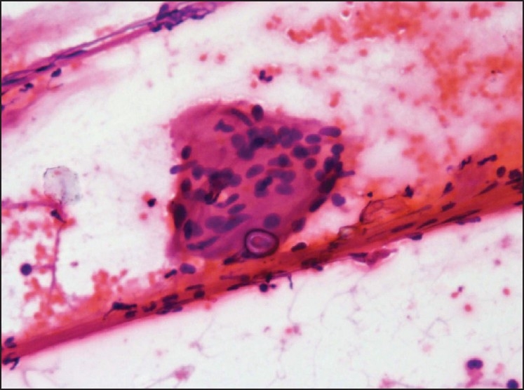
A 32-year old male presented to the chest clinic with history of cough, low grade fever and weight loss for the last 3 months. Chest radiograph revealed bilateral hilar and paratracheal lymphadenopathy with interstitial shadows. Although Montoux test was negative, the patient was given empirical anti-tuberculous therapy for 4 months. Computed tomography scan revealed mediastinal lymphadenopathy with paratracheal as well as hilar lymph node enlargement varying in size from 1-2 cm. Bronchoscopic transbronchial fine needle aspiration cytology (FNAC) was performed from right paratracheal lymph node. The smears were paucicellular with occasional epithelioid cell granulomas and two giant cells. One of the giant showed an intra-cytoplasmic body [Figure 1]. Differential diagnosis based on cytomorphology included sarcoidosis, tuberculosis or fungal infection causing granulomatous reaction. Transbronchial lung biopsy (TBLB) also revealed non-caseating epithelioid cell granulomas with giant cells, although no inclusions could be identified on TBLB. Acetyl cholinesterase (ACE) level was raised confirming the diagnosis of sarcoidosis.
Figure 1.

Microphotograph showing a giant cell with intra-cytoplasmic Schaumann body in a case of sarcoidosis (H and E, ×40)
Sarcoidosis was first identified over 100 years ago by two dermatologists working independently, Dr. Jonathan Hutchinson in England and Dr. Caesar Boeck in Norway. Sarcoidosis was originally called Hutchinson's disease or Boeck's disease. Sarcoidosis is a granulomatous disease of unclear etiology.[1] Its presenting features are protean, ranging from asymptomatic but abnormal findings on chest radiography in many patients to progressive multi-organ failure in an unfortunate minority. Because the lungs and thoracic lymph nodes are almost always involved, most patients report acute or insidious respiratory problems, variably accompanied by symptoms affecting the skin, eyes, or other organs.[2] The disease presents diagnostic challenges both clinically and histologically. Diagnosis of sarcoidosis is often a matter of exclusion. It is characterized by its pathological hallmark, the non-caseating granulomas. A variety of inclusions may be found within the cytoplasm of giant cells. Asteroid bodies are stellate inclusions with numerous rays radiating from a central core. Crystalline inclusions are colorless refractile crystals composed predominantly of calcium oxalate are frequently found in the giant cells of granulomas of sarcoidosis and other diseases. These may serve as the nidus for deposition of calcium leading to formation of Schaumann (conchoidal) bodies, which are large concentric calcifications often containing refractile calcium oxalate crystals. Although these inclusions are seen in cytology smears and are non-pathognomonic for sarcoidosis, these may help in clinching the diagnosis. There are a few case reports in the literature illustrating asteroid bodies and schaumann bodies in cases of sarcoidosis in cytological material.[3–5]
References
- 1.Lagana SM, Parwani AV, Nichols LC. Cardiac sarcoidosis: a pathology-focused review. Arch Pathol Lab Med. 2010;134:1039–46. doi: 10.5858/2009-0274-RA.1. [DOI] [PubMed] [Google Scholar]
- 2.Newman LS, Rose CS, Maier LA. Sarcoidosis. N Engl J Med. 1997;17:1224–34. doi: 10.1056/NEJM199704243361706. [DOI] [PubMed] [Google Scholar]
- 3.Pérez-Guillermo M, Sola Pérez J, Espinosa Parra FJ. Asteroid bodies and calcium oxalate crystals: two infrequent findings in fine-needle aspirates of parotid sarcoidosis. Diagn Cytopathol. 1992;8:248–52. doi: 10.1002/dc.2840080312. [DOI] [PubMed] [Google Scholar]
- 4.Jorns JM, Knoepp SM. Asteroid bodies in lymph node cytology: infrequently seen and still mysterious. Diagn Cytopathol. 2011;39:35–6. doi: 10.1002/dc.21301. [DOI] [PubMed] [Google Scholar]
- 5.Das DK, Muqim AA, Sheikh ZA, Al-Kandari M, Junaid TA. Sarcoidosis diagnosed on transbronchial fine needle aspiration smears: a case report with new information on asteroid bodies. Acta Cytol. 2010;54:225–8. doi: 10.1159/000325016. [DOI] [PubMed] [Google Scholar]


