Introduction
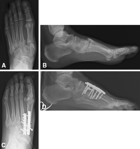
Lisfranc injuries are injuries to the tarsometatarsal joints which separate the midfoot from the forefoot. These injuries can be purely ligamentous or involve the osseous structures in the foot, in which case they are classified as fracture-dislocations. They are usually high-energy injuries and occur most often when an axial load or rotational force is brought on a plantar-flexed foot (Fig. 1). Less commonly, a direct blow to the midfoot can cause disruption of the joint complex.
Fig. 1A−D.
(A) AP and (B) lateral radiographs are shown for a patient with a Lisfranc fracture-dislocation. There is lateral and dorsal displacement of the base of the second metatarsal with respect to the middle cuneiform. Postoperative (C) AP and (D) lateral radiographs of the same patient after ORIF are shown. A combination of dorsal plating and independent screw fixation was used to restore proper anatomic alignment to the midfoot in the coronal and sagittal planes
Injury to the Lisfranc joint commonly affects males, frequently during the third decade of life, and often as a result of a fall from a height or a motor vehicle accident [2]. The majority of patients who sustain such an injury are polytraumatized, therefore, a high index of suspicion must be maintained so that subtle injuries are not missed [16].
Structure and Function
The Lisfranc joint is described in three longitudinal columns. The medial column is composed of the medial cuneiform and the base of the first metatarsal; the middle column is made up of the middle and lateral cuneiforms and the second and third metatarsals; finally, the lateral column is comprised of the cuboid and the fourth and fifth metatarsals. When viewed in the coronal plane, the transverse arch of the midfoot forms an inherently stable construct [12]. Although multiple interosseous ligaments add to this stability, the Lisfranc ligament, or ligament between the medial cuneiform and the base of the second metatarsal, is the largest and strongest.
Diagnosis and Classification
Diagnosing Lisfranc injuries starts with a history and physical examination. Specifics regarding the mechanism of injury and the amount of time elapsed between injury and presentation can be helpful in selecting between alternative treatments. Swelling throughout the foot or point tenderness in the midfoot should raise the level of suspicion for a Lisfranc injury even before images are obtained. Plantar ecchymosis can be pathognomic of these injuries [14]. Patients with tarsometatarsal injuries, which are the result of a high-energy injury or result in considerable swelling, should be monitored for signs and symptoms of compartment syndrome [3].
Anteroposterior, oblique, and lateral views of the foot should be obtained initially. Several anatomic associations should be evaluated radiographically. First, on the AP film, the medial border of the middle cuneiform should line up with the medial border of the second metatarsal. Next, on the 30° oblique view, the medial border of the lateral cuneiform should align with the medial edge of the base of the third metatarsal and the medial border of the cuboid should line up with the medial edge of the fourth metatarsal on the same oblique film. Finally, on the lateral view, no metatarsal should lie more superior or inferior than its respective tarsal bone. Disruption of any of these associations represents an injury to the Lisfranc joint [5].
The “fleck” sign represents an avulsion fracture of the Lisfranc ligament from the base of the second metatarsal. This radiographic finding is diagnostic for a Lisfranc joint injury [11]. When purely ligamentous injuries are present or the injury pattern is subtle, weightbearing or stress films can be helpful. Often, secondary to pain, such radiographs are not possible, therefore, MRI or CT should be the next imaging modality used to clarify the presence or severity of an injury.
Lisfranc injuries can be osseous, ligamentous, or a combination of the two. The original classification system by Quenu and Kuss [13] described injuries as homolateral, isolated, or divergent based on the direction of the displaced metatarsals. Hardcastle et al. further categorized Lisfranc injuries into Type A, B, or C based on displacement and incongruity with a system that they thought would dictate treatment [4]. Myerson followed with modifications to this system based on direction of dislocation [10]. Despite these multiple classification schemes, outcome and treatment do not reliably correlate with any injury type [11].
Treatment
For injuries to the Lisfranc joint for which there is no displacement with confirmed stress or weightbearing radiographs, cast immobilization for 6 to 12 weeks is recommended [16]. We believe injuries with displacement should be treated operatively to avoid posttraumatic midfoot arthritis or further displacement of the dislocation or fracture. If there is gross displacement or dislocation in the joint complex at the time of injury, closed reduction and immobilization are recommended to regain gross alignment. If instability exists or if a reduction cannot be maintained, provisional percutaneous K-wire fixation may be needed. If reduction cannot be obtained, interposed soft tissue or fracture fragments may be present [10]. Definitive management should be delayed until the soft tissue swelling has resolved.
Surgical treatment of Lisfranc injuries has varied, however, current recommendations emphasize screw fixation over smooth K-wire fixation for the medial and middle column [8]. Typically, screw fixation between the medial cuneiform and the second metatarsal, to recreate the stability of the Lisfranc ligament, is accomplished. Fixation between the first metatarsal and the medial cuneiform, and between the second metatarsal and middle cuneiform also are regularly obtained. The fourth and fifth metatarsals of the lateral column can be reduced and pinned to the cuboid effectively with K-wires [15].
External fixation can be used temporarily for reduction if the soft tissues are not amenable to surgery right away because of excessive swelling. Bioabsorbable screws and dorsal plates also have been used in the definitive fixation of Lisfranc injuries [1, 17].
Primary arthrodesis has been suggested for severely comminuted or displaced fracture dislocations, and for purely ligamentous-type injuries [16]. Long-term clinical data have yet to define the role of primary arthrodesis in these injuries.
Outcomes
The rates of posttraumatic arthritis are related to the quality of the anatomic reduction obtained and the amount of damage to the articular surface of the tarsometatarsal joints. Myerson et al. reported that quality of initial surgical reduction was the primary determinant of improved functional scores [11]. Displacement greater than 2 mm, or a nonanatomic reduction, is associated with increased rates of symptomatic and radiographic arthritis in purely ligamentous and combined ligamentous and osseous injuries [7]. The development of degenerative changes also can increase when the severity of injury is not appreciated or when the injury is partially treated. Regarding arthrodesis, in a prospective randomized trial by Ly and Coetzee of ORIF versus primary arthrodesis for purely ligamentous Lisfranc injuries, AOFAS midfoot scores were higher in the primary arthrodesis group, indicating that fusion may have a role outside that of a salvage procedure for posttraumatic arthritis [9]. However, another recent study comparing primary arthrodesis and ORIF (for all types of Lisfranc injuries) reported no differences in patient satisfaction rates or functional scores (SF-36 and SMFA) [6]. Therefore, additional studies are needed before any definitive conclusions can be made.
Pearls
Maintain a high index of suspicion for a Lisfranc injury in a patient with midfoot swelling and/or tenderness. Many Lisfranc injuries are missed initially.
Weightbearing radiographs, when possible, or MRI are essential for diagnosing subtle or purely ligamentous injuries.
The “fleck” sign, which represents an avulsion off the base of the Lisfranc ligament, the largest and strongest interosseous ligament, is radiographically evident in approximately 90% of Lisfranc injuries.
There is no role for nonoperative treatment in Lisfranc injuries which show displacement.
Initial anatomic reduction predicts outcome and decreases the rate of posttraumatic arthritis and the need for salvage arthrodesis.
Footnotes
Each author certifies that he has no commercial association (eg, consultancies, stock ownership, equity interest, patent, licensing arrangements, etc) that might pose a conflict of interest in connection with the submitted article.
References
- 1.Alberta FG, Aronow MS, Barrero M, Diaz-Doran V, Sullivan RJ, Adams DJ. Ligamentous Lisfranc joint injuries: a biomechanical comparison of dorsal plate and transarticular screw fixation. Foot Ankle Int. 2005;26:462–473. doi: 10.1177/107110070502600607. [DOI] [PubMed] [Google Scholar]
- 2.Desmond EA, Chou LB. Current concepts review: Lisfranc injuries. Foot Ankle Int. 2006;27:653–660. doi: 10.1177/107110070602700819. [DOI] [PubMed] [Google Scholar]
- 3.Fakhouri AJ, Manoli A., 2nd Acute foot compartment syndromes. J Orthop Trauma. 1992;6:223–228. doi: 10.1097/00005131-199206000-00015. [DOI] [PubMed] [Google Scholar]
- 4.Hardcastle PH, Reschauer R, Kutscha-Lissberg E, Schoffmann W. Injuries to the tarsometatarsal joint: incidence, classification and treatment. J Bone Joint Surg Br. 1982;64:349–356. doi: 10.1302/0301-620X.64B3.7096403. [DOI] [PubMed] [Google Scholar]
- 5.Heckman JD. Fractures and dislocations of the foot. In: Rockwood CA Jr, Green DP, Bucholz RW, editors. Fractures in Adults. 3. Philadelphia, PA: JB Lippincott; 1991. pp. 2041–2182. [Google Scholar]
- 6.Henning JA, Jones CB, Sietsema DL, Bohay DR, Anderson JG. Open reduction internal fixation versus primary arthrodesis for Lisfranc injuries: a prospective randomized study. Foot Ankle Int. 2009;30:913–922. doi: 10.3113/FAI.2009.0913. [DOI] [PubMed] [Google Scholar]
- 7.Kuo RS, Tejwani NC, Digiovanni CW, Holt SK, Benirschke SK, Hansen ST, Jr, Sangeorzan BJ. Outcome after open reduction and internal fixation of Lisfranc joint injuries. J Bone Joint Surg Am. 2000;82:1609–1618. doi: 10.2106/00004623-200011000-00015. [DOI] [PubMed] [Google Scholar]
- 8.Lee CA, Birkedal JP, Dickerson EA, Vieta PA, Jr, Webb LX, Teasdall RD. Stabilization of Lisfranc joint injuries: a biomechanical study. Foot Ankle Int. 2004;25:365–370. doi: 10.1177/107110070402500515. [DOI] [PubMed] [Google Scholar]
- 9.Ly TV, Coetzee JC. Treatment of primarily ligamentous Lisfranc joint injuries: primary arthrodesis compared with open reduction and internal fixation. A prospective, randomized study. J Bone Joint Surg Am. 2006;88:514–520. doi: 10.2106/JBJS.E.00228. [DOI] [PubMed] [Google Scholar]
- 10.Myerson MS. The diagnosis and treatment of injury to the tarsometatarsal joint complex. J Bone Joint Surg Br. 1999;81:756–763. doi: 10.1302/0301-620X.81B5.10369. [DOI] [PubMed] [Google Scholar]
- 11.Myerson MS, Fisher RT, Burgess AR, Kenzora JE. Fracture dislocations of the tarsometatarsal joints: end results correlated with pathology and treatment. Foot Ankle. 1986;6:225–242. doi: 10.1177/107110078600600504. [DOI] [PubMed] [Google Scholar]
- 12.Peicha G, Labovitz J, Seibert FJ, Grechenig W, Weiglein A, Preidler KW, Quehenbreger F. The anatomy of the joint as a risk factor for Lisfranc dislocation and fracture-dislocation: an anatomical and radiological case control study. J Bone Joint Surg Br. 2002;84:981–985. doi: 10.1302/0301-620X.84B7.12587. [DOI] [PubMed] [Google Scholar]
- 13.Quenu E, Kuss GE. Etude sur les luxations du metatarse (Luxations metatarso-tarsiennes). Du diastasis entre le 1er et le 2e metatarsien. Rev Chir. 1909;39:1–72. [Google Scholar]
- 14.Ross G, Cronin R, Hauzenblas J, Juliano P. Plantar ecchymosis sign: a clinical aid to diagnosis of occult Lisfranc tarsometatarsal injuries. J Orthop Trauma. 1996;10:119–122. doi: 10.1097/00005131-199602000-00008. [DOI] [PubMed] [Google Scholar]
- 15.Sangeorzan BJ, Benirschke SK, Gould MT. Tarsometatarsal Lisfranc injuries: evaluation and management. In: Wiss D, editor. Master Techniques of Orthopaedic Surgery: Fractures. 2. Philadelphia, PA: Lippincott-Williams & Wilkins; 2006. pp. 605–617. [Google Scholar]
- 16.Thompson MC, Mormino MA. Injury to the tarsometatarsal joint complex. J Am Acad Orthop Surg. 2003;11:260–267. doi: 10.5435/00124635-200307000-00005. [DOI] [PubMed] [Google Scholar]
- 17.Thordarson DB, Hurvitz G. PLA screw fixation of Lisfranc injuries. Foot Ankle Int. 2002;23:1003–1007. doi: 10.1177/107110070202301106. [DOI] [PubMed] [Google Scholar]


