Abstract
Three advances necessary to bringing dolastatin 16 (1) into full-scale preclinical development as an anticancer drug have been accomplished. The X-ray crystal structure of dolastatin 16 has been solved, which allowed stereoselective syntheses of its two new amino acid units, dolamethylleuine (Dml) and dolaphenvaline (Dpv), to be completed as summarized in reaction Schemes 1 and 3, respectively. The X-ray crystal structures of synthetic Z-Dml and TFA-Dpv have also been completed.
Very early in the discovery of the biologically remarkable and structurally unique peptides from the sea hare Dolabella auricularia, which we designated dolastatins, it became clear that certain members (e.g. 10–15) exhibited a variety of important properties that include anticancer2 and antifungal activities.3 Indeed, dolastatin 10 and three structural modifications are currently in human cancer phase II and phase III clinical trials.2a Two derivatives of dolastatin 15 are also in cancer clinical trials (phase I–II).2a
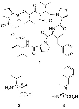
When we extended our field collections of D. auricularia from the Indian Ocean to the Western Pacific (Papua New Guinea and the Philippines), we were able to expand the dolastatin series to 16–19.4 Dolastatin 16 (1)4a especially proved to be an exceptionally potent inhibitor of cancer cell growth and a candidate for further development. However, the latter important initiative has been delayed by the need for unequivocal configurational assignments and a practical total synthesis of dolastatin 16. We are pleased to report herein the X-ray crystal structure of dolastatin 16 (1) and syntheses of the new amino acid units dolamethylleuine (2) and dolaphenvaline (3).
Other options for obtaining certain dolastatin members appeared likely some 35 years ago when we considered2d that Dolabella species derived nutrition by consuming marine microalgae and that such exogenous sources might be providing the dolastatins or intermediates. This expectation has been amply realized over the past decade by the isolation of dolastatins 10–164a,5 or close analogues from the cyanobacterium Lyngbya majuscula and other such microalgae. Thus, fermentation methods using marine cyanobacteria may eventually be competitive with total syntheses for scale-up production of new anticancer drugs in the family. At present, the yields from these initial experiments remain very low, and for the foreseeable future the provision of dolastatin 16 for cancer clinical trial development will require a practical total synthesis for scale-up production. However, the microalgae investigations continue to be very productive and promising for the future.
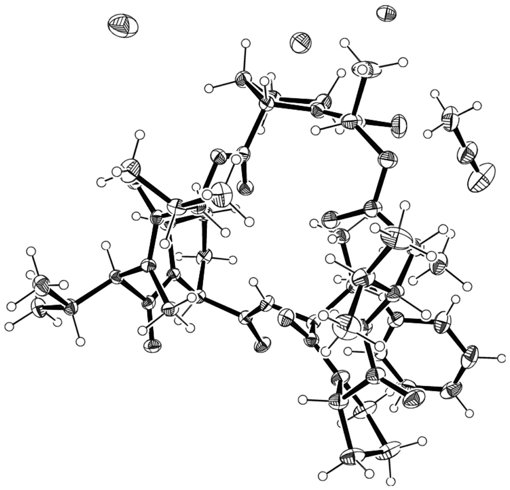
The three most obvious challenges to finding a useful synthesis of dolastatin 16, namely, an X-ray crystal structure to confirm the configuration and convenient stereoselective syntheses of the new amino acid units 2 and 3, have been met as follows. Dolastatin 16 was originally isolated (in 3.1 × 10⁻⁷% yield) as an amorphous powder, and a long period of attempts at crystallization were unsuccessful. Eventually, we found that very slow (over three years) crystal formation from acetonitrile and water provided X-ray quality crystals. Structurally, dolastatin 16 is a cyclodepsipeptide containing two new amino acids, dolamethylleuine (Dml, 2), a β-amino acid, and dolaphenvaline (Dpv, 3). As reported previously,4a the structure of 1 without assignment of the configuration of the novel amino acids was achieved by high-field NMR and tandem MS/MS mass spectroscopic interpretations. X-ray crystallographic analysis of 1 has now confirmed its cyclodepsipeptide structure and permitted the configurational assignments of the novel amino acids as 2R,3R for 2 and 2S,3R for 3 (Figure 1).
Figure 1.
X-ray structure of dolastatin 16 (1). The atoms of this cyclic depsipeptide and solvent (one acetonitrile and three water) molecules are displayed as 30% probability thermal ellipsoids
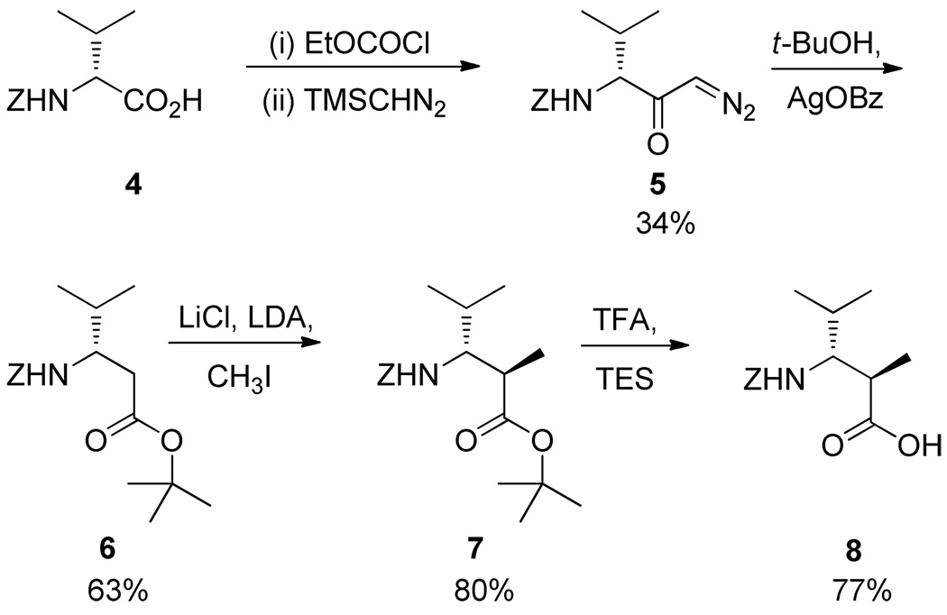
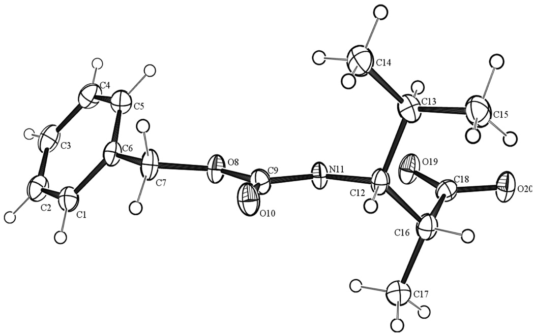
Synthesis of the β-amino acid dolamethylleuine 2 as its Z-protected synthon was carried out in four steps as outlined in Scheme 1 (13% overall yield). With Z-R-valine (4) as substrate, the Arndt-Eistert reaction6 followed by a Wolff rearrangement7 of the resulting diazoketone 5 afforded the protected β-amino acid 6. Methylation at the α-position was accomplished stereoselectively with LDA and iodomethane to afford 7.6,8 Deprotection of the tert-butyl ester by use of trifluoroacetic acid (TFA) and triethylsilane (TES)9 in DCM provided Z-Dml (8). This crystalline acid was subjected to X-ray crystallography, which confirmed the desired configuration (Figure 2).
Scheme 1.
Figure 2.
X-ray structure of N-Z-dolamethylleuine (8). Atoms are displayed as 30% probability thermal ellipsoids.
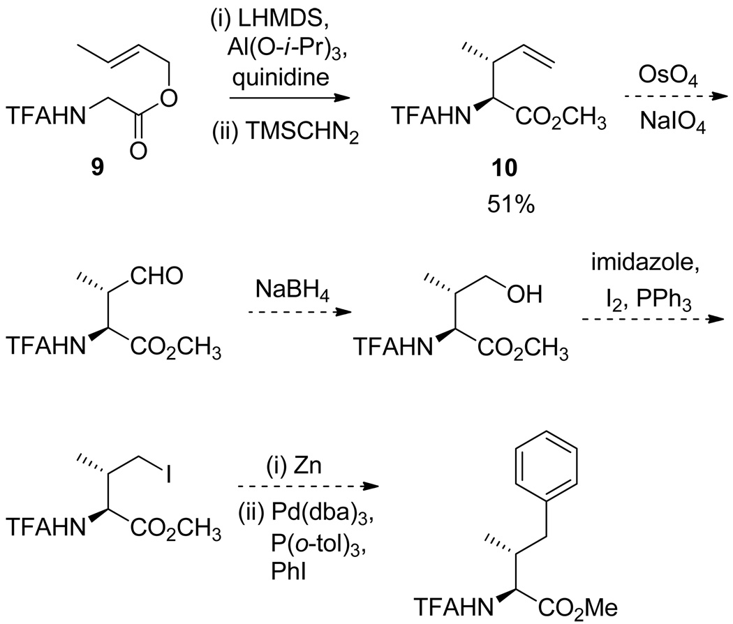
Dolaphenvaline (3) was later reported by Scheuer10 as a constituent of kulokekahilide-1, a cyclodepsipeptide from the cephalaspidean mollusk Philinopsis speciosa. As part of the structure elucidation of kulokekahilide-1, all four diastereoisomers of dolaphenvaline were prepared via a non-stereospecific approach. Since we required a stereocontrolled synthesis, an attractive approach to inducing the required chirality appeared to be the Claisen rearrangement of allylic esters of protected amino acids in the presence of chiral ligands.11,12 The reported rearrangement of allyl ester 9 followed by methylation led to the γ,δ-unsaturated amino acid methyl ester 10 in high yield, with excellent stereoselectivity and reproducibility.12 We repeated that sequence, and 10 seemed to be a viable starting point for the synthesis of N-trifluoroacetyldolaphenvaline, as outlined in Scheme 2.
Scheme 2.
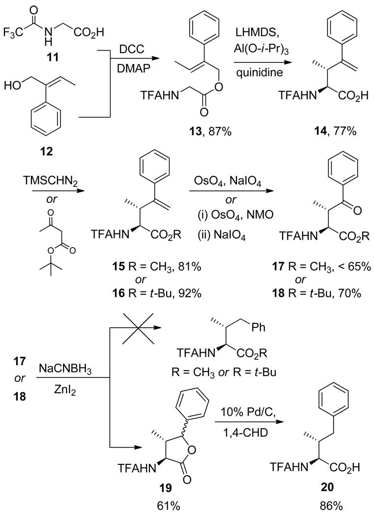
However, while this approach appeared feasible on a pilot scale, it had deficiencies in terms of lack of convergence and potential scale-up problems. Therefore, we explored the more convergent approach outlined in Scheme 3, beginning with the DCC/DMAP-mediated condensation of N-trifluoroacetylglycine (11) with 1213 to provide allylic ester 13 in 87% yield. Claisen rearrangement of 13 with LHMDS in the presence of Al(O-i-Pr)3 and quinidine afforded the γ,δ-unsaturated amino acid 14. After methylation with TMSCHN2, methyl ester 15 was subjected to oxidative cleavage of the double bond by reaction with OsO4 followed by NaIO4 to afford ketone 17 in reasonable yield. However, this material was contaminated by a difficultly separable byproduct tentatively identified as the intermediate diol on the basis of its NMR spectrum. Despite extensive experimentation, it was not feasible either to drive this reaction to completion or to obtain a pure sample of 17. Attempts to selectively remove the ketone group of 17 via the NaCNBH3/ZnI214 procedure did not lead to the desired reduced product but rather a mixture of lactones (19) that presumably arise from cyclization of an intermediate alcohol.
Scheme 3.
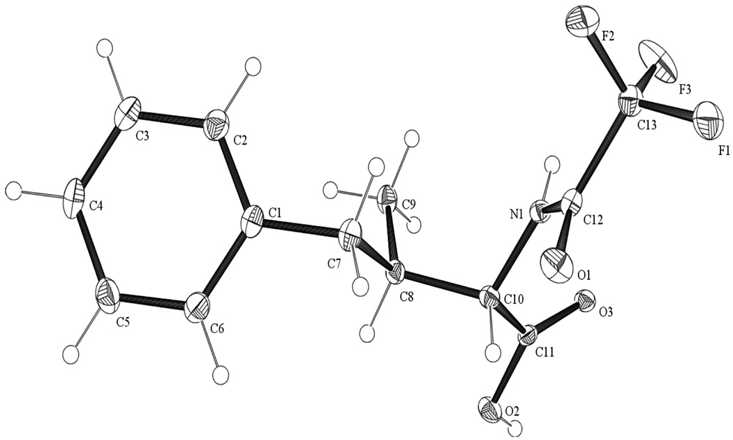
With the intent of avoiding lactone formation, the use of the tert-butyl ester for carboxyl protection was explored. Reaction of 14 with tert-butyl acetoacetate and a catalytic amount of H2SO4 in a sealed vessel15 gave tert-butyl ester 16. An attempt to carry out the oxidative cleavage reaction via the procedure used for 15 failed with 16 as the substrate. However, a two-step procedure using N-methylmorpholine N-oxide (NMO) as cooxidant16 was successful and yielded 18 cleanly in 70% yield. An unexpected bonus of the tert-butyl ester approach is that both 16 and 18 proved to be nicely crystalline solids, whereas methyl esters 15 and 17 were obtained as oils. Interestingly, the NMO-mediated oxidative cleavage approach successful with 16 failed with the methyl ester analogue 15. With the tert-butyl ester ketone 18 in hand, the NaCNBH3/ZnI214 deoxygenation was attempted. Again, as with methyl ester 17, the lactone mixture 19 was the main product. However, since the epimeric lactones (19) were an intermediate reduction product, the reductive process was completed via transfer hydrogenolysis of 19 with 1,4-cyclohexadiene and Pd/C to afford protected dolaphenvaline 20 (22.6% overall yield via 16). This crystalline acid was subjected to X-ray crystallography, which confirmed the desired configuration (Figure 3).
Figure 3.
X-ray structure of N-trifluoroacetyldolaphenvaline (20). Atoms are displayed as 30% probability thermal ellipsoids.
The unequivocally established configuration as well as the preceding stereoselective syntheses of protected Dml and Dpv have allowed our total synthetic approaches to scale-up preparation of dolastatin 16 (1) to proceed nicely, and this will be reported when complete.
Experimental Section
General Experimental Procedures
All starting reagents were used as purchased unless otherwise stated. Reactions were monitored by TLC on Analtech silica gel GHLF uniplates visualized under long- and short-wave UV irradiation and stained with H2SO4/heat, phosphomolybdic acid/heat, or KMnO4/heat. Solvent extracts were dried over anhydrous sodium sulfate. Where appropriate, the crude products were separated by flash chromatography on silica gel (230–400 mesh ASTM) from E. Merck.
Melting points are uncorrected and were determined employing an Electrothermal Mel-Temp apparatus. The 1H and 13C NMR spectra were recorded employing Varian Gemini 300, Varian Unity 400, or Varian Unity 500 instruments in CDCl3 unless otherwise indicated. HRMS data were recorded with a JEOL LCmate or JEOL GCmate mass spectrometer. Elemental analyses were determined by Galbraith Laboratories, Inc., Knoxville, TN. X-ray structure analyses were performed on a Bruker AXS Smart 600 diffractometer. The X-ray data have been submitted as supplemental information.17 Descriptions of the X-ray techniques utilized in our laboratory have been previously described.18
1-Diazo-2-oxo-(3R)-3-benzyloxycarbonylamino-4-methylpentane (5)
A solution of Z-R-valine (4, 1.01 g, 3.98 mmol) and TEA (0.57 mL, 415 mg, 4.11 mmol) in THF (20 mL) under N2 was cooled to −15 °C. Ethyl chloroformate (0.39 mL, 446 mg, 4.11mmol) in THF (4 mL) was added, and the solution stirred at −15 °C for 30 min. The solution was filtered and the precipitate washed with THF (10 mL). The combined filtrate and washings were diluted with acetonitrile (20 mL) and cooled to 0 °C under N2. Trimethylsilyldiazomethane (4 mL of a 2 M solution in hexane, 8 mmol) was added, and the solution stirred at ambient temperature for 18 h. The reaction mixture was diluted with ether (80 mL), washed successively with 10% citric acid (50 mL), saturated NaHCO3 (50 mL), and 5 M NaCl (20 mL), dried, evaporated, and coevaporated with toluene (15 mL). The residue was separated by chromatography on silica gel (30 g, 7:3 hexane–EtOAc) to afford 0.37 g (34%) of 5 as a pale yellow solid: mp 68–69 °C; Rf 0.21 (4:1 hexane–EtOAc); [α]23D +25 (c 1.10, CHCl3); 1H NMR δ 7.33 (5H, m), 5.39 (2H, br s), 5.11 (2H, s), 4.13 (1H, m), 2.09 (1H, heptet), 0.99 (3H, d), 0.89 (3H, d); anal. C 61.31, H 6.56, N 14.93%, calcd for C14H17N3O3, C 61.08, H 6.22, N 15.26%.
tert-Butyl (3S)-3-Z-amino-4-methylpentanoate (6)
Diazo-derivative 5 (0.602 g, 2.19 mmol) was dissolved in t-BuOH (9 mL) under N2 at 70 °C. Silver benzoate (80.2 mg, 0.35 mmol) in TEA (0.94 mL, 685 mg, 6.70 mmol) was added dropwise, and the mixture stirred at 70 °C in the dark for 4 h. The mixture was allowed to cool and was filtered through Celite, and the solvent was evaporated. The residue was partitioned between EtOAc (100 mL) and saturated NaHCO3 (20 mL). The organic phase was separated, washed with saturated NaHCO3 (20 mL), H2O (20 mL), and 5 M NaCl (20 mL), and dried, and the solvent was evaporated. The residue was chromatographed (silica gel, 23 g; 9:1 hexane–acetone) to provide 0.443 g (63%) of 6 as a colorless oil: Rf 0.46 (5:1 hexane–acetone); [α]23D +22 (c 1.20, CHCl3); 1H NMR δ 7.34 (5H, m), 5.12 (1H, d), 5.09 (2H, s), 3.81 (1H, qt), 2.45 (1H, dd, J = 5, 15 Hz), 2.37 (1H, dd, J = 7, 15 Hz), 1.81 (1H, m), 1.42 (9H, s), 0.93 (3H, d, J = 3 Hz), 0.91 (3H, d, J = 3 Hz); 13C NMR δ 170.5, 155.5, 136.2, 127.9, 127.5, 80.4, 66.0, 53.3, 37.9, 31.5, 27.5, 18.7, 18.0; HRMS m/z 322.2041 [M + H]+ (calcd for C18H28NO4, 322.2018); anal. C 67.05, H 8.44, N 4.40%, calcd for C18H27NO4, C 67.26, H 8.47, N 4.36%.
tert-Butyl (2R,3R)-3-N-Z-amino-2,4-dimethylpentanoate (7)
To a stirred mixture of dipyridyl indicator, LiCl (0.77 g, 18 mmol), and diisopropylamine (2.0 mL, 14 mmol), in THF (30 mL) at −78 °C under N2 was added BuLi (1.6 M soln in hexane, 8.75 mL, 14 mmol) dropwise until the mixture turned a wine-red color. The mixture was stirred at −78 °C for 15 min, and 6 (1.90 g, 5.9 mmol) in THF (15 mL) was added. The reaction mixture was stirred for 1 h, followed by the addition of iodomethane (1.9 mL, 31 mmol). Stirring was continued for 21 h at ambient temperature. The reaction was terminated by the addition of saturated NH4Cl (30 mL), and the mixture was extracted with EtOAc (150 mL). The extract was washed with 10% Na2S2O3 (30 mL), and the washing was back-extracted with EtOAc (100 mL). The organic solutions were combined, dried and evaporated. The residue was further separated by chromatography on silica gel (60 g, 9:1 hexane–acetone) to yield 1.60 g (80%) of 7 as a colorless oil: Rf 0.44 (9:1 hexane–acetone); [α]23D +22 (c 0.70, CHCl3); 1H NMR δ 7.33 (5H, m), 5.62 (1H, d, J = 7.2 Hz), 5.10 (2H, s), 3.44 (1H, m), 2.66 (1H, m), 1.71 (1H, m), 1.42 (9H, s), 1.18 (3H, d, J = 7.2 Hz), 0.96 (3H, d, J = 6.6 Hz), 0.92 (3H, d, J = 6.6 Hz); 13C NMR δ 174.6, 156.4, 136.4, 127.9, 80.3, 65.9, 59.0, 40.7, 31.4, 27.5, 19.3, 18.7, 15.3; HRMS m/z 336.2155 [M + H]+ (calcd for C19H30NO4, 336.2175); anal. C 68.16, H 8.91, N 4.47%, calcd for C19H29NO4, C 68.03, H 8.71, N 4.18%.
(2R,3R)-3-Z-amino-2,4-dimethylpentanoic Acid (8)
To a solution of 7 (1.6 g, 4.8 mmol) in DCM (9.9 mL) under N2 was added a mixture of trifluoroacetic acid (4.6 mL, 62 mmol) and triethylsilane (1.9 mL, 12 mmol). Stirring was continued for 4 h at ambient temperature. Solvents were removed and the residue coevaporated with toluene (2 × 30 mL). The residue was dissolved in EtOAc (100 mL) and extracted with 6% NaHCO3 (4 × 40 mL). The aqueous extracts were combined, acidified (pH 2) with 6 N HCl, and extracted with EtOAc (3 × 40 mL). The organic extracts were combined, washed with 5 M NaCl (20 mL), dried, and evaporated to provide a colorless solid that crystallized from 2-propanol–water to provide Z-Dml (8, 1.0 g, 77%) as colorless crystals: mp 135 °C; Rf 0.59 (50:50:1 hexane–acetone–HOAc); [α]25D +35 (c 0.86, CHCl3); 1H NMR δ 7.34 (5H, m), 5.61 (1H, d, J = 10.5 Hz), 5.11 (2H, s), 3.46 (1H, m), 2.83 (1H, m), 1.77 (1H, m), 1.25 (3H, d, J = 7.2 Hz), 0.96 (3H, d, J = 6.6 Hz), 0.93 (3H, d, J = 6.6 Hz); 13C NMR δ 179.4, 156.6, 136.2, 127.9, 127.5, 127.4, 66.1, 58.8, 39.8, 19.4, 18.8, 15.3; HRMS m/z 280.1558 [M + H]+ (calcd for C15H22NO4, 280.1549); anal. C 64.69, H 7.73, N 4.96%, calcd for C15H21NO4, C 64.50, H 7.58, N 5.01%.
(E)-2-Phenylbut-2-enyl 2-(2,2,2-Trifluoroacetamido)acetate (13)
To a suspension of N-trifluoroacetylglycine (11, 3.68 g, 21.50 mmol) and (E)-2-phenyl-2-buten-1-ol (12, 2.68 g, 19.32 mmol) in DCM (60 mL) at −40 °C under N2 was added via cannula a solution of dicyclohexylcarbodiimide (4.43 g, 21.50 mmol) and 4-dimethylaminopyridine (0.269g, 2.15 mmol) in DCM (60 mL). The solution was stirred at ambient temperature for 18 h and filtered, and the precipitate was washed with DCM (2 × 40 mL). The combined filtrate and washing was washed with 10% citric acid (2 × 25 mL), H2O (10 mL), 6% NaHCO3 (2 × 25 mL), and 5 M NaCl (20 mL), and dried, and the solvent was evaporated. The residue was chromatographed (silica gel,150 g; 4:1 hexane–EtOAc) to afford 5.06 g (87%) of 13 as a pale yellow oil that solidified on standing: mp 49–50 °C; Rf 0.66 (4:1 hexane–EtOAc); 1H NMR δ 7.37 (2H, t, J = 7.5 Hz), 7.30 (1H, t, J = 7.2 Hz), 7.18 (2H, d, J = 7.6 Hz), 6.75 (1H, br s), 5.95 (1H, q, J = 6.9 Hz), 4.91 (2H, s), 4.06 (2H, d, J = 4.9 Hz), 1.66 (3H, d, J = 6.9 Hz); 13C NMR δ 167.9, 156.7 (m), 137.3, 135.4, 135.4, 128.5, 128.4, 127.4, 116.9, 70.8, 41.4, 14.6; MS APCI+ m/z 302.1026 [M + H]+ (calcd for C14H15F3NO3, 302.1004); anal. C 55.73, H 4.93, N 4.67%, calcd for C14H14F3NO3, C 55.82, H 4.68, N 4.65%.
3-Methyl-4-phenyl-2-(2,2,2-trifluoroacetamido)-(2S,3R)-pent-4-enoic Acid (14)
To a solution of hexamethyldisilazane (6.19 g, 8.0 mL, 38.5 mmol) in THF (20 mL at −20 °C under N2) was added BuLi (1.6 M solution in hexane, 20 mL, 32 mmol). The solution was stirred at −20 °C for 20 min and added via cannula to a suspension of 13 (2.00 g, 6.64 mmol), quinidine (4.30 g, 13.27 mmol), and aluminum isopropoxide (2.04 g, 10.0 mmol) in THF (70 mL) at −78 °C. The solution was allowed to come to ambient temperature, and stirring was continued for 18 h. The mixture was next diluted with EtOAc (250 mL) and washed with 1 N HCl (3 × 75 mL). The combined washings were extracted with EtOAc (50 mL). The organic solutions were combined and extracted with 6% NaHCO3 (7 × 50 mL). The aqueous extracts were combined, cooled in an ice bath, acidified (pH 1) with 6 N HCl, and extracted with EtOAc (4 × 50 mL). The organic extracts were combined, washed with 5 M NaCl (20 mL), dried, and evaporated to give 1.54 g (77%) of 14 as a pale yellow semi-solid: Rf 0.76 (95:5:1 DCM–CH3OH–HOAc); 1H NMR δ 9.10 (1H, br), 7.33 (5H, m), 6.42 (1H, d, J = 8.2 Hz), 5.37 (1H, m), 5.15 (1H, s), 4.68 (1H, dd, J = 8.6 and 3.4 Hz), 3.57 (1H, m), 1.31 (3H, d, J = 7.2 Hz); 13C NMR δ 174.8, 156.6 (m), 148.6, 140.8, 128.6, 128.2, 126.7, 115.1, 55.0, 39.5, 14.0; MS APCI+ m/z 302.1010 [M + H]+, (calcd for C14H15F3NO3, 302.1004).
Methyl 3-Methyl-4-phenyl-2-(2,2,2-trifluoroacetamido)-(2S,3R)-pent-4-enoate (15)
Carboxylic acid 14 (544.4mg, 1.81 mmol) was placed in 1:1 CH3OH–toluene (6 mL) under N2, and trimethylsilyldiazomethane (2 M solution in hexane, 4.0 mL, 8.0 mmol) was added. The solution was stirred at ambient temperature for 16 h. The solvent was evaporated, and the residue was chromatographed (silica gel, 17 g; 9:1 hexane–EtOAc) to yield 0.46 g (81%) of 15 as a colorless oil: Rf 0.54 (4:1 hexane–EtOAc); 1H NMR δ 7.33 (5H, m), 6.48 (1H, br d), 5.34 (1H, s), 5.11 (1H, d, J = 0.8 Hz), 4.64 (1H, dd, J = 8.7 and 4.0 Hz), 3.76 (3H, s), 3.48 (1H, m), 1.26 (3H, d, J = 7.1 Hz).
tert-Butyl 3-Methyl-4-phenyl-2-(2,2,2-trifluoroacetamido)-(2S,3R)-pent-4-enoate (16)
To carboxylic acid 14 (1.54 g, 5.12 mmol) in a 50 mL round-bottom flask was added tert-butyl acetoacetate (5.8 mL, 5.53 g, 34.97 mmol) and H2SO4 (43.1 mg, 0.44 mmol). The flask was tightly stoppered, and the solution was stirred at ambient temperature under N2 for 20 h. The mixture was cooled (ice) before dilution with EtOAc (100 mL). The organic solution was washed with 6% NaHCO3 (4 × 20 mL) and 5 M NaCl (10 mL), and dried, and the solvent was evaporated. The NaHCO3 washings were combined, acidified (pH 1) with 6 N HCl, and extracted with EtOAc (3 × 15 mL). The extracts were combined, washed with 5 M NaCl (10 mL), and dried, and the solvent was evaporated to afford 0.65 g (42%) of 14. The neutral residue was chromatographed (silica gel, 30 g, 95:5 hexane–EtOAc) and led to 1.06 g (58%, 100% based on recovered starting material) of 16 as a colorless solid: mp 108 °C; Rf 0.50 (95:5 hexane–EtOAc); [α]23D 25.2 (c 1.04, CH3OH); 1H NMR δ 7.34 (5H, m), 6.48 (1H, br d, J = 6.8 Hz), 5.33 (1H, s), 5.10 (1H, s), 4.52 (1H, dd, J = 8.4 and 3.5 Hz), 3.47 (1H, m), 1.49 (9H, s), 1.27 (3H, d, J = 7.0 Hz); 13C NMR δ 168.8, 156.5 (m), 149.0, 141.3, 128.5, 127.9, 126.8, 114.7, 83.3, 55.6, 39.9, 28.0, 14.3; MS APCI+ m/z 358.1681 (0.6) [M + H]+ (calcd for C18H23F3NO3, 358.1630), 302.0990 (100) [M + H - C4H8]+ (calcd for C14H15F3NO3, 302.1004); anal. C 60.14, H 6.41, N 3.96%, calcd for C18H22F3NO3, C 60.50, H 6.21, N 3.92%.
tert-Butyl 3-Methyl-4-oxo-4-phenyl-2-(2,2,2-trifluoroacetamido)-(2S,3R)-butanoate (18)
To olefin 16 (903.4 mg, 2.53 mmol) in THF (25 mL) under N2 was added NMO (60% wt solution in H2O, 0.90 mL, 5.06 mmol) and OsO4 (4% wt solution in H2O, 1.50 mL, 0.25 mmol). The solution was stirred at ambient temperature for 16 h, and NaIO4 (2.16 g, 10.12 mmol) was then added, followed by H2O (2.7 mL). Stirring was continued for 4 h, and the reaction mixture was then diluted with EtOAc (200 mL) and washed with 10% Na2S2O3 (4 × 50 mL). The combined washings were extracted with EtOAc (50 mL). The organic solutions were combined and washed with 5 M NaCl (20 mL) and dried, and the solvent was evaporated. The residue was separated by chromatography (silica gel, 30 g; 9:1 hexane–EtOAc) and led to 0.632 g (70%) of 18 as a colorless solid: mp 127–128 °C; Rf 0.19 (95:5 hexane–EtOAc); [α]23D −39.0 (c 1.05, CH3OH); 1H NMR δ 7.92 (2H, dd, J = 7.5 and 1.3 Hz), 7.60 (1H, tt, J = 7.5 and 1.3 Hz), 7.49 (2H, t, J = 7.6 Hz), 7.03 (1H, br d, J = 6.7 Hz), 4.72 (1H, dd, J = 7.4 and 4.8 Hz), 4.13 (1H, m), 1.49 (9H, s), 1.36 (3H, d, J = 7.2 Hz); 13C NMR δ 200.0, 168.3, 156.9 (q), 135.5, 133.6, 128.8, 128.4, 83.8, 55.0, 42.9, 27.8, 14.2; MS APCI+ m/z 360.1417 [M + H]+ (calcd for C17H21F3NO4, 360.1423); anal. C 56.39%, H 5.60%, N 3.96%, calcd for C17H20F3NO4, C 56.82%, H 5.61%, N 3.90%.
3-N-(2′,2′,2′-Trifluoroacetamido)-4-methyl-2-oxo-5-phenyl-(3S,4S)-tetrahydrofuran (19)
To ketone 18 (331.2 mg, 0.92 mmol) in 1,2-dichloroethane (5.0 mL) under N2 were added ZnI2 (440.2 mg, 1.38 mmol) and NaCNBH3 (434.7 mg, 6.90 mmol). The mixture was stirred at 75 °C for 16 h, quenched with 9:1 saturated NH4Cl–6 N HCl (20 mL), and extracted with EtOAc (3 × 15 mL). The extracts were combined, washed successively with 6% NaHCO3 (2 × 15 mL) and 5 M NaCl (10 mL), and dried. After evaporation of solvent, the residue was chromatographed (silica gel, 10 g; 4:1 hexane–EtOAc) to afford 0.123 g (47%) of 19 as a white, waxy solid: Rf 0.32 (4:1 hexane–EtOAc); 1H NMR δ 7.37 (5H, m), 7.14 (1H, br d), 5.61 (0.33H, d, J = 8.4 Hz), 4.99 (0.67H, d, J = 10.2 Hz), 4.68 (1H, m), 2.99 (0.33 H, m), 2.59 (0.67H, m), 1.22 (0.67H, d, J = 6.6 Hz), 0.87 (0.33H, d, J = 6.9 Hz); HRMS (APCI+) m/z 288.0851 [M + H]+ (calcd for C13H13F3NO3, 288.0848).
(2S,3R)-2-(2,2,2-Trifluoroacetamido)-3-methyl-4-phenyl-2-butanoic Acid (20)
To lactone 19 (74.7 mg, 0.26 mmol) in CH3OH (2.0 mL) under N2 (cooled to 0 °C) was added 10% Pd/C (75 mg), followed by 1,4-cyclohexadiene (0.21 g, 2.60 mmol), and the mixture was stirred at ambient temperature under N2 for 4 h. The reaction mixture was diluted with CH3OH (10 mL), and the solution was filtered through Celite. The filter cake was washed with CH3OH (10 mL). The solvent was evaporated from the combined filtrate and washings, and the residue was dissolved in EtOAc (20 ml) and extracted with 6% NaHCO3 (3 × 15 mL). The organic phase was washed with 5 M NaCl (10 mL) and dried, and removal of solvent gave 48.5 mg of recovered lactone 19. The NaHCO3 extracts were combined, acidified (pH 1) with 6 N HCl, and extracted with EtOAc (3 × 15 mL). The extracts were combined, washed with 5 M NaCl (10 mL), and dried, and the solvent was evaporated to afford 22.7 mg (86% based on recovered starting material) of 20 as a white solid: mp 122 °C; Rf 0.38 (97.5:2.5:0.5 DCM–CH3OH–HOAc); [α]D25 +24.2 (c 1.49, CH3OH); 1H NMR δ 9.78 (1H, br), 7.31 (2H, t, J = 7.3 Hz), 7.23 (1H, t, J = 7.3 Hz), 7.17 (2H, d, J = 7.1 Hz), 6.64 (1H, d, J = 8.4 Hz), 4.75 (1H, dd, J = 8.8 and 3.2 Hz), 2.76 (1H, dd, J = 12.9 and 6.0 Hz), 2.55 (1H, m), 2.49 (1H, dd, J = 13.1 and 8.0 Hz), 0.99 (3H, d, J = 6.6 Hz); 13C NMR δ 175.23, 157.17 (m), 138.58, 128.96, 128.64, 126.71, 115.60 (m), 55.73, 39.64, 37.85, 14.84; MS APCI− m/z 288.0835 [M − H]− (calcd for C13H13F3NO3, 288.0845); anal. C 53.15, H 4.69, N 4.70%, calcd for C13H14F3NO3•0.2 H2O, C 53.32, H 4.96, N 4.78%.
Supplementary Material
Acknowledgment
We are pleased to acknowledge the very necessary financial support provided by grants RO1 CA 90441-02-05, 2R56-CA 090441-06A1, and 5RO1 CA 090441-07 from the Division of Cancer Treatment and Diagnosis, NCI, DHHS; The Arizona Disease Control Research Commission; Dr. Alec D. Keith; J. W. Kieckhefer Foundation; Margaret T. Morris Foundation; the Robert B. Dalton Endowment Fund; and Dr. William Crisp and Mrs. Anita Crisp. For other very helpful assistance, we thank Dr. Fiona Hogan.
Footnotes
In memory of Academician Georgy B. Elyakov (1929–2005), a pioneering expert in the chemistry of marine organism constituents who is deeply missed.
Supporting Information Available: X-ray crystal structure data for dolastatin 16 (1), Z-Dml (8), and trifluoroacetyl-Dpv (20). This material is available without charge via the Internet at http://pubs.acs.org.
References and Notes
- 1.For contribution 589 consult Prokopiou EM, Cooper PA, Pettit GR, Bibby MC, Shnyder SD. Molecular Medicine Reports. 2010;3:309–313. doi: 10.3892/mmr_00000256.
- 2.(a) Pettit GR. In: International Oncology Updates: Marine anticancer compounds in the era of targeted therapies. Chabner B, Cortés-Funes H, editors. Barcelona: Permanyer Publications; 2009. [Google Scholar]; (b) Kingston DGI. J. Nat. Prod. 2009;72:507–515. doi: 10.1021/np800568j. [DOI] [PMC free article] [PubMed] [Google Scholar]; (c) Singh R, Sharma M, Joshi P, Rawat DS. Anti-Cancer Agents Med. Chem. 2008;8:603–617. [PubMed] [Google Scholar]; (d) Pettit GR. In: Progress in the Chemistry of Organic Natural Products. Herz W, Kirby GW, Moore RE, Steglich W, Tamm C, editors. Vol. 70. Vienna: Springer; 1997. pp. 1–79. [Google Scholar]
- 3.(a) Woyke T, Roberson RW, Pettit GR, Winkelmann G, Pettit RK. Antimicrob. Agents Chemother. 2002;46:3802–3808. doi: 10.1128/AAC.46.12.3802-3808.2002. [DOI] [PMC free article] [PubMed] [Google Scholar]; (b) Woyke T, Berens ME, Hoelzinger DB, Pettit GR, Winkelmann G, Pettit RK. Antimicrob. Agents Chemother. 2004;48:561–567. doi: 10.1128/AAC.48.2.561-567.2004. [DOI] [PMC free article] [PubMed] [Google Scholar]
- 4.(a) Pettit GR, Xu J-p, Hogan F, Williams MD, Doubek DL, Schmidt JM, Cerny RL, Boyd MR. J. Nat. Prod. 1997;60:752–754. doi: 10.1021/np9700230. [DOI] [PubMed] [Google Scholar]; (b) Paterson I, Findlay AD. Aust. J. Chem. 2009;62:624–638. [Google Scholar]; (c) Paterson I, Findlay AD. Pure Appl. Chem. 2008;80:1773–1782. [Google Scholar]
- 5.(a) Luesch H, Moore RE, Paul VJ, Mooberry SL, Corbett TH. J. Nat. Prod. 2001;64:907–910. doi: 10.1021/np010049y. [DOI] [PubMed] [Google Scholar]; (b) Harrigan GG, Yoshida WY, Moore RE, Nagle DG, Park PU, Biggs J, Paul VJ, Mooberry SL, Corbett TH, Valeriote FA. J. Nat. Prod. 1998;61:1221–1225. doi: 10.1021/np9801211. [DOI] [PubMed] [Google Scholar]; (c) Nogle LM, Williamson RT, Gerwick WH. J. Nat. Prod. 2001;64:716–719. doi: 10.1021/np000634j. [DOI] [PubMed] [Google Scholar]; (d) Gunasekera SP, Miller MW, Kwan JC, Luesch H, Paul VJ. J. Nat. Prod. 2010;73:459–462. doi: 10.1021/np900603f. [DOI] [PubMed] [Google Scholar]; (e) Adams B, Pörzgen P, Pittman E, Yoshida WY, Westenburg HE, Horgen FD. J. Nat. Prod. 2008;71:750–754. doi: 10.1021/np070346o. [DOI] [PubMed] [Google Scholar]; (f) Taori K, Liu Y, Paul VJ, Luesch H. ChemBioChem. 2009;10:1634–1639. doi: 10.1002/cbic.200900192. [DOI] [PubMed] [Google Scholar]; (g) Nogle LM, Gerwick WH. J. Nat. Prod. 2002;65:21–24. doi: 10.1021/np010348n. [DOI] [PubMed] [Google Scholar]
- 6.Podlech J, Seebach D. Liebigs Ann. 1995:1217–1228. [Google Scholar]
- 7.Hirai Y, Yokota K, Momose T. Heterocycles. 1994;39:603–612. [Google Scholar]
- 8.Seebach D, Estermann H. Tetrahedron Lett. 1987;28:3103–3106. [Google Scholar]
- 9.Mehta A, Jaouhari R, Benson TJ, Douglas KT. Tetrahedron Lett. 1992;33:5441–5444. [Google Scholar]
- 10.Kimura J, Takada Y, Inayoshi T, Nakao Y, Goetz G, Yoshida WY, Scheuer PJ. J. Org. Chem. 2002;67:1760–1767. doi: 10.1021/jo010176z. [DOI] [PubMed] [Google Scholar]
- 11.Mues H, Kazmaier U. Synthesis. 2001:487–498. [Google Scholar]
- 12.Kazmaier U, Mues H, Krebs A. Chem. —Eur. J. 2002;8:1850–1855. doi: 10.1002/1521-3765(20020415)8:8<1850::AID-CHEM1850>3.0.CO;2-Q. [DOI] [PubMed] [Google Scholar]
- 13.Duboudin JG, Jousseaume B, Saux A. J. Organomet. Chem. 1979;168:1–11. [Google Scholar]
- 14.Lau CK, Dufresne C, Belanger PC, Piétré S, Scheigetz J. J. Org. Chem. 1986;51:3038–3043. [Google Scholar]
- 15.Taber DF, Gerstenhaber DA, Zhao X. Tetrahedron Lett. 2006;47:3065–3066. [Google Scholar]
- 16.Keck GE, Giles RL, Cee VJ, Wager CA, Yu T, Kraft MB. J. Org. Chem. 2008;73:9675–9691. doi: 10.1021/jo802215v. [DOI] [PMC free article] [PubMed] [Google Scholar]
- 17.CCDC 801992 (1), 801990 (8), and 801991 (20) contain the supplementary crystallographic data for this paper. These data can be obtained free of charge from The Cambridge Crystallographic Data Centre via www.ccdc.cam.ac.uk/data_request/cif.
- 18.Pettit GR, Knight JC, Herald DL, Pettit RK, Hogan F, Mukku VJRV, Hamblin JS, Dodson MJ, II, Chapuis J-C. J. Nat. Prod. 2009;72:366–371. doi: 10.1021/np800603u. [DOI] [PMC free article] [PubMed] [Google Scholar]
Associated Data
This section collects any data citations, data availability statements, or supplementary materials included in this article.


