Abstract
Desulfurococcus mucosus Zillig and Stetter 1983 is the type species of the genus Desulfurococcus, which belongs to the crenarchaeal family Desulfurococcaceae. The species is of interest because of its position in the tree of life, its ability for sulfur respiration, and several biotechnologically relevant thermostable and thermoactive extracellular enzymes. This is the third completed genome sequence of a member of the genus Desulfurococcus and already the 8th sequence from a member the family Desulfurococcaceae. The 1,314,639 bp long genome with its 1,371 protein-coding and 50 RNA genes is a part of the Genomic Encyclopedia of Bacteria and Archaea project.
Keywords: hyperthermophile, anaerobic, organotroph, sulfur respiration, spheroid-shaped, non-motile, extracellular enzymes, Desulfurococcaceae, GEBA
Introduction
Strain O7/1T (= DSM 2162 = ATCC 35584 = JCM 9187) is the type strain of the species Desulfurococcus mucosus [1], which is the type species of its genus Desulfurococcus. The genus currently consists of five species with a validly published name [2]. For the genus name the Neo-Latin 'desulfo-' meaning 'desulfuricating, is used to characterize the dissimilatory sulfate-reducing feature of this spheroid-shaped 'coccus' [2]. The species epithet is derived from the Latin word 'mucosus' (slimy) [2]. Strain O7/1T was isolated from an acidic hot spring in Askja, Iceland and the name of the species was effectively published by Zillig et al. in 1982 [1]; valid publication of the name followed in 1983 [3]. The strain was an early target for phylogenetic studies of the domain Archaea (at that time termed ‘Archaebacteria’) via DNA-rRNA cross-hybridizations [4,5], as well as studies on the archaeal DNA-dependent RNA polymerase structure [6] and Archaea-specific quinones [7]. Subsequently, strain O7/1T was used for studies on thermostable extracellular enzymes such as proteinase [8] and pullulanase [9]. Here we present a summary classification and a set of features for D. mucosus strain O7/1T, together with a description of the complete genome sequencing and annotation.
Classification and features
The single genomic 16S rRNA sequence of strain O7/1T was compared using NCBI BLAST under default settings (e.g., considering only the high-scoring segment pairs (HSPs) from the best 250 hits) with the most recent release of the Greengenes database [10] and the relative frequencies, weighted by BLAST scores, of taxa and keywords (reduced to their stem [11]) were determined. The five most frequent genera were Sulfolobus (27.8%), Aeropyrum (11.3%), Desulfurococcus (11.3%), Ignicoccus (6.5%) and Vulcanisaeta (6.2%) (100 hits in total). Regarding the five hits to sequences from other members of the genus, the average identity within HSPs was 96.7%, whereas the average coverage by HSPs was 97.4%. Among all other species, the one yielding the highest score was Desulfurococcus mobilis, which corresponded to an identity of 100.0% and an HSP coverage of 100.0%. The highest-scoring environmental sequence was AB462558 ('Microbial production and energy source hyperthermophilic prokaryotes geothermal hot spring pool clone DDP-A01'), which showed an identity of 95.8% and a HSP coverage of 98.2%. The five most frequent keywords within the labels of environmental samples which yielded hits were 'spring' (9.2%), 'microbi' (6.8%), 'hot' (6.2%), 'nation/park/yellowston' (5.4%) and 'popul' (4.8%) (150 hits in total), indicating a good fit to the original habitat of D. mucosus. Environmental samples which yielded hits of a higher score than the highest scoring species were not found.
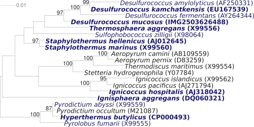
Figure 1 shows the phylogenetic neighborhood of D. mucosus in a 16S rRNA based tree. A 16S rRNA reference sequence for D. mucosus has not been previously published.
Figure 1.
Phylogenetic tree highlighting the position of D. mucosus relative to the other type strains within the family Desulfurococcaceae. The tree was inferred from 1,334 aligned characters [12,13] of the 16S rRNA gene sequence under the maximum likelihood criterion [14] and rooted in accordance with the current taxonomy. The branches are scaled in terms of the expected number of substitutions per site. Numbers above branches are support values from 1,000 bootstrap replicates [15] if larger than 60%. Lineages with type strain genome sequencing projects registered in GOLD [16] are shown in blue, Staphylothermus hellenicus CP002051 and published genomes in bold [17-22].
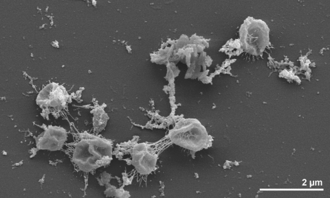
The non-motile cells of strain O7/1T are spheroid with diameters of 0.3 to 2.0 µm [1] (Figure 2), sometimes up to 10 µm [23], surrounded by a slimy mucoid layer, which covers the envelope and consists of neutral sugars and a small fraction of amino sugars [24] (Figure 2). In growing cultures, cells of strain O7/1T were often found in pairs [2] (Table 1). Cells of strain O7/1T can be differentiated from those of D. mobilis, the closest relative of D. mucosus, which are mobile by monopolar polytrichous flagella and devoid of the mucous polymer surrounding the D. mucosus cells [1,23]. Strain O7/1T can utilize yeast extract and casein or its tryptic digests, but not casamino acids as the sole carbon source, by sulfur respiration with the production of H2S and CO2, or by fermentation [1]. Growing cultures synthesize a strong smelling uncharacterized product [1]. Cultures require little or no NaCl in growth media [1,23]. The temperature range for growth of strain O7/1T is 76 to 93ºC, with an optimum at 85ºC [1,23]. At the optimal growth temperature, the generation time of strain O7/1T was about four hours [1]. The pH range is 4.5 to 7.0, with an optimum at 6.0 [1,23]. Sugars, starch, glycogen, alcohols and intermediary metabolites are also not utilized [1]. Strain O7/1T lacks an intron in the 23S RNA gene, which has been described for its close relative D. mobilis [35].
Figure 2.
Scanning electron micrograph of D. mucosus strain O7/1T
Table 1. Classification and general features of D. mucosus 07/1T according to the MIGS recommendations [25].
| MIGS ID | Property | Term | Evidence code |
|---|---|---|---|
| Current classification | Domain Archaea | TAS [26] | |
| Phylum Crenarchaeota | TAS [27,28] | ||
| Class Thermoprotei | TAS [27,29] | ||
| Order Desulfurococcales | TAS [27,30] | ||
| Family Desulfurococcaceae | TAS [2,3,31] | ||
| Genus Desulfurococcus | TAS [1,3,32] | ||
| Species Desulfurococcus mucosus | TAS [1,3] | ||
| Type strain O7/1 | TAS [1] | ||
| Gram stain | negative | TAS [1] | |
| Cell shape | spheroid, often in pairs | TAS [1] | |
| Motility | non-motile | TAS [1] | |
| Sporulation | none | NAS | |
| Temperature range | 76°C-93°C | TAS [23] | |
| Optimum temperature | 85°C | TAS [1,23] | |
| Salinity | around 0 | TAS [23] | |
| MIGS-22 | Oxygen requirement | strictly anaerobic | TAS [1] |
| Carbon source | yeast extract, casein or its tryptic digest | TAS [1] | |
| Energy metabolism | organotroph | TAS [1] | |
| MIGS-6 | Habitat | fresh water, sulfur spring | TAS [1] |
| MIGS-15 | Biotic relationship | free living | TAS [1] |
| MIGS-14 | Pathogenicity | none | NAS |
| Biosafety level | 1 | TAS [33] | |
| Isolation | acidic hot spring | TAS [1] | |
| MIGS-4 | Geographic location | Askja, Iceland | TAS [1] |
| MIGS-5 | Sample collection time | 1981 or before | TAS [1] |
| MIGS-4.1 | Latitude | 65.05 | NAS |
| MIGS-4.2 | Longitude | -16.8 | NAS |
| MIGS-4.3 | Depth | not reported | NAS |
| MIGS-4.4 | Altitude | approx. 1,053 m | NAS |
Evidence codes - IDA: Inferred from Direct Assay (first time in publication); TAS: Traceable Author Statement (i.e., a direct report exists in the literature); NAS: Non-traceable Author Statement (i.e., not directly observed for the living, isolated sample, but based on a generally accepted property for the species, or anecdotal evidence). These evidence codes are from of the Gene Ontology project [34]. If the evidence code is IDA, then the property was directly observed by one of the authors or an expert mentioned in the acknowledgements.
Chemotaxonomy
According to Zillig et al. 1982 [1], the cell envelope of the strain O7/1T is flexible and probably composed of two layers of which at least the outer one appears to consist of subunits perpendicular to the surface [1]. Scarce information is available regarding the lipid composition of D. mucosus. The lipids in the strain O7/1T are composed of phytanol and C40 polyisoprenoid dialcohols [1]. The polar lipid profile of the closely related D. mobilis has been studied and the structure of its three complex lipids has been elucidated in detail [36].
Genome sequencing and annotation
Genome project history
This organism was selected for sequencing on the basis of its phylogenetic position [37], and is part of the Genomic Encyclopedia of Bacteria and Archaea project [38]. The genome project is deposited in the Genomes On Line Database [16] and the complete genome sequence is deposited in GenBank. Sequencing, finishing and annotation were performed by the DOE Joint Genome Institute (JGI). A summary of the project information is shown in Table 2.
Table 2. Genome sequencing project information.
| MIGS ID | Property | Term |
|---|---|---|
| MIGS-31 | Finishing quality | Finished |
| MIGS-28 | Libraries used | Three genomic libraries: one 454 pyrosequence standard library, one 454 PE library (13 kb insert size), one Illumina library |
| MIGS-29 | Sequencing platforms | Illumina GAii, 454 GS FLX Titanium |
| MIGS-31.2 | Sequencing coverage | 75.7 × Illumina; 44.8 × pyrosequence |
| MIGS-30 | Assemblers | Newbler version 2.5-internal-10Apr08-1-threads, Velvet, phrap |
| MIGS-32 | Gene calling method | Prodigal 1.4, GenePRIMP |
| INSDC ID | CP002363 | |
| Genbank Date of Release | January 20, 2011 | |
| GOLD ID | Gc02914 | |
| NCBI project ID | 48641 | |
| Database: IMG-GEBA | 2503538025 | |
| MIGS-13 | Source material identifier | DSM 2162 |
| Project relevance | Tree of Life, GEBA |
Growth conditions and DNA isolation
D. mucosus strain 07/1T, DSM 2162, was grown anaerobically in DSMZ medium 184 (Desulfurococcus medium) [39] at 85°C. DNA was isolated from 0.5-1 g of cell paste using Qiagen Genomic 500 DNA kit (Qiagen 10262) following the standard protocol as recommended by the manufacturer, with no modification. DNA is available through the DNA Bank Network [40].
Genome sequencing and assembly
The genome was sequenced using a combination of Illumina and 454 sequencing platforms. All general aspects of library construction and sequencing can be found at the JGI website [41]. Pyrosequencing reads were assembled using the Newbler assembler version 2.5-internal-10Apr08-1-threads (Roche). The initial Newbler assembly consisting of three contigs in one scaffold was converted into a phrap assembly [42] by making fake reads from the consensus, to collect the read pairs in the 454 paired end library. Illumina GAii sequencing data (99.5 Mb) were assembled with Velvet [43] and the consensus sequences were shredded into 1.5 kb overlapped fake reads and assembled together with the 454 data. The 454 draft assembly was based on 546.5 Mb 454 draft data and all of the 454 paired end data. Newbler parameters are -consed -a 50 -l 350 -g -m -ml 20. The Phred/Phrap/Consed software package [42] was used for sequence assembly and quality assessment in the subsequent finishing process. After the shotgun stage, reads were assembled with parallel phrap (High Performance Software, LLC). Possible mis-assemblies were corrected with gapResolution [41], Dupfinisher [44], or sequencing cloned bridging PCR fragments with subcloning or transposon bombing (Epicentre Biotechnologies, Madison, WI). Gaps between contigs were closed by editing in Consed, by PCR and by Bubble PCR primer walks (J.-F.Chang, unpublished). A total of 12 additional reactions were necessary to close gaps and to raise the quality of the finished sequence. Illumina reads were also used to correct potential base errors and increase consensus quality using a software Polisher developed at JGI [45]. The error rate of the completed genome sequence is less than 1 in 100,000. Together, the combination of the Illumina and 454 sequencing platforms provided 120.5 × coverage of the genome. The final assembly contained 264,988 pyrosequence and 1,310,055 Illumina reads.
Genome annotation
Genes were identified using Prodigal [46] as part of the Oak Ridge National Laboratory genome annotation pipeline, followed by a round of manual curation using the JGI GenePRIMP pipeline [47]. The predicted CDSs were translated and used to search the National Center for Biotechnology Information (NCBI) nonredundant database, UniProt, TIGR-Fam, Pfam, PRIAM, KEGG, COG, and InterPro databases. Additional gene prediction analysis and functional annotation were performed within the Integrated Microbial Genomes - Expert Review (IMG-ER) platform [48].
Genome properties
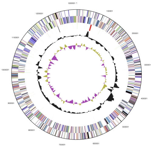
The genome consists of a 1,314,639 bp long chromosome with a G+C content of 53.1% (Table 3 and Figure 3). Of the 1,421 genes predicted, 1,371 were protein-coding genes, and 50 RNAs; 26 pseudogenes were also identified. The majority of the protein-coding genes (65.5%) were assigned with a putative function while the remaining ones were annotated as hypothetical proteins. The distribution of genes into COGs functional categories is presented in Table 4.
Table 3. Genome Statistics.
| Attribute | Value | % of Total |
|---|---|---|
| Genome size (bp) | 1,314,639 | 100.00% |
| DNA coding region (bp) | 1,186,810 | 90.28% |
| DNA G+C content (bp) | 698,621 | 53.14% |
| Number of replicons | 1 | |
| Extrachromosomal elements | 0 | |
| Total genes | 1,421 | 100.00% |
| RNA genes | 50 | 3.52% |
| rRNA operons | 1 | |
| Protein-coding genes | 1,371 | 96.48% |
| Pseudo genes | 26 | 1.83% |
| Genes with function prediction | 931 | 65.52% |
| Genes in paralog clusters | 103 | 7.25% |
| Genes assigned to COGs | 1,001 | 70.44% |
| Genes assigned Pfam domains | 1,010 | 71.08% |
| Genes with signal peptides | 146 | 10.27% |
| Genes with transmembrane helices | 296 | 20.83% |
| CRISPR repeats | 3 |
Figure 3.
Graphical circular map of genome. From outside to the center: Genes on forward strand (color by COG categories), Genes on reverse strand (color by COG categories), RNA genes (tRNAs green, rRNAs red, other RNAs black), GC content, GC skew.
Table 4. Number of genes associated with the general COG functional categories.
| Code | value | %age | Description |
|---|---|---|---|
| J | 148 | 13.9 | Translation, ribosomal structure and biogenesis |
| A | 2 | 0.2 | RNA processing and modification |
| K | 50 | 4.7 | Transcription |
| L | 62 | 5.8 | Replication, recombination and repair |
| B | 1 | 0.1 | Chromatin structure and dynamics |
| D | 7 | 0.7 | Cell cycle control, cell division, chromosome partitioning |
| Y | 0 | 0.0 | Nuclear structure |
| V | 10 | 0.9 | Defense mechanisms |
| T | 14 | 1.3 | Signal transduction mechanisms |
| M | 37 | 3.5 | Cell wall/membrane/envelope biogenesis |
| N | 4 | 0.4 | Cell motility |
| Z | 0 | 0.0 | Cytoskeleton |
| W | 0 | 0.0 | Extracellular structures |
| U | 10 | 0.9 | Intracellular trafficking, secretion, and vesicular transport |
| O | 45 | 4.2 | Posttranslational modification, protein turnover, chaperones |
| C | 97 | 9.1 | Energy production and conversion |
| G | 52 | 4.9 | Carbohydrate transport and metabolism |
| E | 77 | 7.2 | Amino acid transport and metabolism |
| F | 39 | 3.7 | Nucleotide transport and metabolism |
| H | 45 | 4.2 | Coenzyme transport and metabolism |
| I | 14 | 1.3 | Lipid transport and metabolism |
| P | 81 | 7.6 | Inorganic ion transport and metabolism |
| Q | 3 | 0.3 | Secondary metabolites biosynthesis, transport and catabolism |
| R | 170 | 16.0 | General function prediction only |
| S | 96 | 9.0 | Function unknown |
| - | 420 | 29.6 | Not in COGs |
Acknowledgements
We would like to gratefully acknowledge the help of Olivier D. Ngatchou-Djao (HZI) in preparing the manuscript. This work was performed under the auspices of the US Department of Energy Office of Science, Biological and Environmental Research Program, and by the University of California, Lawrence Berkeley National Laboratory under contract No. DE-AC02-05CH11231, Lawrence Livermore National Laboratory under Contract No. DE-AC52-07NA27344, and Los Alamos National Laboratory under contract No. DE-AC02-06NA25396, UT-Battelle and Oak Ridge National Laboratory under contract DE-AC05-00OR22725, as well as German Research Foundation (DFG) INST 599/1-2.
References
- 1.Zillig W, Stetter KO, Prangishvilli D, Schäfer W, Wunderl S, Janekovic D, Holz I, Palm P. Desulfurococcaceae, the second family of the extremely thermophilic, anaerobic, sulfur-respiring Thermoproteales. Zentralbl Bakteriol 1982; 3:304-317 [Google Scholar]
- 2.Garrity G. NamesforLife. BrowserTool takes expertise out of the database and puts it right in the browser. Microbiol Today 2010; 37:9 [Google Scholar]
- 3.List Editor Validation List no. 10. Validation of the publication of new names and new combinations previously effectively published outside the IJSB. Int J Syst Bacteriol 1983; 33:438-440 10.1099/00207713-33-2-438 [DOI] [Google Scholar]
- 4.Tu JK, Prangishvilli D, Huber H, Wildgruber G, Zillig W, Stetter KO. Taxonomic relations between Archaebacteria including 6 novel genera examined by cross hybridization of DNAs and 16S rRNAs. Mol Evol 1982; 18:109-114 10.1007/BF01810829 [DOI] [PubMed] [Google Scholar]
- 5.Klenk HP, Haas B, Schwass V, Zillig W. Hybridization homology: a new parameter for the analysis of phylogenetic relations, demonstrated with the urkingdom of the Archaebacteria. J Mol Evol 1986; 24:167-173 10.1007/BF02099964 [DOI] [Google Scholar]
- 6.Prangishvilli D, Zillig W, Gierl A, Biesert L, Holz I. DNA-dependent RNA polymerase of thermoacidophilic archaebacteria. Eur J Biochem 1982; 122:471-477 10.1111/j.1432-1033.1982.tb06461.x [DOI] [PubMed] [Google Scholar]
- 7.Thurl S, Witke W, Buhrow I, Schäfer W. Quinones from archaebacteria, II. Different types of quinones from sulphur-dependent archaebacteria. Biol Chem Hoppe Seyler 1986; 367:191-197 10.1515/bchm3.1986.367.1.191 [DOI] [PubMed] [Google Scholar]
- 8.Cowan DA, Smolenski KA, Daniel RM, Morgan HW. An extracellular proteinase from a strain of the archaebacterium Desulfurococcus growing at 88 degrees C. Biochem J 1987; 247:121-133 [DOI] [PMC free article] [PubMed] [Google Scholar]
- 9.Duffner F, Bertoldo C, Andersen JT, Wagner K, Antranikian G. A new thermoactive pullulanase from Desulfurococcus mucosus: cloning, sequencing, purification, and characterization of the recombinant enzyme ofter expression in Bacillus subtilis. J Bacteriol 2000; 182:6331-6338 10.1128/JB.182.22.6331-6338.2000 [DOI] [PMC free article] [PubMed] [Google Scholar]
- 10.DeSantis TZ, Hugenholtz P, Larsen N, Rojas M, Brodie E, Keller K, Huber T, Dalevi D, Hu P, Andersen G. Greengenes, a chimera-checked 16S rRNA gene database and workbench compatible with ARB. Appl Environ Microbiol 2006; 72:5069-5072 10.1128/AEM.03006-05 [DOI] [PMC free article] [PubMed] [Google Scholar]
- 11.Porter MF. An algorithm for suffix stripping. Program: electronic library and information systems 1980; 14:130-137 10.1108/eb046814 [DOI] [Google Scholar]
- 12.Castresana J. Selection of conserved blocks from multiple alignments for their use in phylogenetic analysis. Mol Biol Evol 2000; 17:540-552 [DOI] [PubMed] [Google Scholar]
- 13.Lee C, Grasso C, Sharlow MF. Multiple sequence alignment using partial order graphs. Bioinformatics 2002; 18:452-464 10.1093/bioinformatics/18.3.452 [DOI] [PubMed] [Google Scholar]
- 14.Stamatakis A, Hoover P, Rougemont J. A rapid bootstrap algorithm for the RAxML Web servers. Syst Biol 2008; 57:758-771 10.1080/10635150802429642 [DOI] [PubMed] [Google Scholar]
- 15.Pattengale ND, Alipour M, Bininda-Emonds ORP, Moret BME, Stamatakis A. How many bootstrap replicates are necessary? Lect Notes Comput Sci 2009; 5541:184-200 10.1007/978-3-642-02008-7_13 [DOI] [PubMed] [Google Scholar]
- 16.Liolios K, Chen IM, Mavromatis K, Tavernarakis N, Hugenholtz P, Markowitz VM, Kyrpides NC. The Genomes On Line Database (GOLD) in 2009: status of genomic and metagenomic projects and their associated metadata. Nucleic Acids Res 2009; 38:D346-D354 10.1093/nar/gkp848 [DOI] [PMC free article] [PubMed] [Google Scholar]
- 17.Ravin NV, Mardanov AV, Beletsky AV, Kublanov IV, Kolganova TV, Lebedinsky AV, Chernyh NA, Bonch-Osmolovskaya EA, Skryabin KG. Complete genome sequence of the anaerobic, protein-degrading hyperthermophilic crenarchaeon Desulfurococcus kamchatkensis. J Bacteriol 2009; 191:2371-2379 10.1128/JB.01525-08 [DOI] [PMC free article] [PubMed] [Google Scholar]
- 18.Anderson IJ, Sun H, Lapidus A, Copeland A, Glavina Del Rio T, Tice H, Dalin E, Lucas S, Barry K, Land M, et al. Complete genome sequence of Staphylothermus marinus Stetter and Fiala 1986 type strain F1. Stand Genomic Sci 2009; 1:183-188 10.4056/sigs.30527 [DOI] [PMC free article] [PubMed] [Google Scholar]
- 19.Brügger K, Chen L, Stark M, Zibat A, Redder P, Ruepp A, Awayez M, She Q, Garrett RA, Klenk HP. The genome of Hyperthermus butylicus: a sulfur-reducing, peptide fermenting, neutrophilic crenarchaeote growing up to 108 °C. Archaea 2007; 2:127-135 10.1155/2007/745987 [DOI] [PMC free article] [PubMed] [Google Scholar]
- 20.Göker M, Held B, Lapidus A, Nolan M, Spring S, Yasawong M, Lucas S, Glavina Del Rio T, Tice H, Cheng JF, et al. Complete genome sequence of Ignisphaera aggregans type strain (AQ1.S1T). Stand Genomic Sci 2010; 3:66-75 10.4056/sigs.1072907 [DOI] [PMC free article] [PubMed] [Google Scholar]
- 21.Podar M, Anderson I, Makarova KS, Elkins JG, Ivanova N, Wall MA, Lykidis A, Mavromatis K, Sun H, Hudson ME, et al. A genomic analysis of the archaeal system Ignicoccus hospitalis-Nanoarchaeum equitans. Genome Biol 2008; 9:R158 10.1186/gb-2008-9-11-r158 [DOI] [PMC free article] [PubMed] [Google Scholar]
- 22.Spring S, Rachel R, Lapidus A, Davenport K, Tice H, Copeland A, Cheng JF, Lucas S, Chen F, Nolan M, et al. Complete genome sequence of Thermosphaera aggregans type strain (M11TLT). Stand Genomic Sci 2010; 2:245-259 10.4056/sigs.821804 [DOI] [PMC free article] [PubMed] [Google Scholar]
- 23.Huber H, Stetter KO. 2006. Desulfurococcales In: M Dworkin, S Falkow, E Rosenberg, KH Schleifer E Stackebrandt (eds), The Prokaryotes, 3. ed, vol. 3. Springer, New York, p. 52-68. [Google Scholar]
- 24.Stetter KO, Zillig W. 1985. Thermoplasma and the thermophilic sulfur-dependent archaebacteria. In: Woese CR, and Wolfe RS (eds) The Bacteria. Academic Press. New York, NY. 8:100-201. [Google Scholar]
- 25.Field D, Garrity G, Gray T, Morrison N, Selengut J, Sterk P, Tatusova T, Thomson N, Allen MJ, Angiuoli SV, et al. The minimum information about a genome sequence (MIGS) specification. Nat Biotechnol 2008; 26:541-547 10.1038/nbt1360 [DOI] [PMC free article] [PubMed] [Google Scholar]
- 26.Woese CR, Kandler O, Wheelis ML. Towards a natural system of organisms: proposal for the domains Archaea, Bacteria, and Eucarya. Proc Natl Acad Sci USA 1990; 87:4576-4579 10.1073/pnas.87.12.4576 [DOI] [PMC free article] [PubMed] [Google Scholar]
- 27.Validation list 85: Validation of publication of new names and new combinations previously effectively published outside the IJSEM. Int J Syst Evol Microbiol 2002; 52:685-690 10.1099/ijs.0.02358-0 [DOI] [PubMed] [Google Scholar]
- 28.Garrity GM, Holt JG. 2001. Phylum AI. Crenarchaeota phy. nov. In: Garrity GM, Boone DR, Castenholz RW (eds), Bergey's Manual of Systematic Bacteriology, Second Edition, Volume 1, Springer, New York, p. 169-210. [Google Scholar]
- 29.Reysenbach AL. 2001. Class I. Thermoprotei class. nov. In: Garrity GM, Boone DR, Castenholz RW (eds), Bergey's Manual of Systematic Bacteriology, Second Edition, Volume 1, Springer, New York, p.169. [Google Scholar]
- 30.Huber H, Stetter O. 2001. Order II. Desulfurococcales ord. nov. In: Garrity GM, Boone DR, Castenholz RW (eds), Bergey's Manual of Systematic Bacteriology, Second Edition, Volume 1, Springer, New York, p. 179-180. [Google Scholar]
- 31.Burggraf S, Huber H, Stetter KO. Reclassification of the crenarchael orders and families in accordance with 16S rRNA sequence data. Int J Syst Bacteriol 1997; 47:657-660 10.1099/00207713-47-3-657 [DOI] [PubMed] [Google Scholar]
- 32.Perevalova AA, Svetlichny VA, Kublanov IV, Chernyh NA, Kostrikina NA, Tourova TP, Kuznetsov BB, Bonch-Osmolovskaya EA. Desulfurococcus fermentans sp. nov., a novel hyperthermophilic archaeon from a Kamchatka hot spring, and emended description of the genus Desulfurococcus. Int J Syst Evol Microbiol 2005; 55:995-999 10.1099/ijs.0.63378-0 [DOI] [PubMed] [Google Scholar]
- 33.Classification of. Bacteria and Archaea in risk groups. www.baua.de TRBA 466.
- 34.Ashburner M, Ball CA, Blake JA, Botstein D, Butler H, Cherry JM, Davis AP, Dolinski K, Dwight SS, Eppig JT, et al. Gene Ontology: tool for the unification of biology. Nat Genet 2000; 25:25-29 10.1038/75556 [DOI] [PMC free article] [PubMed] [Google Scholar]
- 35.Kjems J, Garrett RA. Novel splicing mechanism for the ribosomal RNA intron in the archaebacterium Desulfurococcus mobilis. Cell 1988; 54:693-703 10.1016/S0092-8674(88)80014-X [DOI] [PubMed] [Google Scholar]
- 36.Lanzotti V, De Rosa M, Trincone A, Basso AL, Gambacorta A, Zillig W. Complex lipids from Desulfurococcus mobilis, a sulfur-reducing archaebacterium. Biochim Biophys Acta 1987; 922:95-102 [Google Scholar]
- 37.Klenk HP, Göker M. En route to a genome-based classification of Archaea and Bacteria? Syst Appl Microbiol 2010; 33:175-182 10.1016/j.syapm.2010.03.003 [DOI] [PubMed] [Google Scholar]
- 38.Wu D, Hugenholtz P, Mavromatis K, Pukall R, Dalin E, Ivanova NN, Kunin V, Goodwin L, Wu M, Tindall BJ, et al. A phylogeny-driven genomic encyclopaedia of Bacteria and Archaea. Nature 2009; 462:1056-1060 10.1038/nature08656 [DOI] [PMC free article] [PubMed] [Google Scholar]
- 39.List of growth media used at DSMZ: http://www.dsmz.de/microorganisms/media_list.php
- 40.Gemeinholzer B, Dröge G, Zetzsche H, Haszprunar G, Klenk HP, Güntsch A, Berendsohn WG, Wägele JW. The DNA Bank Network: the start from a German initiative. Biopreservation and Biobanking 2011; 9:51-55 10.1089/bio.2010.0029 [DOI] [PubMed] [Google Scholar]
- 41.DOE Joint Genome Institute. http://www.jgi.doe.gov
- 42.Phrap and Phred for Windows, MacOS, Linux, and Unix. http://www.phrap.com
- 43.Zerbino DR, Birney E. Velvet: algorithms for de novo short read assembly using de Bruijn graphs. Genome Res 2008; 18:821-829 10.1101/gr.074492.107 [DOI] [PMC free article] [PubMed] [Google Scholar]
- 44.Han C, Chain P. 2006. Finishing repeat regions automatically with Dupfinisher. In: Proceeding of the 2006 international conference on bioinformatics & computational biology. Edited by Hamid R. Arabnia & Homayoun Valafar, CSREA Press. June 26-29, 2006: 141-146. [Google Scholar]
- 45.Lapidus A, LaButti K, Foster B, Lowry S, Trong S, Goltsman E. POLISHER: An effective tool for using ultra short reads in microbial genome assembly and finishing. AGBT, Marco Island, FL, 2008. [Google Scholar]
- 46.Hyatt D, Chen GL, LoCascio PF, Land ML, Larimer FW, Hauser LJ. Prodigal: prokaryotic gene recognition and translation initiation site identification. BMC Bioinformatics 2010; 11:119 10.1186/1471-2105-11-119 [DOI] [PMC free article] [PubMed] [Google Scholar]
- 47.Pati A, Ivanova NN, Mikhailova N, Ovchinnikova G, Hooper SD, Lykidis A, Kyrpides NC. GenePRIMP: a gene prediction improvement pipeline for prokaryotic genomes. Nat Methods 2010; 7:455-457 10.1038/nmeth.1457 [DOI] [PubMed] [Google Scholar]
- 48.Markowitz VM, Ivanova NN, Chen IMA, Chu K, Kyrpides NC. IMG ER: a system for microbial genome annotation expert review and curation. Bioinformatics 2009; 25:2271-2278 10.1093/bioinformatics/btp393 [DOI] [PubMed] [Google Scholar]


