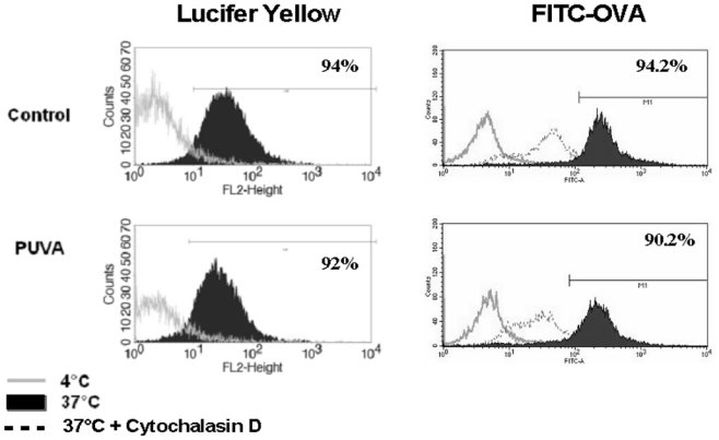
Figure 2. Endocytic capacity of monocytes is not impaired following PUVA treatment.

Endocytosis assays were performed 24h after PUVA treatment in the presence of either Lucifer Yellow (left panel) or FITC-OVA (right panel) at 37°C for 3 hours (black histogram). Negative controls were performed at 4°C (empty histogram) or at 37°C in presence of 10μg/ml of Cytochalasin D. One experiment histogram is a representative of 2 performed with 2 donors.
