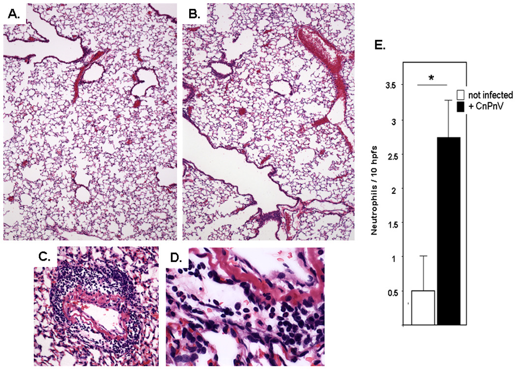
Figure 4. Lung histopathology.

A. Normal mouse lung tissue. B. Lung tissue (day 7) from mouse inoculated with CnPnV (35 TCID50 units). C. An example of one of the few focal perivascular inflammatory infiltrates with D. neutrophils as the predominant cell type. E. Neutrophils, albeit few in number, are also detected in bronchoalveolar lavage (BAL) fluid; hpf, high power (40X) field; n = 5 mice per point.
