Abstract
Objective:
Cholecystectomy is one of the most common general surgical procedures performed today. The laparoscopic approach is beneficial to patients in terms of length of stay, postoperative pain, return to work, and cosmesis. Some drawbacks are associated with the minimal access form of cholecystectomy, including an increased incidence of common bile duct injuries. In addition, when the gallbladder is inadvertently perforated during laparoscopic cholecystectomy, retrieval of dropped gallstones may be difficult. We present a case in which gallstones spilled during cholecystectomy, causing near circumferential, extraluminal common hepatic duct compression, and clinical jaundice 1 year later.
Methods:
The patient experienced jaundice and pruritus 12 months after laparoscopic cholecystectomy. A computed tomographic scan was interpreted as cholelithiasis, but otherwise was normal (despite a previous cholecystectomy). Endoscopic retrograde cholangiopancreatography was performed and a stent placed across a stenotic common hepatic duct.
Results:
The results of brush biopsies were negative. The stent rapidly occluded and surgical intervention was undertaken. At exploratory laparotomy, an abscess cavity containing multiple gallstones was encountered. This abscess had encircled the common hepatic duct, causing compression and fibrosis. The stones were extracted and a hepaticojejunostomy was tailored. The patient's bilirubin level slowly decreased and she recovered without complication.
Conclusions:
Gallstones lost within the peritoneal cavity usually have no adverse sequela. Recently, however, numerous reports have surfaced describing untoward events. This case is certainly one to be included on the list. A surgeon should make every attempt to retrieve spilled gallstones due to the potential later complications described herein.
Keywords: Jaundice, Gallstones, Laparoscopy, Cholecystectomy
INTRODUCTION
At present, laparoscopic cholecystectomy (LC) is the preferred method for removing the gallbladder in patients with cholelithiasis. It is estimated that during LC, gallstones are inadvertently spilled into the peritoneal cavity in 10% to 40% of cases.1 Although some authors have reported no adverse consequences from spilled gallstones remaining in the peritoneal cavity,2 others have described a variety of unusual complications, including intraabdominal abscess,3 empyema,4 pneumonia,5 colobiliary-cutaneous fistula,5 intrahepatic abscess,6 small bowel obstruction,7 incarcerated direct inguinal hernia,8 and urinary bladder fistula.9 We have added to the growing number of adverse events associated with “lost” gallstones. In this case, spilled gallstones resulted in subhepatic abscess formation with near circumferential compression of the common hepatic duct and resultant obstructive jaundice 1 year after LC.
CASE REPORT
A 68-year-old woman with a past medical history significant for coronary artery disease and coronary artery bypass grafting, chronic obstructive pulmonary disease, and depression was hospitalized for an episode of gallstone pancreatitis. After fluid resuscitation and a period of observation, she underwent LC prior to discharge. No mention was made of spilled or dropped gallstones in the operative report.
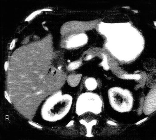
Twelve months later, she had severe pruruitis, nausea, and painless jaundice. She also reported a 30-pound weight loss and acholic stools. On examination, her abdomen was soft and all her incisions were well healed. Laboratory evaluation demonstrated a total bilirubin of 16.0 mg/dL, aspartate aminotransferase (AST) of 37 IU/L,alanine aminotransferase (ALT) of 44 IU/L, and alkaline phosphatase of 513 IU/L. Computed tomographic scan of the abdomen was interpreted as cholelithiasis and cholecystitis (despite a previous cholecystectomy), but was otherwise normal (Figure 1). Some mild intrahepatic bile duct dilation was present. On magnetic resonance cholangiopancreatography, thickening of the bile ducts was observed, which was suspicious for cholangiocarcinoma. Endoscopic retrograde cholangiopancreatography demonstrated a stenosis of the common hepatic duct and a stent was placed. Results of brush biopsies were negative for cholangiocarcinoma. The stent occluded within a few days of placement, and the patient's jaundice was not resolved. She was then transferred to our institution for surgical care.
Figure 1.
Computed tomography scan interpreted as cholelithiasis, despite a previous cholecystectomy. Double arrows point to extrabiliary gallstones.
At exploratory laparotomy, an abscess cavity containing multiple small gallstones was encountered. The abscess encircled the common hepatic duct approximately 1 cm from the bifurcation of the left and right hepatic ducts. Significant fibrosis and compression of the common hepatic duct were present. Biopsy of the duct was abnormal with marked fibrosis but no evidence of neoplasm. The stones were extracted and a hepaticojejunostomy with an end-to-side anastomosis was performed. Postoperatively, the patient's bilirubin and alkaline phosphatase levels gradually returned to normal, and she recovered without complication.
DISCUSSION
Spillage of gallstones during LC is a common occurrence. In a laboratory model, spilled gallstones that remain in the peritoneal cavity significantly increase the rate of adhesion and abscess formation.10 Reports have been published of other complications occurring due to these lost stones, including thoracic empyema,4 small bowel obstruction,7 hernias,8 and fistulas.9 We present here a case of obstructive jaundice that occurred 1 year after LC that was caused by retained dropped gallstones and resulted in a common hepatic duct stricture. Although some studies11 have suggested that spilled gallstones are not associated with postoperative sequela, we believe this report and others demonstrate that lost gallstones can be responsible for severe postoperative complications. Every effort should be made to retrieve dropped gallstones at the time of operation.
Stones are spilled when the wall of the gallbladder is inadvertently perforated during LC. This commonly occurs during separation of the gallbladder from the liver surface, with traction on the neck of an inflamed gall-bladder during Triangle of Calot dissection, or during extraction through a port incision. Prevention of the gall-bladder perforation obviously will eliminate complications associated with spilled stones.
Apart from being fully aware of the amount of tension being placed on the gallbladder during manipulation, a few steps can be taken to prevent the gallbladder from being perforated. First, during retrieval, especially of a gangrenous, thin-walled, or inflamed gallbladder, placement of the gallbladder into a retrieval bag prior to attempting extraction is advisable. Second, atraumatic graspers should be used when manipulating the gall-bladder to minimize small lacerations. Finally, 2-handed dissection by the operating surgeon will help eliminate a “tug of war” situation that leads to excessive force being placed on the gallbladder wall.
If a perforation does occur, repositioning the atraumatic grasper to re-approximate the 2 sides of the cholecystotomy can prevent the escaping stone. Although the stones will then remain within the gallbladder, some bile will likely spill. Alternatively, a clip may be applied to seal the defect, but we believe this is less often helpful. When graspers are used, it is important not to move them during the remainder of the procedure or to enlarge the perforation.
When stones are inadvertently spilled, a number of well-known techniques can be used to aid in retrieval. Use of a 30-degree laparoscope (which we routinely use in all LC cases) will increase the size and depth of the visual field. This laparoscope, when coupled with systematically changing the orientation of the operating room table, will help locate most recesses where stones may be “hiding.” The methodical use of the 10-mm stone scooper (Karl Storz, Tuttlingen, Germany) to retrieve individual stones is effective and safe.
A more difficult decision is whether a procedure should be converted to an open cholecystectomy for recovery of gallstones that cannot be located laparoscopically. We do not routinely do this. Instead, we try to minimize the risk of spillage by attempting to avoid perforation of the gall-bladder at operation and utilizing a bag device for gall-bladder removal in case of inadvertent rupture. Variables that are to be considered when making the decision to convert to an open procedure include number and size of gallstones that are unable to be located, acute inflammation of the gallbladder, age, overall health, and comorbidities of the patient. Further studies may be useful to quantify the risk of lost gallstones and to determine when the increased morbidity of converting to an open procedure is justified.
Footnotes
Presented as a poster at the 10th International Congress and Endo Expo (SLS Annual Meeting), New York, NY, USA, December 5-8, 2001.
References:
- 1. Peters JH, Gibbons GD, Innes JT, et al. Complications of laparoscopic cholecystectomy. Surgery. 1991;110:769–778 [PubMed] [Google Scholar]
- 2. Assaf Y, Matter I, Sabo E, et al. Laparoscopic cholecystectomy for acute cholecystitis and the consequences of gallbladder perforation, bile spillage, and “loss” of stones. Eur J Surg. 1998;164:425–431 [DOI] [PubMed] [Google Scholar]
- 3. Gallinaro RN, Miller FB. The lost gallstone. Complication after laparoscopic cholecystectomy. Surg Endoscopy. 1994;8:913–914 [DOI] [PubMed] [Google Scholar]
- 4. Willekes CL, Widmann WD. Empyema from lost gallstones: a thoracic complication of laparoscopic cholecystectomy. J Laparoendosc Surg. 1996;6(2):123–126 [DOI] [PubMed] [Google Scholar]
- 5. McDonald MP, Munson JL, Sanders L, Tsao J, Buyske J. Consequences of lost gallstone. Surg Endosc. 1997;11:774–777 [DOI] [PubMed] [Google Scholar]
- 6. Steerman PH, Steerman SN. Unretrieved gallstones presenting as a Streptococcus bovis liver abscess. JSLS. 2000;4(3):263–265 [PMC free article] [PubMed] [Google Scholar]
- 7. Huynh T, Mercer D. Early postoperative small bowel obstruction caused by spilled gallstones during laparoscopic cholecystectomy. Surgery. 1996;119:352–353 [DOI] [PubMed] [Google Scholar]
- 8. Bebawi M, Wassef S, Ramcharan A, Bapat K. Incarcerated indirect inguinal hernia: a complication of spilled gallstones. JSLS. 2000;4(3):267–269 [PMC free article] [PubMed] [Google Scholar]
- 9. Castro MG, Alves AS, Oliveira CA, Vieira Junior A, Vianna JL, Costa RF. Elimination of biliary stones through the urinary tract: a complication of the laparoscopic cholecystectomy. Rev Hosp Clin Fac Med Sao Paulo. 1999;54(6):209–212 [DOI] [PubMed] [Google Scholar]
- 10. Agalar F, Sayek I, Agalar C, Cakmakci M, Hayran M, Kavuklu B. Factors that may increase morbidity in a model of intraabdominal contamination caused by gallstones lost in the peritoneal cavity. Eur J Surg. 1997;163(12):909–914 [PubMed] [Google Scholar]
- 11. Memon MA, Deeik RK, Maffi TR, et al. The outcome of unretrieved gallstones in the peritoneal cavity during laparoscopic cholecystectomy: a prospective analysis. Surg Endosc. 1999;13:848–857 [DOI] [PubMed] [Google Scholar]


