Abstract
Sera from 1,448 adult ewes in 91 flocks, representing all 26 counties in the Republic of Ireland, were examined for pestivirus antibodies using a commercially available ELISA which detected IgG1 antibody to border disease virus. Eighty-one sheep (5.6%) in 42 flocks (46.0%) were antibody-positive. Within infected flocks, the mean seroprevalence level was 11.4% with a range of 6.3% to 30.0%. The highest antibody prevalence was detected in sheep from central lowland counties of Ireland. Comparative neutralisation testing of 42 ELISA-positive sera detected geometric mean antibody titres of 136 to the NADL strain of bovine viral diarrhoea virus (BVDV), 92 to the Moredun strain of border disease virus and 21 to the 137/4 strain of border disease virus. These results suggest that BVDV may be the major ruminant pestivirus infecting sheep in Ireland. Although there are high numbers of infected flocks, many sheep within such flocks remain antibody-negative and are at risk of giving birth to lambs with congenital border disease.
Keywords: Sheep, Pestivirus, Border disease, Republic of Ireland, Antibody
Introduction
The primary members of the genus Pestivirus are classical swine fever virus (CSFV), bovine viral diarrhoea virus (BVDV) types I and II, and border disease virus (BDV). CSFV is host specific under natural circumstances (i.e., it infects pigs only); BDV affects sheep, goats and pigs; and BVDV genotypes I and II affect cattle, sheep, goats, deer and pigs [11]. Consequently, current recommendations are that the virus genome, rather than species of origin, be used as the basis for a revised genus classification [32].
Border disease virus spreads naturally by horizontal and vertical transmission. Oro-nasal infection in healthy adults or neonates causes mild or inapparent disease. The consequences of infection are primarily reproductive - barren ewes, abortions, stillbirths and stunted, weak lambs with variable degrees of nervous dysfunction [19,24]. Other occasional effects include 'hairy' and malpigmented wool, skeletal abnormalities, and immunosuppression with subsequent secondary bacterial infection [12,24]. However, border disease is an uncommon clinical finding, in contrast to the pervasive consequences of BVDV infection in the cattle population in the Republic of Ireland [8]. When they do occur, outbreaks of clinical BDV can significantly reduce productivity in individual flocks [2]. Genomic, antigenic and serological studies have shown that BDV is more closely related to CSFV than to BVDV [33]. BDV infection of pigs complicates the serological diagnosis of CSFV, a Class A notifiable disease [23], and is a potential cause of antibody false-positives with serious implications for animal movement and trade. In addition, congenital infection of pigs with BDV can mimic clinical infection with low virulence strains of CSFV [21]. The objectives of the present study were to assess the serological prevalence of border disease virus in the Irish sheep population, and to explore the relationship and cross-reactions between antibodies to BDV and the other important pestivirus, BVDV. Similar studies have been performed in the UK [27], Norway [16], Sweden [17], Denmark [30] and Northern Ireland [7].
Materials and methods
Animals
Sera from 1,448 adult ewes, collected from 91 flocks, were examined by ELISA for BDV antibodies. Flocks were tested from every county in the Republic of Ireland, the number selected for each county was weighted to represent the respective proportion of the national flock farmed there - according to the 2001 National Sheep Census figures [6]. The flocks selected had a mean size of 172; the largest had 744 sheep and the smallest had 18 sheep. Table 1 shows how these sheep flocks were distributed, both by size and by county.
Table 1.
Numbers of sheep flocks tested, categorised on size and county of origin
| Flock size | 0 - 49 | 50 - 99 | 100 - 199 | 200 - 299 | 300+ |
|---|---|---|---|---|---|
| No. of flocks | 11 | 20 | 36 | 16 | 8 |
| No. of different counties | 7 | 14 | 18 | 12 | 6 |
Sixteen sera per flock were tested with the exception of one flock in Co Kildare, with eight sera only. The blood samples were collected as part of the surveillance for foot-and-mouth disease (FMD) virus antibodies over a six-week period: May 17, 2001 to June 26, 2001. At this time serum was collected, centrifuged and stored at -20°C. Eight nonadjacent flocks were identified as having high seroprevalence to pestivirus (≥20%) and underwent a further targeted round of testing, using all available sera.
Tests
Border disease ELISA
A commercially available solid-phase indirect ELISA (BDV-Ab, SVANOVA Biotech, Sweden) was used according to the manufacturer's instructions. Results were read at 450 nm. Corrected optical density (COD) values of less than 0.25 were regarded as negative.
Bovine viral diarrhoea virus serum neutralisation test
The BVDV serum neutralisation test (SNT), using the cytopathic NADL strain [9], was performed as described previously [15] on 42 BDV-ELISA-positive sera as identified above, at dilutions 1/8 to 1/1,024.
Border disease serum neutralisation tests
Serum neutralisation tests, using the Moredun strain of BDV, were performed in a sheep thymus cell line, SFT-R, while tests with the 137/4 BDV strain were performed using secondary foetal bovine kidney (FBK) cell cultures. Sera were tested over a dilution range of 1/8 to 1/1,024. After incubation for three days, the monolayers were fixed at 80°C, overlaid with porcine polyclonal pestivirus antiserum followed by a commercial HRPO-conjugated rabbit anti-pig immunoglobulin. Virus growth was indicated by the presence of reddish brown intracytoplasmic staining on microscopic examination [13].
Results
A total of 81 (5.6%, CI95 ± 1.2%) of the 1,448 sheep were positive for pestivirus antibody. Positive sheep were detected in 42 (46%, CI95 ± 10.2%) of the 91 sheep flocks tested (Table 2). The average pestivirus antibody prevalence among the positiveonly flocks was 11.4%, with a maximum of 30.0% and a minimum 6.3%. The mean COD of the positive sera was 0.59, with a minimum of 0.25 and a maximum of 1.39.
Table 2.
Numbers and percentages of flocks and individual sheep seropositive to pestivirus by ELISA per county in the Republic of Ireland
| County | Number of flocks tested | Number of sheep tested | Number positive | Percentage positive | ||
|---|---|---|---|---|---|---|
| Flocks | Sheep | Flocks | Sheep | |||
| Carlow | 3 | 48 | 3 | 4 | 100 | 8 |
| Cavan | 2 | 32 | 2 | 4 | 100 | 13 |
| Clare | 1 | 16 | 1 | 2 | 100 | 13 |
| Cork | 5 | 80 | 3 | 7 | 60 | 9 |
| Donegal | 8 | 128 | 3 | 3 | 38 | 2 |
| Dublin | 1 | 16 | 0 | 0 | 0 | 0 |
| Galway | 10 | 160 | 4 | 8 | 40 | 5 |
| Kerry | 7 | 112 | 3 | 4 | 43 | 4 |
| Kildare | 3 | 40 | 3 | 3 | 100 | 8 |
| Kilkenny | 3 | 48 | 1 | 1 | 33 | 2 |
| Laois | 3 | 48 | 0 | 0 | 0 | 0 |
| Leitrim | 2 | 32 | 0 | 0 | 0 | 0 |
| Limerick | 1 | 16 | 0 | 0 | 0 | 0 |
| Longford | 2 | 32 | 1 | 2 | 50 | 6 |
| Louth | 2 | 32 | 1 | 1 | 50 | 3 |
| Mayo | 7 | 112 | 0 | 0 | 0 | 0 |
| Meath | 3 | 48 | 3 | 11 | 100 | 23 |
| Monaghan | 2 | 32 | 1 | 6 | 50 | 19 |
| Offaly | 3 | 48 | 3 | 11 | 100 | 23 |
| Roscommon | 4 | 64 | 3 | 4 | 75 | 6 |
| Sligo | 2 | 32 | 0 | 0 | 0 | 0 |
| Tipperary | 4 | 64 | 2 | 3 | 50 | 5 |
| Waterford | 2 | 32 | 1 | 2 | 50 | 6 |
| Westmeath | 2 | 32 | 1 | 1 | 50 | 3 |
| Wexford | 4 | 64 | 1 | 1 | 25 | 2 |
| Wicklow | 5 | 80 | 2 | 3 | 40 | 4 |
| Totals | 91 | 1448 | 42 | 81 | ||
The second focused phase of testing concentrated on 139 samples taken from eight nonadjacent flocks identified as having high seroprevalence. Twenty-eight (20.1%) of these second phase samples proved positive. The mean overall intraflock antibody prevalence among this specific group of eight flocks was 23.9% (CI95 ± 5.1%) with a range of 15.7% to 30.0%; the mean positive COD among this subset of samples remained virtually unchanged at 0.61.
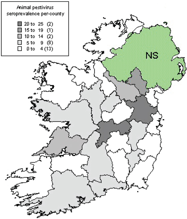
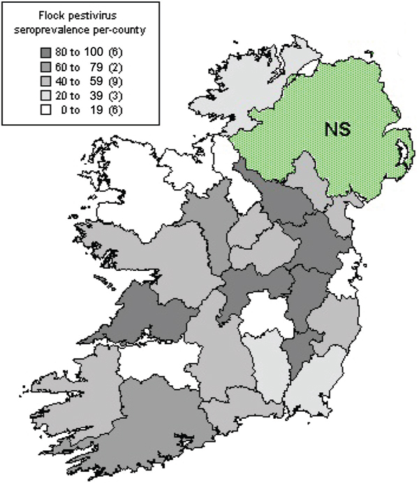
The per-county geographic pattern of seroprevalence among individual animals and among individual flocks is illustrated in Figures 1 and 2.
Figure 1.
Percentage of individual sheep that were seropositive to pestivirus in each county in the Republic of Ireland. NS: not sampled.
Figure 2.
Percentage of sheep flocks that were seropositive to pestivirus in each county in the Republic of Ireland. NS: not sampled.
The results of testing 42 pestivirus-antibody ELISA-positive sera by comparative neutralisation tests are shown in Table 3.
Table 3.
Results of comparative serum neutralisation testing of 42 ovine sera that were seropositive on screening using pestivirus-specific ELISA
| Virus strain (pestivirus designation) | No. of positive sera (≥1/8) | Reciprocal geometric mean antibody titre |
|---|---|---|
| NADL (BVDV) | 39 | 136 |
| Moredun (BDV) | 39 | 92 |
| 137/4 (BDV) | 36 | 21 |
Thirty-nine sera contained neutralising antibody to the BVDV strain, NADL and the BDV strain, Moredun, while 36 sera contained neutralising antibody to BDV strain 137/4. Three ELISA-positive sera (mean COD = 0.29) had neutralising titres less than 1/8 in all SNTs. The highest geometric mean antibody titres were obtained against the NADL strain of BVDV followed by the Moredun strain, with strain 137/4 having lowest geometric mean antibody titres. Among those that tested positive to the NADL strain, the reciprocal geometric mean titre was 136, with a minimum of 16 and a maximum of 512. For the other SNTs, Moredun reciprocal antibody titres ranged between 8 and 1,024, while strain 137/4 antibody titres were limited to between 8 and 64. Thirty-four (81%) sera had fourfold or greater (≥2 dilutions) antibody titres to BVDV than to BDV strain 137/4, while 16 (38%) sera had four-fold or greater antibody titres to BVDV than to BDV strain Moredun. Higher antibody titres to BDV strain 137/4 compared to BVDV were not detected, although three sera had four-fold or greater antibody titres to BDV strain Moredun than to BVDV.
Discussion
The level of seroprevalence for pestivirus antibodies found in the initial phase of this survey approximated well with the 5.3% found in a study of the Northern Ireland (NI) flock [7]. As all sampled sheep were of breeding age, it was assumed that maternal antibodies no longer persisted and that antibody indicated direct exposure to pestivirus. The detected seroprevalence may underestimate pestivirus activity in infected flocks. While the antibody has persisted for up to 485 days in ewes, infected when pregnant [10], it may become undetectable earlier in other sheep [28]. Out of the 91 flocks tested, 42 (46%) were found to contain sheep that were seropositive for pestivirus. This flock seroprevalence level was considerably higher than the 30.4% reported in the NI study by Graham et al. [7] and the 32.6% flock prevalence for indigenous sheep reported in an earlier NI study [1]. This disparity may be because a distinction was not made in the present survey for flocks with higher numbers of non-indigenous breeds and because of the larger per-flock sample size used in this study (sixteen).
A survey of antibodies to pestivirus in England and Wales found 10.8% of individual animals and 37.4% of flocks seropositive [27]. The most recent European work suggests an individual animal seroprevalence of 8.3% among Danish sheep [30]. In Switzerland, the influence of flock management and sheep breed was clearly illustrated [29] as 20% of pedigree flocks were infected compared to 65% of other flock types. Five of six French flocks showed positive serology [3], while an animal seroprevalence of 17.9% was reported in Spanish sheep [18]. A pestivirus seroprevalence level of 4.5% among individual animals was documented in Norwegian sheep with 18% of sheep flocks showing exposure to the virus [16]. Seroprevalences of 25% and 66% to pestivirus were reported in two Irish flocks with recent histories of clinical border disease [9].
There was considerable geographical variation in levels of flock seroprevalence. Higher numbers of infected flocks were in the east and midlands, with many apparently uninfected flocks being found in the south and west of the country. The influence of stocking density and farm management practices on regional variation in pestivirus antibody prevalence has been discussed previously [27,14,16,7]. Figures 1 and 2 illustrate these geographical differences and suggest that the highest pestivirus flock seroprevalences are in the central lowland counties of Ireland. Higher stocking densities and husbandry systems associated with more intensive sheep production may explain these higher levels of pestivirus seroconversion. Substantial regional variations in the use of particular sheep breeds and variable breed susceptibility to pestivirus may confound these management effects [29].
All flock disease problems were considered significant during the state of high alert of the foot-and-mouth disease crisis, so it may be assumed that the vast majority of seropositive sheep did not have clinical signs of oral/digital lesions, pyrexia, or depression. Similarly, abortion storms were reportable events during this period, and none was reported in the 91 flocks sampled.
In the current study, the sample collection period was early to mid-summer, some months after the normal lambing season. As pestiviruses are commonly released in foetal fluids at lambing, high seroprevalence levels would be expected when sampling at this time [19]. Beyond the lambing season, the virus seems to spread rather slowly among sheep at grass. Only four out of 22 sheep seroconverted after three months of mixing with known persistently-infected animals [2]. Trough-feeding, housing and other intensive farm management features are considered to increase the pestivirus transmission rates among sheep [19].
More targeted testing of antibody-positive flocks was undertaken to evaluate disease dynamics within such flocks. None of these seropositive flocks had levels of infection above 30%, and the mean level was less than 25%. Cattle were present on all of these higher incidence farms. Levels of seroprevalence of this order would suggest medium-level pestivirus transmission, probably due to the presence of persistently-infected animals within these flocks or transmission from cattle to sheep on these farms.
Persistently-infected animals can show quite marked temporal changes in levels of pestivirus antibodies [26]. There has been little research as to the probability of one or more animals, persistently infected with border disease virus, acting as the source of above-average infection rates in particular sheep flocks, although a comparable situation exists with BVDV in cattle [5]. Examination of the ELISA-positive sera by comparative serum neutralisation tests found that the highest antibody titres were to the NADL strain of BVDV. Similar findings were obtained in the NI sheep and it is suggested that interspecies pestiviral transmission in Ireland may be predominantly from cattle to sheep, mainly due to BVDV [7]. This is supported by the detection of four-fold or greater antibody titres to BVDV than to BDV strain 137/4, in over 80% of samples tested and, similarly, fourfold or greater antibody titres to BVDV than to BDV strain Moredun, in almost 40% of samples tested.
Cattle were present on a very high proportion (92%) of the farms tested, so this study proved unsuitable to test fully the relationship between the concurrent presence of cattle and levels of ovine pestivirus seroconversion. Interestingly, only 2.7% of samples collected from sheep farms where cattle were not present, were pestivirus antibody-positive, in contrast to 5.8% seropositive on farms where cattle were present. Transmission of BVDV from cattle to sheep has been reported before [4] but pestiviruses are not thought to spread commonly from sheep to cattle [22,25].
The high serum neutralising antibody titres to the Moredun strain of BDV detected in the present study could be due to cross reactions with BVDV but may also indicate the local occurrence of BDV strains related to subtype B [33]. Three sera had four-fold or greater antibody titres to BDV strain Moredun than to BVDV. Low antibody titres to BDV strain 137/4 in both the Republic of Ireland and Northern Ireland [7] suggest that BDV strains of subtype A may not be present in Ireland. While infection of pigs in Ireland with ruminant pestiviruses is uncommon [20,7], the strains circulating in sheep have the potential to cross-react in serological surveillance and diagnostics for CSFV in pigs in Ireland, as well as mimicking chronic or pre-natal CSFV infection clinically [31,21].
This study highlights the apparently uneven distribution of pestivirus infection among Irish sheep, with some flocks apparently naïve to the viruses involved and, as a result, highly vulnerable to outbreaks of pestivirus-related disease.
Abbreviations
BDV: Border disease virus; BVDV: Bovine viral diarrhoea virus; CI95: 95% confidence interval; COD: Corrected optical density; CSFV: Classical swine fever virus; ELISA: Enzyme-linked immunosorbent assay; FBK: Foetal bovine kidney; FMD: Foot-and-mouth disease; HRPO: Horseradish peroxidase; NI: Northern Ireland; SNT: Serum neutralisation test
Acknowledgements
The authors thank Dr R. Riebe, Federal Research Institute of Virus Diseases of Animals, Isle of Riems, Germany, for provision of the SFT-R cell line and B. Wilson, P. Dillon and V. Geraghty for providing FBK cultures and the NADL strain of BVDV.
References
- Adair BM, McFerran JB, McKillop ER, McCullough SJ. Survey for antibodies to respiratory viruses in two groups of sheep in Northern Ireland. Veterinary Record. 1984;115:403–406. doi: 10.1136/vr.115.16.403. [DOI] [PubMed] [Google Scholar]
- Bonniwell MA, Nettleton PF, Gardiner AC, Barlow RM, Gilmour JS. Border disease without nervous signs or fleece changes. Veterinary Record. 1987;120:246–249. doi: 10.1136/vr.120.11.246. [DOI] [PubMed] [Google Scholar]
- Brugere-Picoux J. Border disease in France. Veterinary Record. 1987;120:374–374. doi: 10.1136/vr.120.15.374-a. [DOI] [PubMed] [Google Scholar]
- Carlsson U. Border disease in sheep caused by transmission of virus from cattle persistently infected with bovine virus diarrhoea virus. Veterinary Record. 1991;128:145–147. doi: 10.1136/vr.128.7.145. [DOI] [PubMed] [Google Scholar]
- Carlsson U, Belak K. Border disease virus transmitted to sheep and cattle by a persistently infected ewe: epidemiology and control. Acta Veterinaria Scandinavica. 1994;35:79–88. doi: 10.1186/BF03548357. [DOI] [PMC free article] [PubMed] [Google Scholar]
- Costelloe JA, Gaynor MC, Gaynor S, McAteer WJ, O'Reilly PJ. Control of foot-and-mouth disease: lessons from the experience of Ireland. International Office of Epizootics Scientific and Technical Review. 2002;21:739–750. doi: 10.20506/rst.21.3.1369. [DOI] [PubMed] [Google Scholar]
- Graham DA, Calvert V, German A, McCullough SJ. Pestiviral infections in sheep and pigs in Northern Ireland. Veterinary Record. 2001;148:69–71. doi: 10.1136/vr.148.3.69. [DOI] [PubMed] [Google Scholar]
- Gunn M. BVD in cattle: Continuing education. Irish Veterinary Journal. 1996;49:434–435. [Google Scholar]
- Hamilton AF, Timoney PJ. Bovine virus diarrhoeamucosal disease virus and border disease. Research in Veterinary Science. 1973;15:265–267. [PubMed] [Google Scholar]
- Huck RA, Evans DH, Woods DG, King AA, Stuart P. Border disease of sheep. Comparison of the results of serological testing using complement fixation, immunodiffusion, neutralisation and immunofluorescent techniques. British Veterinary Journal. 1975;131:427–435. doi: 10.1016/s0007-1935(17)35238-7. [DOI] [PubMed] [Google Scholar]
- Hurtado A, Garcia-Perez AL, Aduriz G, Juste RA. Genetic diversity of ruminant pestiviruses from Spain. Virus Research. 2003;92:67–73. doi: 10.1016/S0168-1702(02)00315-5. [DOI] [PubMed] [Google Scholar]
- Jeffrey M, Roeder PL. Variable nature of border disease on a single farm: clinical and pathological description of affected sheep. Research in Veterinary Science. 1987;43:22–27. [PubMed] [Google Scholar]
- Jensen MH. Detection of antibodies against hog cholera virus and bovine viral diarrhea virus in porcine serum. A comparative examination using CF, PLA and NPLA assays. Acta Veterinaria Scandinavica. 1981;22:85–98. doi: 10.1186/BF03547210. [DOI] [PMC free article] [PubMed] [Google Scholar]
- Lamontagne L, Roy R. Presence of antibodies to bovine viral diarrhea-mucosal disease virus (border disease) in sheep and goat flocks in Quebec. Canadian Journal of Comparative Medicine. 1984;48:225–227. [PMC free article] [PubMed] [Google Scholar]
- Lenihan P, Collery P. Hog cholera/Classical Swine Fever and African Swine Fever. Vol. 5904. Publication ref. EUR; 1977. Bovine viral diarrhoea infection in pigs in Ireland: a serological survey and an epidemiological study; pp. 314–322. [Google Scholar]
- Loken T, Krogsrud J, Larsen IL. Pestivirus infections in Norway. Serological investigations in cattle, sheep and pigs. Acta Veterinaria Scandinavica. 1991;32:27–34. doi: 10.1186/BF03546994. [DOI] [PMC free article] [PubMed] [Google Scholar]
- Lunden A, Carlsson U, Naslund K. Toxoplasmosis and border disease in 54 Swedish sheep flocks. Seroprevalence and incidence during one gestation period. Acta Veterinaria Scandinavica. 1992;33:175–184. doi: 10.1186/BF03547324. [DOI] [PMC free article] [PubMed] [Google Scholar]
- Mainar-Jaime RC, Vazquez-Boland JA. Associations of veterinary services and farmer characteristics with the prevalences of brucellosis and border disease in small ruminants in Spain. Preventative Veterinary Medicine. 1999;40:193–205. doi: 10.1016/S0167-5877(99)00027-6. [DOI] [PubMed] [Google Scholar]
- Nettleton PF, Gilray JA, Russo P, Dlissi E. Border disease of sheep and goats. Veterinary Research. 1998;29:327–340. [PubMed] [Google Scholar]
- O'Connor M, Lenihan P, Dillon P. Pestivirus antibodies in pigs in Ireland. Veterinary Record. 1991;129:269. doi: 10.1136/vr.129.12.269. [DOI] [PubMed] [Google Scholar]
- Paton DJ, Done SH. Congenital infection of pigs with ruminant-type pestiviruses. Journal of Comparative Pathology. 1994;111:151–163. doi: 10.1016/S0021-9975(05)80047-7. [DOI] [PubMed] [Google Scholar]
- Paton DJ, Sharp G, Ibata G. Foetal cross-protection experiments between type 1 and type 2 bovine viral diarrhoea virus in pregnant ewes. Veterinary Microbiology. 1999;64:185–196. doi: 10.1016/S0378-1135(98)00269-7. [DOI] [PubMed] [Google Scholar]
- Pearson JE. Hog cholera diagnostic techniques. Comparative Immunology, Microbiology and Infectious Diseases. 1992;15:213–219. doi: 10.1016/0147-9571(92)90094-8. [DOI] [PubMed] [Google Scholar]
- Pratelli A, Bollo E, Martella V, Guarda F, Chiocco D, Buonavoglia C. Pestivirus infection in small ruminants: virological and histopathological findings. New Microbiology. 1999;22:351–356. [PubMed] [Google Scholar]
- Pratelli A, Martella V, Cirone F, Buonavoglia D, Elia G, Tempesta M, Buonavoglia C. Genomic characterization of pestiviruses isolated from lambs and kids in southern Italy. Journal of Virological Methods. 2001;94:81–85. doi: 10.1016/S0166-0934(01)00277-4. [DOI] [PubMed] [Google Scholar]
- Roeder PL, Jeffrey M, Drew TW. Variable nature of border disease on a single farm: the infection status of affected sheep. Research in Veterinary Science. 1987;43:28–33. [PubMed] [Google Scholar]
- Sands JJ, Harkness JW. The distribution of antibodies to Border disease virus among sheep in England and Wales. Research in Veterinary Science. 1978;25:241–242. [PubMed] [Google Scholar]
- Sawyer MM, Schore CE, Menzies PI, Osburn BI. Border disease in a flock of sheep: epidemiologic, laboratory, and clinical findings. Journal of American Veterinary Medical Association. 1986;189:61–65. [PubMed] [Google Scholar]
- Schaller P, Vogt HR, Strasser M, Nettleton PF, Peterhans E, Zanoni R. Seroprevalence of maedi-visna and border disease in Switzerland. Schweizer Arciv für Tierheilkunde. 2000;142:145–153. [PubMed] [Google Scholar]
- Tegtmeier C, Stryhn H, Uttentha I, Kjeldsen AM, Nielsen TK. Seroprevalence of border disease in Danish sheep and goat herds. Acta Veterinaria Scandinavica. 2000;41:339–344. doi: 10.1186/BF03549624. [DOI] [PMC free article] [PubMed] [Google Scholar]
- Terpstra C, Wensvoort G. Natural infections of pigs with bovine viral diarrhoea virus associated with signs resembling swine fever. Research in Veterinary Science. 1988;45:137–142. [PubMed] [Google Scholar]
- Vilcek S, Drew TW, McGoldrick A, Paton DJ. Genetic typing of bovine pestiviruses from England and Wales. Veterinary Microbiology. 1999;69:227–237. doi: 10.1016/S0378-1135(99)00111-X. [DOI] [PubMed] [Google Scholar]
- Vilcek S, Nettleton PF, Paton DJ, Belak S. Molecular characterization of ovine pestiviruses. Journal of General Virology. 1997;78:725–735. doi: 10.1099/0022-1317-78-4-725. [DOI] [PubMed] [Google Scholar]


