Abstract
APPL1 is an adaptor protein that binds to both AKT and adiponectin receptors and is hypothesised to mediate the effects of adiponectin in activating downstream effectors such as AMP-activated protein kinase (AMPK). We aimed to establish whether APPL1 plays a physiological role in mediating glycogen accumulation and insulin sensitivity in muscle and the signalling pathways involved. In vivo electrotransfer of cDNA- and shRNA-expressing constructs was used to over-express or silence APPL1 for 1 week in single tibialis cranialis muscles of rats. Resulting changes in glucose and lipid metabolism and signalling pathway activation were investigated under basal conditions and in high-fat diet (HFD)- or chow-fed rats under hyperinsulinaemic–euglycaemic clamp conditions. APPL1 over-expression (OE) caused an increase in glycogen storage and insulin-stimulated glycogen synthesis in muscle, accompanied by a modest increase in glucose uptake. Glycogen synthesis during the clamp was reduced by HFD but normalised by APPL1 OE. These effects are likely explained by APPL1 OE-induced increase in basal and insulin-stimulated phosphorylation of IRS1, AKT, GSK3β and TBC1D4. On the contrary, APPL1 OE, such as HFD, reduced AMPK and acetyl-CoA carboxylase phosphorylation and PPARγ coactivator-1α and uncoupling protein 3 expression. Furthermore, APPL1 silencing caused complementary changes in glycogen storage and phosphorylation of AMPK and PI3-kinase pathway intermediates. Thus, APPL1 may provide a means for crosstalk between adiponectin and insulin signalling pathways, mediating the insulin-sensitising effects of adiponectin on muscle glucose disposal. These effects do not appear to require AMPK. Activation of signalling mediated via APPL1 may be beneficial in overcoming muscle insulin resistance.
Introduction
Resistance of skeletal muscle to the action of insulin is an essential pre-requisite for the development of type 2 diabetes and is frequently associated with obesity (Zimmet et al. 2001). This insulin resistance has been characterised by lipid-induced impairments in glucose disposal, glycogen synthesis and lipid use in muscle and in the associated signalling pathways (Hegarty et al. 2003). The identification of key molecular mediators of normal metabolism and obesity-associated insulin resistance is thus essential to underpin the development of novel pharmacological approaches to the treatment of diabetes.
Adiponectin (AdipoQ, Acrp30) is an adipokine that is secreted in a mixture of trimeric, hexameric (low molecular weight) and 12–18 mer (high molecular weight, HMW) forms (Tsao et al. 2003). It is now well established that there is an inverse relationship between total plasma adiponectin, or more particularly HMW adiponectin, and degree of adiposity or whole-body insulin resistance (Pajvani et al. 2004, Lara-Castro et al. 2006). However, adiponectin circulates at a concentration of 5–10 μg/ml (Weyer et al. 2001), orders of magnitude greater than the levels of other insulin-sensitising adipokines, such as leptin (Segal et al. 1996). Thus, it is uncertain whether the relatively modest variations in the circulating level are of physiological significance or whether the primary regulation of adiponectin action occurs at the target tissue.
A direct role for adiponectin to increase both fatty acid oxidation and glucose disposal into skeletal muscle (Yamauchi et al. 2002, Yoon et al. 2006) and additionally to improve insulin sensitivity (Yamauchi et al. 2001) has been proposed, effects that have been shown to be impaired as a result of obesity (Bruce et al. 2005, Chen et al. 2005, Mullen et al. 2009), implying adiponectin resistance. These altered metabolic end points have been shown to be primarily mediated through activation of the energy sensing molecule AMP-activated protein kinase (AMPK; Tomas et al. 2002, Yamauchi et al. 2002, Yoon et al. 2006). AMPK activation is reported to occur after binding of adiponectin to a pair of adiponectin receptors (AdipoRs) located on the plasma membrane, with AdipoR1 being the predominant isoform expressed in muscle (Yamauchi et al. 2003). Although until recently the signalling pathway linking receptor binding to activation of effector molecules has been poorly characterised, recently published data implicate a ceramidase activity of the receptors in the improvement of insulin sensitivity through a reduction in intracellular ceramide accumulation (Holland et al. 2010).
Adaptor protein containing pleckstrin homology domain, phosphotyrosine-binding domain and leucine zipper motif (APPL1) has been proposed on the basis of in vitro studies to be a key node linking adiponectin binding to AdipoRs with activation of downstream effector molecules (Mao et al. 2006). APPL1 is thought to act as a dynamic scaffold for endosomal signalling proteins (Miaczynska et al. 2004, Chial et al. 2008) and was first identified as an interacting partner for the serine/threonine kinase AKT2 (Mitsuuchi et al. 1999). AKT2 is required for the full effect of insulin on glucose disposal and glycogen synthesis in muscle (Cho et al. 2001, Cleasby et al. 2007) and an interaction with APPL1 is necessary for this process (Saito et al. 2007). APPL1 was also shown to interact with AdipoRs in response to adiponectin, causing increased AMPK phosphorylation and translocation of GLUT4 to the plasma membrane of C2C12 myotubes (Mao et al. 2006). Furthermore, the synergistic effect of adiponectin and insulin treatment on AKT activation was abolished by APPL1 down-regulation in these cells (Mao et al. 2006). These data suggest that APPL1 may provide a means for crosstalk between adiponectin and insulin signalling pathways and thus a potential mechanism for the insulin-sensitising effect of adiponectin. However, the role of APPL1 in determining glucose disposal and storage as glycogen in muscle and the signalling pathways involved have not been investigated in a physiologically relevant in vivo setting.
In this study, we aimed to establish whether APPL1 has a physiological role in skeletal muscle metabolism in vivo, whether increasing APPL1 expression would be sufficient to overcome high-fat diet (HFD)-induced insulin resistance and the signalling pathways implicated in this.
Materials and Methods
Materials
General reagents were supplied by Sigma–Aldrich and molecular reagents by Promega, Invitrogen or New England Biolabs (Genesearch, Arundel, Australia). The APPL1 antibody used was as described previously (Cheng et al. 2007). The APPL2 antibody was from Abnova (Heidelberg, Germany), pY608-IRS1 antibody was from Biosource International (Camarillo, CA, USA), uncoupling protein (UCP) 3 antibody from Thermo Scientific (Rockford, IL, USA), total IRS1 and anti-peroxisome proliferator-activated receptor gamma coactivator (PGC) 1α from Calbiochem (Merck Chemicals), total GSK3α/β and pT642-TBC1D4 antibodies from Millipore (Billerica, MA, USA), GFP antibody from Molecular Probes (Leiden, The Netherlands), anti-FLAG and actin antibodies from Sigma, and all others from Cell Signaling Technology (Beverley, MA, USA).
Vector construction
The Gateway-converted (Invitrogen) skeletal muscle-specific mammalian expression vector EH114-GW (Cleasby et al. 2007), the Donor vector pDONR201-BSIIMCS (Cleasby et al. 2007), EH114-EGFP (Cleasby et al. 2005) and the FLAG-tagged APPL1 cDNA contained within pEGFP (Cheng et al. 2007) have previously been described. To obtain EH114-APPL1, pEGFP-APPL1 was digested with HindIII and KpnI, the 2·1 kb APPL1 cDNA fragment ligated into similarly digested pDONR201-BSIIMCS and recombination into EH114-GW performed by LR Clonase II. Correct insertion was confirmed by restriction digest and sequencing; two shRNA sequences targeting distinct regions of the APPL1 mRNA and a scrambled sequence cloned into the pSUPER vector (OligoEngine, Seattle, WA, USA) were obtained from Dr Paul Pilch (Saito et al. 2007). The second shRNA clone, which was not previously described, has targeting sequence GCTGGATACCTAAATGCTAGG.
Animals
All experimental procedures were approved by the Garvan Institute Animal Experimentation ethics committee and were in accordance with the National Health and Medical Research Council of Australia Guidelines on Animal Experimentation or were carried out under UK Home Office licence to comply with the Animals (Scientific Procedures) Act 1986.
Male Wistar rats (∼150 g) were obtained from the Animal Resources Centre (Perth, Australia) or Charles River (Margate, UK). Animals were maintained at 22±0·5 °C under a 12 h light : 12 h darkness cycle and were fed a standard maintenance chow diet (Gordon's Specialty Feeds, Yanderra, Australia or Lillico, Betchworth, UK) (8% fat, 21% protein and 71% carbohydrate as a percentage of total dietary energy), made available ad libitum. Where a HFD was employed, half of the subject animals were fed a homemade diet contributing 45% of calories from fat (lard), 20% from protein and 35% from carbohydrate (4·7 kcal/g) for 5 weeks, made available ad libitum, a period demonstrated to generate muscle insulin resistance in the absence of overt obesity (Bruce et al. 2008). Before 1 week of hyperinsulinaemic–euglycaemic clamp (HEC) study, both jugular veins of rats were cannulated, as described previously (10). Rats were subsequently single-housed and acclimatised to handling daily for the following week. Isoflurane anaesthesia was supplemented with local bupivacaine irrigation (0·5 mg/100 g) and 5 mg/kg s.c. ketoprofen or carprofen to provide analgesia. Only those rats that had recovered their pre-surgery weight were studied. At the end of each study, rats were killed by injection of pentobarbitone and their muscles rapidly dissected and freeze-clamped.
In vivo electrotransfer
In vivo electrotransfer (IVE) was carried out under anaesthesia and simultaneously with cannulation surgery where applicable. Preparation and injection of DNA and electrotransfer was carried out as described previously (Cleasby et al. 2005), with 2 times 0·25 ml injections of 120 IU/ml hyaluronidase (Sigma) administered to each muscle 2 h before IVE. Tibialis cranialis muscles (TCMs) were injected percutaneously with six spaced 50 μl aliquots of DNA prepared in endotoxin-free sterile saline (Qiagen kit) at 0·5 mg/ml. For over-expression (OE) studies, right muscles were injected with EH114-APPL1 and left muscles with EH114-EGFP vector as within-animal control. To silence APPL1 expression, 0·5 mg/ml of each APPL1 shRNA vector and 1 mg/ml of scrambled shRNA vector were injected into right and left TCMs respectively. Immediately afterwards, one 800 V/cm 100 μs electrical pulse and four 80 V/cm 100 ms pulses at 1 Hz were administered across the distal limb via tweezer electrodes attached to an ECM-830 electroporator (BTX, Holliston, MA, USA).
Assessment of in vivo glucose metabolism in rats under HEC conditions
Conscious rats were studied after 5–7 h fasting. Jugular cannulae were connected to infusion and sampling lines between 0830 and 0930 h and the rats were left to acclimatise for 30–40 min. HECs were conducted as described (Kraegen et al. 1983), using a constant insulin infusion rate of 1·25 U/h, commensurate with the generation of normal postprandial plasma levels. Blood was withdrawn every 10 min for the determination of glucose using a YSI 2300 glucose/lactate analyser (YSI Life Sciences, Yellow Springs, OH, USA) and the infusion rate of 30% glucose adjusted to maintain euglycaemia. A combined bolus injection of 2-deoxy-d-[2,6-3H]glucose and d-[U-14C]glucose (GE Healthcare, Little Chalfont, UK) was administered once euglycaemia was achieved, 45 min before the end of the clamp. Plasma glucose tracer disappearance was used to calculate whole-body glucose disposal (Rd). Endogenous glucose output (EGO) was derived from the difference between Rd and the net glucose infusion rate (GIR). The area under the tracer disappearance curve of 2-deoxy-d-[2,6-3H]glucose, together with the disintegrations per minute of phosphorylated [3H]deoxyglucose from individual muscles, was used to calculate insulin-stimulated glucose metabolic index (Rg′), an estimate of tissue glucose uptake (Kraegen et al. 1985).
Biochemical assays
During clamps, plasma was immediately obtained from withdrawn blood by centrifugation and frozen for subsequent plasma insulin determination by RIA (Linco Research, Inc., St Charles, MO, USA). Triglyceride was extracted from TCMs (Bligh & Dyer 1959) and measured using a Peridochrom Triglyceride GPO-PAP kit (Boehringer Mannheim). Muscle glycogen was extracted and analysed as described previously (Chan & Exton 1976). Rate of synthesis of glycogen and triglycerides was determined from the d-[U-14C]glucose tracer disappearance curve and counts of [14C] in the extracted muscle glycogen and triglycerides as described previously (Kraegen et al. 1985). Ceramide content of muscles was assessed using the method of Preiss et al. (1986).
Muscle lysates, SDS-PAGE and immunoblotting
Protein expression and phosphorylation of molecules present in muscle were assessed by SDS-PAGE and quantification of western blots of a minimum of n=7 tissue lysates, typically in duplicate. Whole muscle lysates were prepared from muscle powdered under liquid nitrogen by manual homogenisation in RIPA buffer (65 mmol/l Tris, 150 mmol/l NaCl, 5 mmol/l EDTA, pH 7·4, 1% (v/v) NP-40 detergent, 0·5% (w/v) sodium deoxycholate, 0·1% (w/v) SDS, 10% (v/v) glycerol, containing 25 μg/ml leupeptin, 10 μg/ml aprotinin, 2 mmol/l sodium orthovanadate, 10 mmol/l NaF and 1 mmol/l polymethylsulphonyl fluoride) followed by rotation for 90 min at 4 °C and centrifugation for 10 min at 16 000 g. Protein content of supernatants was quantified using the Bradford method (Bio-Rad Laboratories), normalised to the lowest concentration and aliquots containing 20–80 μg protein denatured in Laemmli buffer for 10 min at 65 °C. Proteins were resolved by SDS-PAGE, electrotransferred and immunoblotted as described previously (Cleasby et al. 2004). Specific bands were detected using chemiluminescence (Western Lightning Plus, Perkin Elmer, Seer Green, UK) on Fuji Super RX film (Bedford, UK), scanned and quantified using Quantity One software (Bio-Rad). Anti-β actin blots for each set of muscle lysates were used to confirm the lack of differences in total protein content between treatment groups.
Statistical analysis
Data are obtained as mean±s.e.m. Comparisons between treated and control muscles in the same animal were made using paired Student's t-tests and between chow- and HFD-fed rats using unpaired t-tests. Comparisons between HFD- and chow-fed rats subsequently electroporated were made by two-way ANOVA with one repeated measure (comparison between limbs). Post hoc analysis was undertaken using the Holm-Sidak test, together, where appropriate, with the paired or unpaired Student's t-test to elucidate interactions. Analyses were conducted using Sigma Stat v3.00 (SPSS, Inc., Chicago, IL, USA), with P<0·05 regarded as significant.
Results
APPL1 OE and silencing in rat TCMs in vivo
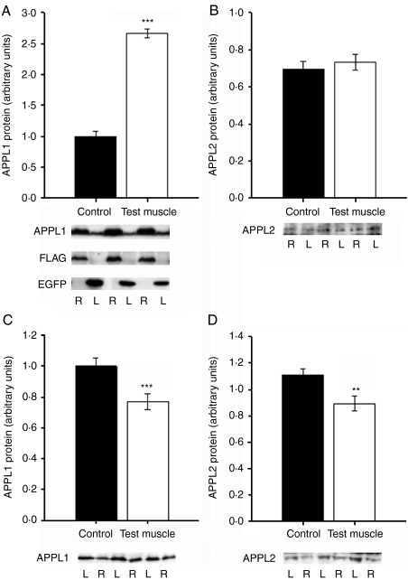
IVE of EH114-GW-APPL1 was used to over-express APPL1 in the right TCMs of cohorts of chow-fed young adult rats, with EH114-EGFP administered to the left TCMs as paired control. After 1 week, immunoblotting of muscle lysates confirmed that APPL1 protein was successfully over-expressed by a mean 188% (n=10; P≪0·001; Fig. 1A). Immunoblotting for EGFP and the FLAG tag attached to the APPL1 cDNA confirmed the expression of control and exogenous APPL1 protein in the appropriate muscles. APPL2 protein levels were unaffected by this manipulation (Fig. 1B).
Figure 1.
Modulation of APPL1 protein levels in single tibialis cranialis muscles of rats by in vivo electrotransfer (IVE). IVE of APPL1 cDNA-containing or APPL1-targeting shRNA constructs was used to over-express or silence APPL1 expression in tibialis cranialis muscles (TCMs) of chow-fed rats. After 1 week, successful (A) over-expression of APPL1 (n=10) was demonstrated by immunoblotting of tissue lysates derived from test and control TCMs. (B) APPL2 protein expression was unaffected by this manipulation. (C) Successful down-regulation (n=8) of APPL1 as a result of shRNA electroporation. (D) APPL2 protein was also reduced by APPL1 shRNA administration. Typical immunoblots for APPL1, APPL2, the FLAG tag attached to the APPL1 cDNA and EGFP in test (right, R, open bars) and paired control (left, L, solid bars) muscles are shown. Data are mean±s.e.m. Paired t-testing: **P<0·01, ***P<0·001 versus control.
IVE of APPL1-targeting shRNAs into TCMs of a parallel cohort of chow-fed rats was used to examine the effects of knocking down this protein in muscle. As shown in Fig. 1C, we achieved a mean 22·5% reduction in APPL1 expression in test versus control muscles (n=15; P<0·001). However, this was accompanied by a 19·4% reduction in APPL2 protein (P=0·0026; Fig. 1D), despite the specificity of the APPL1 targeting sequences used in the shRNA constructs and the apparent lack of cross-reactivity of the antibody.
One week after IVE, no differences in muscle mass or cross-sectional area of fibres in transverse section were detected, implying no effect of this level of APPL1 silencing on cellular growth or proliferation (data not shown).
OE and silencing of APPL1 expression results in parallel changes in glycogen storage in muscles
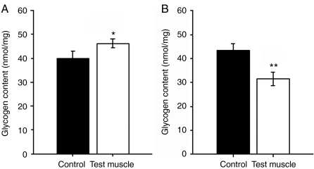
To establish whether altering expression of APPL1 would have an effect on net energy storage in muscle, we measured glycogen content of pairs of TCMs removed from APPL1 cDNA- and shRNA-electroporated rats. APPL1 OE increased glycogen content in TCMs by 19% (P=0·048; Fig. 2A), reflecting an increase in net glycogen accumulation during the week of the experiment. Consistent with this change, we also identified a 26% reduction in glycogen content of muscles in which APPL1 expression had been knocked down (P=0·003; Fig. 2B). Thus, APPL1 positively impacts on glycogen stores in skeletal muscle.
Figure 2.
APPL1 over-expression and silencing result in complementary changes in muscle glycogen storage. After 1 week of IVE, glycogen content was measured in TCMs showing (A) APPL1 over-expression (n=10) and (B) APPL1 down-regulation (n=8). Data are mean±s.e.m. Glycogen was significantly increased by APPL1 over-expression and reduced by APPL1 silencing. Panels show results for test (open bars) and paired control (solid bars) muscles. Paired t-testing: *P<0·05, **P<0·01 versus control.
APPL1 OE also increases phosphorylation of key PI3-kinase signalling pathway intermediates in the absence of an effect on ceramide content
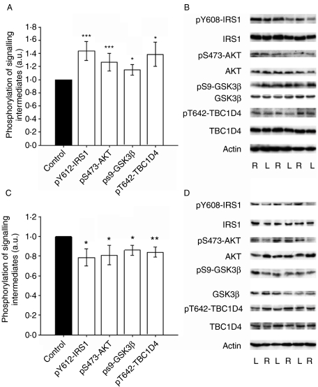
We hypothesised that activation of the PI3-kinase pathway may mediate the observed effects of APPL1 on glycogen accumulation in muscle (Mao et al. 2006, Saito et al. 2007, Schenck et al. 2008, Cheng et al. 2009) in vivo. Thus, we examined the expression and phosphorylation of key intermediates in the PI3-kinase pathway in lysates from APPL1 over-expressing and control muscles. APPL1 OE caused a mean 50% (P<0·001) increase in pY608-IRS1 (the rat equivalent of human pY612), together with a 25% increase (P<0·001) in Ser473 phosphorylation of AKT, a 15% increase (P=0·012) in pS9-GSK3β and a 47% increase in Thr642-phosphorylation of TBC1D4 (AS160; P=0·028), respectively, in test versus control muscles (Fig. 3A and B). These increases in phosphorylation are consistent with increased flux through the PI3-kinase pathway, culminating in increased translocation of GLUT4-containing vesicles into sarcolemma (Larance et al. 2005) and disinhibition of glycogen synthesis. Small but significant increases in expression of IRS1 and TBC1D4 proteins were also detected in APPL1 OE muscles, although there was no difference in expression of β-actin between groups and the upward regulatory trend on a phosphorylation/expression ratio basis was preserved in each case (Fig. 3B).
Figure 3.
APPL1 over-expression and silencing result in a respective increase and decrease in phosphorylation of PI3-kinase pathway signalling intermediates under basal insulin conditions. Phosphorylation and expression of signalling molecules were measured in lysates prepared from paired TCMs removed from APPL1 over-expressing (n=10) and silenced (n=8–15) chow-fed rats. Data are mean±s.e.m.; solid bars, left muscles; open bars, right muscles. (A) Summary phosphorylation data from APPL1 over-expressing test muscles normalised to control for Tyr612-insulin receptor substrate-1, Ser473-AKT, Ser9-glycogen synthase kinase-3β and Thr642-TBC1D4. Phosphorylation of all these intermediates was increased by the manipulation. (B) Representative immunoblots of phosphorylated and total protein from APPL1 over-expressing (R) and control (L) muscles. Protein expression of IRS1 and TBC1D4 was also increased by APPL1 over-expression. (C) Summary phosphorylation data from APPL1 knock-down test muscles normalised to control for Tyr612-insulin receptor substrate-1, Ser473-AKT, Ser9-glycogen synthase kinase-3β and Thr642-TBC1D4. Phosphorylation of all these intermediates was decreased by the manipulation. (D) Representative immunoblots of phosphorylated and total protein from APPL1 knockdown (R) and control (L) muscles. Protein expression of IRS1 was also decreased by APPL1 over-expression. Sample actin blots are also shown demonstrating no differences in loading between sets of lysates. Paired t-testing: *P<0·05, **P<0·01, ***P<0·001 versus control muscle.
In an attempt to corroborate these findings, we compared phosphorylation and expression of the same PI3-kinase pathway intermediates in APPL1-silenced TCMs with paired controls and found consistent reductions in phosphorylation (pY612-IRS1, 19% reduction, P=0·035; pS473-AKT, 21% reduction, P=0·021; pS9-GSK3β, 12% reduction, P=0·023; Fig. 3C and D), which were thus complementary to the effects of OE. Again, despite no difference in gel loading, as indicated by a β-actin blot, a significant decrease in protein levels of IRS1 was detected (Fig. 3D). These consistent changes in IRS1 expression may reflect effects on proteasomal degradation rate resulting from altered serine and tyrosine phosphorylation induced by the manipulation (Haruta et al. 2000, Pirola et al. 2003, Cleasby et al. 2007). However, the clear regulation by APPL1 expression levels of phosphorylation of PI3-kinase pathway intermediates and thus flux through the pathway down to the level of GSK3β could be sufficient to explain the reduced glycogen storage observed.
In the light of the recent demonstration that adiponectin mediates its insulin-sensitising effects through activation of a receptor intrinsic ceramidase activity (Holland et al. 2010), we measured the levels of ceramides in APPL1 over-expressing and silenced TCMs. Ceramide content was unchanged in APPL1 OE versus control muscles (72·6±8·7 vs 69·9±13·3 nmol/g), whereas there was a non-significant trend towards APPL1 silencing resulting in increased ceramide levels (89·8±10·1 vs 69·7±10·0 nmol/g; P=0·11). Thus, these data are not suggestive that the effects of altered APPL1 expression on PI3-kinase signalling are mediated through altered ceramide metabolism.
APPL1 OE increases insulin-stimulated muscle glycogen synthesis in vivo and partially ameliorates HFD-induced insulin resistance
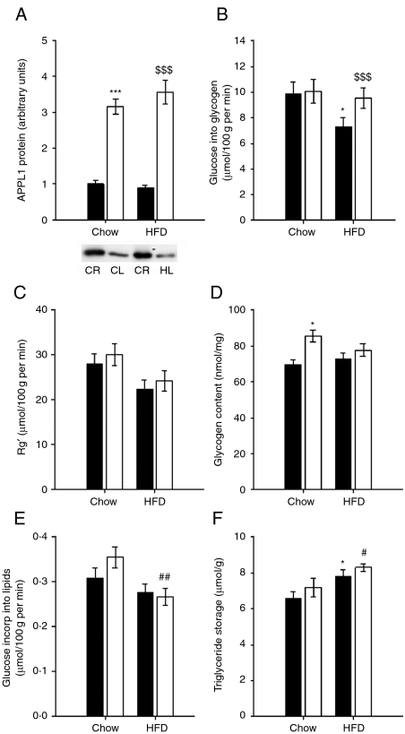
To establish whether the APPL1-mediated increase in muscle glycogen storage would be associated with acute increases in insulin-stimulated glucose uptake and glycogen synthesis, rats were subjected to a HEC combined with administration of radiolabelled glucose and 2-deoxyglucose tracers 1 week after IVE with APPL1 expressing or control vectors. We also aimed to determine whether APPL1 OE was capable of ameliorating muscle insulin resistance by measuring these variables in rats that had been fed a HFD for 5 weeks before clamping and comparing these to chow-fed animals. IVE induced 360% (n=18) and 271% (n=15) increases in APPL1 expression in HFD- and chow-fed rats subsequently subjected to HEC respectively (effect of APPL1 P<0·001; Fig. 4A). However, there was no effect of diet on APPL1 expression (P=0·474).
Figure 4.
APPL1 over-expression in muscle increases local insulin-stimulated glycogen accumulation and partially overcomes high-fat diet (HFD)-induced insulin resistance. APPL1 was over-expressed by IVE in additional cohorts of 5-week HFD (n=18) and chow-fed (n=15) rats for 1 week and then insulin-stimulated disposal and storage of glucose under hyperinsulinaemic–euglycaemic clamp conditions were measured in paired TCMs. Data are mean±s.e.m.; solid bars, left muscles; open bars, right muscles. (A) APPL1 protein expression summary graph and representative blot, demonstrating increased expression in both sets of animals (P<0·001), with no effect of HFD on APPL1 expression. (B) Glucose incorporation into glycogen during the clamp was increased overall by APPL1 over-expression (P=0·016) and there was an interaction between APPL1 expression and diet (P=0·048). (C) Glucose uptake (Rg′) was increased overall in APPL1 over-expressing (OE) muscles (P=0·050) and tended to be reduced by HFD (P=0·064). (D) Glycogen content of APPL1 OE TCMs was increased compared with controls (P=0·022), principally in chow-fed animals. (E) Glucose incorporation into triglyceride during the clamp was reduced overall by HFD feeding (P=0·027). Interaction term P=0·069. (F) Triglyceride storage was increased in HFD versus chow-fed muscles overall (P=0·012), with a tendency for APPL1 OE also to increase triglycerides (P=0·082). CL – chow fed, control; CR – chow fed, test; HL – HFD fed, control; HR – HFD fed, test muscles. Post hoc analyses: *P<0·05, ***P<0·001 versus chow control muscle; $$$P<0·001 versus HFD control muscle; #P<0·05, ##P<0·01 versus chow APPL1 OE muscle.
HFD feeding caused 9·6 and 74% increases in body mass and epididymal fat pad mass as a percentage of body mass respectively (Table 1). Basal (5–7 h starved) plasma glucose and insulin were also increased in HFD-fed rats (Table 1). Clamped plasma glucose levels were similar between groups, although total mean plasma insulin was slightly higher in HFD rats (Table 1), as a result of higher endogenous plasma insulin. HFD rats showed the expected reductions in GIR and Rd and increase in EGO (Table 1), commensurate with the induction of whole-body insulin resistance by the diet.
Table 1.
Chow- and high-fat diet-fed rat characteristics (mean±s.e.m.) under basal and hyperinsulinaemic–euglycaemic clamp conditions
| Chow fed | High-fat diet fed | |||
|---|---|---|---|---|
| Basal | Clamp | Basal | Clamp | |
| Plasma glucose (mM) | 7·5 (±0·4) | 8·0 (±0·1) | 9·1 (±0·6)* | 8·3 (±0·1) |
| Plasma insulin (mU/l) | 28·2 (±2·7) | 124·3 (±8·3) | 65·7 (±5·1)‡ | 155·8 (±6·4)† |
| Body weight (BW) at study (g) | 393 (±8) | 428 (±8)† | ||
| Epididymal fat pad mass (%age BW) | 0·52 (±0·02) | 0·91 (±0·06)‡ | ||
| Glucose infusion rate (mg/kg per min) | 29·4 (±1·8) | 20·8 (±1·3)‡ | ||
| Whole-body glucose disposal (mg/kg per min) | 26·7 (±2·0) | 22·1 (±1·8)* | ||
| Endogenous glucose output (mg/kg per min) | −3·2 (±1·9) | 2·1 (±1·2)* | ||
*P<0·05, †P<0·01, ‡P<0·001 versus equivalent measurement for chow-fed rats.
Glucose tracer incorporation into glycogen was attenuated by HFD feeding in control muscles, but this effect was abrogated by APPL1 OE (effect of IVE P=0·016; interaction P=0·048; Fig. 4B), implying both an acute effect on glycogen synthesis and an amelioration of the defect in insulin-stimulated glycogen synthesis as a result of this manipulation. Glucose uptake (Rg′) into TCMs during the clamp tended to be reduced by HFD feeding (P=0·064) but was increased by a mean 10% across all test versus control muscles (P=0·050; Fig. 4C). Although these were modest effects, it is likely that this attenuated glucose disposal, at least in part, explains the effects of APPL1 on glycogen synthesis. APPL1 OE caused an overall increase in muscle glycogen content (P=0·022), with the principal effect being in chow-fed animals (Fig. 4D), consistent with the effect on acute glycogen synthesis and the changes in glycogen content described under basal insulin conditions earlier.
Effects of APPL1 on lipid accumulation were also assessed in these animals. Glucose tracer incorporation into triglycerides was lower overall in HFD rats during the clamp (P=0·027), mainly due to a specific attenuation of an effect of APPL1 OE to increase this parameter (interaction P=0·069; Fig. 4E). Total content of triglyceride, reflecting longer term effects on lipid turnover, was increased in HFD-fed rats (P=0·012). However, there was also a tendency for APPL1 OE to increase triglyceride storage (P=0·082; Fig. 4F).
APPL1 OE further increases activation of the PI3-kinase pathway in muscle of HEC rats
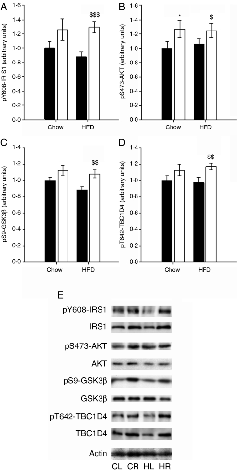
To verify that the observed increase in insulin-stimulated glycogen synthesis was related to up-regulation of the PI3-kinase pathway signalling in this setting, we measured phosphorylation and expression of IRS1, AKT, GSK3β and TBC1D4 in muscle lysates. APPL1 OE resulted in increased Tyr608 phosphorylation on IRS1 (P=0·001), principally due to increased phosphorylation in TCMs from HFD-fed rats (P<0·001; Fig. 5A). This change was accompanied by a similar trend in IRS1 protein (P=0·063). AKT phosphorylation at Ser473 was increased in APPL1 OE muscles (P=0·001; Fig. 5B) and total AKT protein was also increased (P=0·003; chow rats P=0·055, HFD-fed rats P=0·002). Phosphorylation at Ser9 of GSK3β was also increased in APPL1 OE muscles (P<0·001; Fig. 5C), again mainly due to an effect in HFD-fed muscles (P=0·001 and P=0·002 respectively). Both phosphorylation and protein expression of TBC1D4 were increased by APPL1 OE (P=0·009 and P<0·001 respectively; Fig. 5D), again principally due to an effect in the muscles of HFD-fed rats (TBC1D4 interaction term P=0·049; effect of APPL1 OE in HFD-fed TCMs P=0·008 and P<0·001 respectively). These changes occurred in the absence of any differences in β-actin expression between groups (Fig. 5E).
Figure 5.
APPL1 over-expression has an additive effect to high physiological levels of insulin to activate the PI3-kinase pathway. Expression and phosphorylation at key residues of intermediates in the PI3-kinase pathway were measured by immunoblotting of lysates of paired TCMs from chow- (n=12–18) and HFD- (n=10–15) fed hyperinsulinaemic–euglycaemic clamped rats. Data are mean±s.e.m.; solid bars, left muscles; open bars, right muscles. (A) Tyr612 phosphorylation of IRS1 was increased by APPL1 over-expression (OE) (P<0·001). (B) Ser473 phosphorylation of AKT was also increased by APPL1 OE (P=0·001). (C) Ser9 phosphorylation of GSK3β was also greater in APPL1 over-expressing muscles (P<0·001). (D) Thr642 phosphorylation of TBC1D4 was increased by APPL1 OE (P=0·009). (E) Representative immunoblots of phosphorylated and total protein from CL – chow fed, control; CR – chow fed, test; HL – HFD fed, control; HR – HFD fed, test muscles. There was a tendency for IRS1 protein to be increased by APPL1 OE (P=0·063), whereas AKT protein was increased overall by APPL1 OE (P=0·003) and showed a tendency to be decreased by HFD feeding (P=0·085). TBC1D4 protein was also increased (P<0·001), with an interaction between diet and OE effects (P=0·049), showing a larger effect in the HFD-fed animals. However, an actin loading control blot is also shown, demonstrating that loading was not different among samples. Post hoc analyses: *P<0·05 versus chow control muscle; $P<0·05, $$P<0·01, $$$P<0·001 versus HFD control muscle.
These findings are consistent with those observed under basal insulin conditions and together likely explain the demonstrated effect of APPL1 OE to increase glycogen synthesis and thus promote glycogen accumulation in muscle. Furthermore, it seems that APPL1 OE has an additive effect to the high physiological levels of insulin stimulation of the HEC to increase flux through the PI3-kinase pathway. Although in this study impairments in phosphorylation of signalling intermediates in the PI3-kinase pathway were not demonstrated that would fully account for the defects observed in insulin action, the consistent increases in phosphorylation of these signalling intermediates down to the level of GSK3β may nevertheless represent a potential mechanism by which APPL1 may ameliorate insulin resistance in muscle in vivo.
APPL1 OE impairs activation of the AMPK pathway in insulin-stimulated muscle
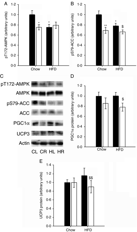
AMPK activation is thought to be the principal mediator of adiponectin action in skeletal muscle (Tomas et al. 2002, Yamauchi et al. 2002, Yoon et al. 2006) and has been shown to be the result of binding of liganded AdipoRs to APPL1 in C2C12 myotubes (Mao et al. 2006). Therefore, to establish whether AMPK activation might also be responsible for some of the effects of APPL1 OE in vivo, paired TCMs from insulin-stimulated HFD- and chow-fed rats were immunoblotted for AMPK protein and phosphorylation at the LKB1 target residue, Thr172 (Hawley et al. 2003). HFD-feeding caused a reduction in phosphorylation of AMPK, as would have been predicted (Liu et al. 2006). However, perhaps unexpectedly (Mao et al. 2006, Zhou et al. 2009), APPL1 OE reduced AMPK phosphorylation in chow-fed rats by a similar magnitude (P=0·041 effect of IVE, P=0·006 interaction; Fig. 6A).
Figure 6.
APPL1 over-expression reduces activation of AMP-activated protein kinase in muscle from hyperinsulinaemic–euglycaemic clamped rats. Phosphorylation and expression of AMPK and downstream substrates were measured by immunoblotting of lysates of paired TCMs from chow- (n=12–18) and HFD- (n=10–15) fed hyperinsulinaemic–euglycaemic clamped rats. Data are mean±s.e.m.; solid bars, left muscles; open bars, right muscles. (A) Thr172 phosphorylation of AMPK was reduced overall by APPL1 over-expression (OE) (P=0·041) to a similar extent to the reduction caused by diet (interaction P=0·006). (B) Serine79 phosphorylation of ACC was reduced overall as a result of both APPL1 OE (P<0·001) and HFD feeding (P=0·048); interaction P=0·075. (C) Representative immunoblots of phosphorylated and total protein from CL – chow fed, control; CR – chow fed, test; HL – HFD fed, control; HR – HFD fed, test muscles. ACC protein expression was slightly increased overall by APPL1 OE (P=0·022). Levels of actin were not different between treatment groups. (D) PGC1α protein was reduced overall by APPL1 OE (P=0·008). (E) UCP3 was also reduced overall by APPL1 OE (P=0·043) but principally due to normalisation of an increase in expression due to HFD feeding (interaction P=0·048). Post hoc analyses: *P<0·05, **P<0·01 versus chow control muscle; $P<0·05, $$P<0·01 versus HFD control muscle.
To establish whether these impairments in AMPK phosphorylation resulted in downstream effects, we examined phosphorylation of the AMPK substrate enzyme acetyl-CoA carboxylase (ACC) at its target residue (Ser79). The results were entirely consistent with the pT172-AMPK data, as pS79-ACC was reduced overall by HFD feeding (P=0·048), but also by APPL1 OE, most significantly in chow-fed animals (interaction P=0·075; Fig. 6B). ACC protein was slightly increased overall as a result of APPL1 OE (P=0·022; sample blot in Fig. 6C), although protein loading, as indicated by β-actin expression, was not different. We also measured protein expression of PGC1α and UCP3, key AMPK-regulated mediators of mitochondrial oxidative metabolism (Ruderman et al. 2003). Both PGC1α (P=0·008; Fig. 6D) and UCP3 (P=0·043; Fig. 6E) were reduced by APPL1 OE; although in the case of UCP3, this appeared to be an effect to reverse an increase in expression in high-fat-fed muscles (interaction P=0·048).
Thus, APPL1 OE in muscle in vivo caused phosphorylation changes implying impaired acute activation of AMPK and consequently increased activity of ACC and attenuated target gene expression, implying longer term impairment of AMPK activity. The likely increase in malonyl-CoA and impaired β-oxidation resulting from these changes (McGarry 2002) is consistent with the observed tendency for increased triglyceride storage in APPL1 OE muscles. Furthermore, AMPK phosphorylation tended to be increased by APPL1 OE under basal insulin conditions and was significantly elevated by APPL1 silencing (data not shown), results consistent with these data.
Discussion
This study strongly implies a role for APPL1 in enhancing glycogen synthesis in skeletal muscle in vivo, mediated via activation of the insulin signalling pathway. Acute local OE of APPL1 in adult muscle resulted in increased insulin-stimulated glycogen synthesis and correction of HFD-induced insulin resistance of this parameter in this critical insulin target tissue. This effect was most likely mediated via up-regulated signalling to glucose transport and glycogen synthesis through the PI3-kinase pathway from the level of IRS1. In addition, we present evidence that APPL1 may be involved in attenuating AMPK-dependent pathways in skeletal muscle, contrary to the results of previous in vitro studies. These findings are supportive of a role for crosstalk between the adiponectin and the insulin signalling pathways at the level of APPL1 in mediating the insulin-sensitising effects of adiponectin in skeletal muscle in vivo.
In this study, the degree of silencing in muscles achieved using APPL1 shRNA seems to be relatively modest compared with the degree of OE induced in a separate group of animals. However, the similarity in downstream effect renders the opposing results readily comparable for interpretation purposes. Our prior experience of manipulating expression of signalling molecules such as AKT in muscle (Cleasby et al. 2007) suggests that IVE of shRNA constructs commonly results in expression changes of lower magnitude than IVE of OE constructs. This may be due to differences in electrotransfer efficiency or expression of the constructs, which are contained within different plasmid vectors. It is likely that relatively small reductions in expression are capable of adversely affecting the signalling threshold of the PI3-kinase pathway, although large degrees of OE have relatively more limited effects on a percentage change basis, due to limitations in the dynamic range of the pathway (Hoehn et al. 2008, Janes et al. 2008). The effect of APPL1 shRNA administration to also reduce APPL2 protein is intriguing. Given the lack of effect on APPL2 after APPL1 OE, antibody cross-reactivity would not seem to be the explanation. In addition, a BLAST search for the APPL1 shRNA target sequences used confirms their specificity. Instead, this would seem to be a secondary effect of APPL1 silencing and may help to explain the magnitude of the effect of IVE of the APPL1 shRNA construct on downstream signalling. A possible explanation may be that reduced APPL1 protein availability results in reduced APPL heterodimerisation, exposing APPL2 molecules to proteasomal degradation. However, previously published in vitro data have suggested that APPL1 and APPL2 play antagonistic roles (Wang et al. 2009), which would make it surprising if reduced APPL2 expression had additive effects to silencing of APPL1. This issue is discussed later.
APPL1 was first identified as an adaptor protein that interacts with AKT2 and plays a role in its recruitment to the plasma membrane (Mitsuuchi et al. 1999). The discovery of a functional in vitro interaction between APPL1 and AdipoRs mediating downstream effects of adiponectin (Mao et al. 2006), coupled with a demonstration of the requirement of the APPL1/AKT2 interaction for full insulin-stimulated GLUT4 translocation in adipocytes (Saito et al. 2007), raised the intriguing possibility that APPL1 may provide a key node for interaction between the PI3-kinase pathway and the putative adiponectin signalling pathway (Mao et al. 2006). The increased phosphorylation of key intermediates in the PI3-kinase pathway (IRS1, AKT, TBC1D4 and GSK3) in APPL1 OE muscles under basal and insulin-stimulated conditions and the complementary reductions observed after APPL1 silencing demonstrate that activation of this pathway can be achieved via APPL1, and with additive effects to those of insulin alone, under physiological conditions in muscle. This is consistent with a recent study that demonstrated an additive effect of adiponectin to that of insulin in stimulating IRS1 and AKT phosphorylation in L6 myotubes (Fang et al. 2009). In a previous study, treatment of cultured myotubes with adiponectin enhanced the ability of insulin to activate the PI3-kinase pathway because of disinhibition of IRS1 tyrosine phosphorylation due to reduced AMPK-mediated activation of the serine kinase p70S6 kinase (Wang et al. 2007). However, the opposite effects observed on the activation of AMPK imply that an alternative mechanism is operative in muscle in vivo. Nevertheless, in our study, altered APPL1 expression also had effects on the PI3-kinase pathway from the level of IRS1, and in addition, we were unable to demonstrate a quantifiable interaction between AKT and APPL1 by co-immunoprecipitation of muscle lysates (data not shown), suggesting that APPL1 may interact with a more proximal binding partner in the pathway in this context.
In this study, we were unable to demonstrate that the effects of altered APPL1 expression on signalling were mediated by alterations in ceramide content, the recently proposed mechanism for the effects of ligand binding by AdipoRs (Holland et al. 2010). This would be consistent with the effects of altered APPL1 expression being mediated through binding to downstream signalling molecules rather than AdipoRs. APPL1 OE also caused changes in protein expression of some of the signalling intermediates measured. This may reflect translational or pre-translational effects of APPL1, perhaps exerted by regulation of transcription factors, analogous to the effects of APPL1 OE recently observed on β-catenin/TCF (Rashid et al. 2009) or in the case of IRS1, and is likely to be the result of altered proteasomal degradation rates, secondary to changes in phosphorylation (Haruta et al. 2000, Pirola et al. 2003, Cleasby et al. 2007). The lack of a complementary down-regulatory effect on TBC1D4 protein in the shRNA-electroporated muscles is likely to reflect relatively lower sensitivity of this mechanism to silencing than that of the PI3-kinase phosphorylation cascade. Nevertheless, it seems likely that APPL1 OE represents a state of increased flux through the proximal adiponectin signalling pathway and thus these findings implicate APPL1 as a key node for crosstalk between pathways and hence potentially for the insulin-sensitising effect of adiponectin in muscle.
Although PI3-kinase signalling was not significantly impaired in the moderate duration HFD-fed rat model used in this study, OE of APPL1 largely had its stimulatory effects in the muscles of HFD-fed animals, suggesting that therapeutic insulin sensitisation could be engineered in insulin-resistant states through this molecule. This assertion is supported on a metabolic level by the correction of HFD-impaired insulin-stimulated glycogen synthesis by APPL1 OE, although the improvement in acute glucose uptake is modest and appears to occur irrespective of dietary status. However, a significant potential risk in targeting this molecule therapeutically is likely to be its relative promiscuity in phosphoinositide and protein binding partners and the as yet poorly characterised cellular implications of its activation. Indeed, APPL1 has been shown to interact with APPL2 (Chial et al. 2008), Rab5 (Miaczynska et al. 2004), histone deacetylases (Miaczynska et al. 2004, Rashid et al. 2009), AKT2 (Mitsuuchi et al. 1999, Saito et al. 2007), AdipoRs (Mao et al. 2006) and TrkA and GIPC1 (Lin et al. 2006), thus being implicated in cellular growth and proliferation, in addition to metabolism.
Adiponectin is generally thought to achieve its effects on glucose disposal and fatty acid oxidation through AMPK activation in muscle (Yamauchi et al. 2002, Yoon et al. 2006, Wang et al. 2007, Zhou et al. 2009) and the mechanism has been shown to involve APPL1 in myotubes in culture (Mao et al. 2006, Zhou et al. 2009). However, the role of APPL1 in AMPK activation has not previously been investigated in muscle in vivo. In this study, APPL1 OE resulted in similar significant reductions in phosphorylation of both AMPK and its target ACC during HEC to those caused by an HFD. This finding certainly implies that in this context the beneficial effects of muscle APPL1 OE on glucose disposal and glycogen synthesis are not exerted through AMPK, but more likely through the demonstrated increases in TBC1D4 and GSK3 phosphorylation. However, it is possible that the increased levels of muscle glycogen may contribute to the reduced AMPK phosphorylation (McBride et al. 2009). Nevertheless, the decreases in expression of both PGC1α and UCP3, the tendency for APPL1 OE muscles to accumulate more triglyceride and the complementary effects of APPL1 silencing are consistent with the effects of APPL1 OE on AMPK and ACC activation and suggest that the effect on AMPK is not purely acute in nature and related to inhibition by high physiological levels of insulin during the HEC (Kovacic et al. 2003). Instead, these data suggest that APPL1 may play dual roles to increase glucose disposal into muscle while attenuating AMPK-mediated mitochondrial fatty acid oxidation.
APPL2 has been shown to have a similar role to APPL1 in mediating cell proliferation (Miaczynska et al. 2004) and β-catenin/TCF-dependent transcription (Rashid et al. 2009), but a recent report suggested that APPL2 competes with APPL1 for binding to AdipoR1 in myotubes and thus has an antagonistic effect on adiponectin and insulin signalling (Wang et al. 2009). Supply of additional APPL1 to muscle cells may, therefore, have its beneficial effects due to a shift in the APPL1:APPL2 ratio. This would seem to make it less likely that the reduction in APPL2 protein induced in addition to APPL1 silencing would have an additive effect on PI3-kinase signalling or glycogen accumulation. In addition, it is possible that adiponectin-mediated AMPK activation and consequent fatty acid use may involve APPL2 in muscle, whereas APPL1 mediates an inhibitory effect, analogous to the differential effector activation shown to be mediated through adiponectin binding to AdipoR1 and AdipoR2 in the liver (Yamauchi et al. 2007). It is clear that further work is necessary to establish the metabolic roles of putative adiponectin signalling molecules in insulin target tissues in relevant physiological settings.
In conclusion, APPL1 promotes glucose disposal into skeletal muscle in vivo through activation of the PI3-kinase pathway. This adaptor protein may provide a means of crosstalk between insulin and adiponectin signalling pathways and thus a means for the insulin-sensitising effect of adiponectin in muscle in vivo. Furthermore, OE of this molecule is capable of partially ameliorating muscle insulin resistance in a relevant physiological setting. Further work is required to more fully delineate the signalling networks integrated by this scaffolding protein with a view to establish whether it may provide a suitable target for novel therapeutic interventions aimed at ameliorating type 2 diabetes.
Declaration of interest
The authors declare that there is no conflict of interest that could be perceived as prejudicing the impartiality of the research reported.
Funding
This research was funded by the National Health and Medical Research Council of Australia (NHMRC, #481303), the Royal Veterinary College (RVC) and Diabetes UK (Small Grant #07/0003540). M E C is a Wellcome Trust University Award Fellow. N T is supported by a Career Development Award and E W K and G J C by Research Fellowships from the NHMRC.
Author contribution statement
M E C, G J C and E W K conceived and designed the experiments. M E C, Q L, E P, S A P, S J L and N T collected data. M E C, N T, G J C, A X and E W K analysed and interpreted data. M E C, N T, A X and E W K drafted and revised the manuscript. Experiments were conducted at the Garvan Institute, Sydney, and the Royal Veterinary College, London.
Acknowledgements
We are indebted to Dr Paul Pilch, Harvard University Medical School, for providing the shRNA constructs, to Tracie Reinten (Garvan Institute) for technical assistance, to Prof. David James (Garvan Institute) for critical reading of the manuscript, to Dr Andrew Hoy (Monash University) for advice regarding the ceramide assay and to the staff of the Biological Testing Facility at the Garvan Institute and the Biological Services Unit at the RVC for animal care.
References
- Bligh EG, Dyer WJ. A rapid method of total lipid extraction and purification. Canadian Journal of Biochemistry and Physiology. 1959;37:911–917. doi: 10.1139/o59-099. [DOI] [PubMed] [Google Scholar]
- Bruce CR, Mertz VA, Heigenhauser GJ, Dyck DJ. The stimulatory effect of globular adiponectin on insulin-stimulated glucose uptake and fatty acid oxidation is impaired in skeletal muscle from obese subjects. Diabetes. 2005;54:3154–3160. doi: 10.2337/diabetes.54.11.3154. [DOI] [PubMed] [Google Scholar]
- Bruce CR, Hoy AJ, Turner N, Watt MJ, Allen TL, Carpenter K, Cooney GJ, Febbraio MA, Kraegen EW. Overexpression of carnitine palmitoyltransferase-1 in skeletal muscle is sufficient to enhance fatty acid oxidation and improve high fat diet-induced insulin resistance. Diabetes. 2008;58:550–558. doi: 10.2337/db08-1078. [DOI] [PMC free article] [PubMed] [Google Scholar]
- Chan TM, Exton JH. A rapid method for the determination of glycogen content and radioactivity in small quantities of tissue or isolated hepatocytes. Analytical Biochemistry. 1976;71:96–105. doi: 10.1016/0003-2697(76)90014-2. [DOI] [PubMed] [Google Scholar]
- Chen MB, McAinch AJ, Macaulay SL, Castelli LA, O'Brien PE, Dixon JB, Cameron-Smith D, Kemp BE, Steinberg GR. Impaired activation of AMP-kinase and fatty acid oxidation by globular adiponectin in cultured human skeletal muscle of obese type 2 diabetics. Journal of Clinical Endocrinology and Metabolism. 2005;90:3665–3672. doi: 10.1210/jc.2004-1980. [DOI] [PubMed] [Google Scholar]
- Cheng KK, Lam KS, Wang Y, Huang Y, Carling D, Wu D, Wong C, Xu A. Adiponectin-induced endothelial nitric oxide synthase activation and nitric oxide production are mediated by APPL1 in endothelial cells. Diabetes. 2007;56:1387–1394. doi: 10.2337/db06-1580. [DOI] [PubMed] [Google Scholar]
- Cheng KK, Iglesias MA, Lam KS, Wang Y, Sweeney G, Zhu W, Vanhoutte PM, Kraegen EW, Xu A. APPL1 potentiates insulin-mediated inhibition of hepatic glucose production and alleviates diabetes via Akt activation in mice. Cell Metabolism. 2009;9:417–427. doi: 10.1016/j.cmet.2009.03.013. [DOI] [PubMed] [Google Scholar]
- Chial HJ, Wu R, Ustach CV, McPhail LC, Mobley WC, Chen YQ. Membrane targeting by APPL1 and APPL2: dynamic scaffolds that oligomerize and bind phosphoinositides. Traffic. 2008;9:215–229. doi: 10.1111/j.1600-0854.2007.00680.x. [DOI] [PMC free article] [PubMed] [Google Scholar]
- Cho H, Mu J, Kim JK, Thorvaldsen JL, Chu Q, Crenshaw EB, III, Kaestner KH, Bartolomei MS, Shulman GI, Birnbaum MJ. Insulin resistance and a diabetes mellitus-like syndrome in mice lacking the protein kinase Akt2 (PKB beta) Science. 2001;292:1728–1731. doi: 10.1126/science.292.5522.1728. [DOI] [PubMed] [Google Scholar]
- Cleasby ME, Dzamko N, Hegarty BD, Cooney GJ, Kraegen EW, Ye JM. Metformin prevents the development of acute lipid-induced insulin resistance in the rat through altered hepatic signaling mechanisms. Diabetes. 2004;53:3258–3266. doi: 10.2337/diabetes.53.12.3258. [DOI] [PubMed] [Google Scholar]
- Cleasby ME, Davey JR, Reinten TA, Graham MW, James DE, Kraegen EW, Cooney GJ. Acute bidirectional manipulation of muscle glucose uptake by in vivo electrotransfer of constructs targeting glucose transporter genes. Diabetes. 2005;54:2702–2711. doi: 10.2337/diabetes.54.9.2702. [DOI] [PubMed] [Google Scholar]
- Cleasby ME, Reinten TA, Cooney GJ, James DE, Kraegen EW. Functional studies of Akt isoform specificity in skeletal muscle in vivo; maintained insulin sensitivity despite reduced insulin receptor substrate-1 expression. Molecular Endocrinology. 2007;21:215–228. doi: 10.1210/me.2006-0154. [DOI] [PubMed] [Google Scholar]
- Fang X, Fetros J, Dadson KE, Xu A, Sweeney G. Leptin prevents the metabolic effects of adiponectin in L6 myotubes. Diabetologia. 2009;52:2190–2200. doi: 10.1007/s00125-009-1462-0. [DOI] [PubMed] [Google Scholar]
- Haruta T, Uno T, Kawahara J, Takano A, Egawa K, Sharma PM, Olefsky JM, Kobayashi M. A rapamycin-sensitive pathway down-regulates insulin signaling via phosphorylation and proteasomal degradation of insulin receptor substrate-1. Molecular Endocrinology. 2000;14:783–794. doi: 10.1210/me.14.6.783. [DOI] [PubMed] [Google Scholar]
- Hawley SA, Boudeau J, Reid JL, Mustard KJ, Udd L, Makela TP, Alessi DR, Hardie DG. Complexes between the LKB1 tumor suppressor, STRAD alpha/beta and MO25 alpha/beta are upstream kinases in the AMP-activated protein kinase cascade. Journal of Biology. 2003;2:28. doi: 10.1186/1475-4924-2-28. [DOI] [PMC free article] [PubMed] [Google Scholar]
- Hegarty BD, Furler SM, Ye J, Cooney GJ, Kraegen EW. The role of intramuscular lipid in insulin resistance. Acta Physiologica Scandinavica. 2003;178:373–383. doi: 10.1046/j.1365-201X.2003.01162.x. [DOI] [PubMed] [Google Scholar]
- Hoehn KL, Hohnen-Behrens C, Cederberg A, Wu LE, Turner N, Yuasa T, Ebina Y, James DE. IRS1-independent defects define major nodes of insulin resistance. Cell Metabolism. 2008;7:421–433. doi: 10.1016/j.cmet.2008.04.005. [DOI] [PMC free article] [PubMed] [Google Scholar]
- Holland WL, Miller RA, Wang ZV, Sun K, Barth BM, Bui HH, Davis KE, Bikman BT, Halberg N, Rutkowski JM, et al. Receptor-mediated activation of ceramidase activity initiates the pleiotropic actions of adiponectin. Nature Medicine. 2010;17:55–63. doi: 10.1038/nm.2277. [DOI] [PMC free article] [PubMed] [Google Scholar]
- Janes KA, Reinhardt HC, Yaffe MB. Cytokine-induced signaling networks prioritize dynamic range over signal strength. Cell. 2008;135:343–354. doi: 10.1016/j.cell.2008.08.034. [DOI] [PMC free article] [PubMed] [Google Scholar]
- Kovacic S, Soltys CL, Barr AJ, Shiojima I, Walsh K, Dyck JR. Akt activity negatively regulates phosphorylation of AMP-activated protein kinase in the heart. Journal of Biological Chemistry. 2003;278:39422–39427. doi: 10.1074/jbc.M305371200. [DOI] [PubMed] [Google Scholar]
- Kraegen EW, James DE, Bennett SP, Chisholm DJ. In vivo insulin sensitivity in the rat determined by euglycemic clamp. American Journal of Physiology. 1983;245:E1–E7. doi: 10.1152/ajpendo.1983.245.1.E1. [DOI] [PubMed] [Google Scholar]
- Kraegen EW, James DE, Jenkins AB, Chisholm DJ. Dose–response curves for in vivo insulin sensitivity in individual tissues in rats. American Journal of Physiology. 1985;248:E353–E362. doi: 10.1152/ajpendo.1985.248.3.E353. [DOI] [PubMed] [Google Scholar]
- Lara-Castro C, Luo N, Wallace P, Klein RL, Garvey WT. Adiponectin multimeric complexes and the metabolic syndrome trait cluster. Diabetes. 2006;55:249–259. doi: 10.2337/diabetes.55.01.06.db05-1105. [DOI] [PubMed] [Google Scholar]
- Larance M, Ramm G, Stockli J, van Dam EM, Winata S, Wasinger V, Simpson F, Graham M, Junutula JR, Guilhaus M, et al. Characterization of the role of the Rab GTPase-activating protein AS160 in insulin-regulated GLUT4 trafficking. Journal of Biological Chemistry. 2005;280:37803–37813. doi: 10.1074/jbc.M503897200. [DOI] [PubMed] [Google Scholar]
- Lin DC, Quevedo C, Brewer NE, Bell A, Testa JR, Grimes ML, Miller FD, Kaplan DR. APPL1 associates with TrkA and GIPC1 and is required for nerve growth factor-mediated signal transduction. Molecular and Cellular Biology. 2006;26:8928–8941. doi: 10.1128/MCB.00228-06. [DOI] [PMC free article] [PubMed] [Google Scholar]
- Liu Y, Wan Q, Guan Q, Gao L, Zhao J. High-fat diet feeding impairs both the expression and activity of AMPKa in rats' skeletal muscle. Biochemical and Biophysical Research Communications. 2006;339:701–707. doi: 10.1016/j.bbrc.2005.11.068. [DOI] [PubMed] [Google Scholar]
- Mao X, Kikani CK, Riojas RA, Langlais P, Wang L, Ramos FJ, Fang Q, Christ-Roberts CY, Hong JY, Kim RY, et al. APPL1 binds to adiponectin receptors and mediates adiponectin signalling and function. Nature Cell Biology. 2006;8:516–523. doi: 10.1038/ncb1404. [DOI] [PubMed] [Google Scholar]
- McBride A, Ghilagaber S, Nikolaev A, Hardie DG. The glycogen-binding domain on the AMPK beta subunit allows the kinase to act as a glycogen sensor. Cell Metabolism. 2009;9:23–34. doi: 10.1016/j.cmet.2008.11.008. [DOI] [PMC free article] [PubMed] [Google Scholar]
- McGarry JD. Banting lecture 2001: dysregulation of fatty acid metabolism in the etiology of type 2 diabetes. Diabetes. 2002;51:7–18. doi: 10.2337/diabetes.51.1.7. [DOI] [PubMed] [Google Scholar]
- Miaczynska M, Christoforidis S, Giner A, Shevchenko A, Uttenweiler-Joseph S, Habermann B, Wilm M, Parton RG, Zerial M. APPL proteins link Rab5 to nuclear signal transduction via an endosomal compartment. Cell. 2004;116:445–456. doi: 10.1016/S0092-8674(04)00117-5. [DOI] [PubMed] [Google Scholar]
- Mitsuuchi Y, Johnson SW, Sonoda G, Tanno S, Golemis EA, Testa JR. Identification of a chromosome 3p14.3–21.1 gene, APPL, encoding an adaptor molecule that interacts with the oncoprotein-serine/threonine kinase AKT2. Oncogene. 1999;18:4891–4898. doi: 10.1038/sj.onc.1203080. [DOI] [PubMed] [Google Scholar]
- Mullen KL, Pritchard J, Ritchie I, Snook LA, Chabowski A, Bonen A, Wright D, Dyck DJ. Adiponectin resistance precedes the accumulation of skeletal muscle lipids and insulin resistance in high fat fed rats. American Journal of Physiology. Regulatory, Integrative and Comparative Physiology. 2009;296:R243–R251. doi: 10.1152/ajpregu.90774.2008. [DOI] [PubMed] [Google Scholar]
- Pajvani UB, Hawkins M, Combs TP, Rajala MW, Doebber T, Berger JP, Wagner JA, Wu M, Knopps A, Xiang AH, et al. Complex distribution, not absolute amount of adiponectin, correlates with thiazolidinedione-mediated improvement in insulin sensitivity. Journal of Biological Chemistry. 2004;279:12152–12162. doi: 10.1074/jbc.M311113200. [DOI] [PubMed] [Google Scholar]
- Pirola L, Bonnafous S, Johnston AM, Chaussade C, Portis F, Van Obberghen E. Phosphoinositide 3-kinase-mediated reduction of insulin receptor substrate-1/2 protein expression via different mechanisms contributes to the insulin-induced desensitization of its signaling pathways in L6 muscle cells. Journal of Biological Chemistry. 2003;278:15641–15651. doi: 10.1074/jbc.M208984200. [DOI] [PubMed] [Google Scholar]
- Preiss J, Loomis CR, Bishop WR, Stein R, Niedel JE, Bell RM. Quantitative measurement of sn-1,2-diacylglycerols present in platelets, hepatocytes, and ras- and sis-transformed normal rat kidney cells. Journal of Biological Chemistry. 1986;261:8597–8600. [PubMed] [Google Scholar]
- Rashid S, Pilecka I, Torun A, Olchowik M, Bielinska B, Miaczynska M. Endosomal adaptor proteins APPL1 and APPL2 are novel activators of beta-catenin/TCF-mediated transcription. Journal of Biological Chemistry. 2009;284:18115–18128. doi: 10.1074/jbc.M109.007237. [DOI] [PMC free article] [PubMed] [Google Scholar]
- Ruderman NB, Saha AK, Kraegen EW. Minireview: malonyl CoA, AMP-activated protein kinase, and adiposity. Endocrinology. 2003;144:5166–5171. doi: 10.1210/en.2003-0849. [DOI] [PubMed] [Google Scholar]
- Saito T, Jones CC, Huang S, Czech MP, Pilch PF. The interaction of AKT with APPL1 is required for insulin-stimulated Glut4 translocation. Journal of Biological Chemistry. 2007;282:32280–32287. doi: 10.1074/jbc.M704150200. [DOI] [PubMed] [Google Scholar]
- Schenck A, Goto-Silva L, Collinet C, Rhinn M, Giner A, Habermann B, Brand M, Zerial M. The endosomal protein Appl1 mediates Akt substrate specificity and cell survival in vertebrate development. Cell. 2008;133:486–497. doi: 10.1016/j.cell.2008.02.044. [DOI] [PubMed] [Google Scholar]
- Segal KR, Landt M, Klein S. Relationship between insulin sensitivity and plasma leptin concentration in lean and obese men. Diabetes. 1996;45:988–991. doi: 10.2337/diabetes.45.7.988. [DOI] [PubMed] [Google Scholar]
- Tomas E, Tsao TS, Saha AK, Murrey HE, Zhang CC, Itani SI, Lodish HF, Ruderman NB. Enhanced muscle fat oxidation and glucose transport by ACRP30 globular domain: acetyl-CoA carboxylase inhibition and AMP-activated protein kinase activation. PNAS. 2002;99:16309–16313. doi: 10.1073/pnas.222657499. [DOI] [PMC free article] [PubMed] [Google Scholar]
- Tsao TS, Tomas E, Murrey HE, Hug C, Lee DH, Ruderman NB, Heuser JE, Lodish HF. Role of disulfide bonds in Acrp30/adiponectin structure and signaling specificity. Different oligomers activate different signal transduction pathways. Journal of Biological Chemistry. 2003;278:50810–50817. doi: 10.1074/jbc.M309469200. [DOI] [PubMed] [Google Scholar]
- Wang C, Mao X, Wang L, Liu M, Wetzel MD, Guan KL, Dong LQ, Liu F. Adiponectin sensitizes insulin signaling by reducing p70 S6 kinase-mediated serine phosphorylation of IRS-1. Journal of Biological Chemistry. 2007;282:7991–7996. doi: 10.1074/jbc.M700098200. [DOI] [PubMed] [Google Scholar]
- Wang C, Xin X, Xiang R, Ramos FJ, Liu M, Lee HJ, Chen H, Mao X, Kikani CK, Liu F, et al. "Yin-Yang" regulation of adiponectin signaling by APPL isoforms in muscle cells. Journal of Biological Chemistry. 2009;284:31608–31615. doi: 10.1074/jbc.M109.010355. [DOI] [PMC free article] [PubMed] [Google Scholar]
- Weyer C, Funahashi T, Tanaka S, Hotta K, Matsuzawa Y, Pratley RE, Tataranni PA. Hypoadiponectinemia in obesity and type 2 diabetes: close association with insulin resistance and hyperinsulinemia. Journal of Clinical Endocrinology and Metabolism. 2001;86:1930–1935. doi: 10.1210/jc.86.5.1930. [DOI] [PubMed] [Google Scholar]
- Yamauchi T, Kamon J, Waki H, Terauchi Y, Kubota N, Hara K, Mori Y, Ide T, Murakami K, Tsuboyama-Kasaoka N, et al. The fat-derived hormone adiponectin reverses insulin resistance associated with both lipoatrophy and obesity. Nature Medicine. 2001;7:941–946. doi: 10.1038/90984. [DOI] [PubMed] [Google Scholar]
- Yamauchi T, Kamon J, Minokoshi Y, Ito Y, Waki H, Uchida S, Yamashita S, Noda M, Kita S, Ueki K, et al. Adiponectin stimulates glucose utilization and fatty-acid oxidation by activating AMP-activated protein kinase. Nature Medicine. 2002;8:1288–1295. doi: 10.1038/nm788. [DOI] [PubMed] [Google Scholar]
- Yamauchi T, Kamon J, Ito Y, Tsuchida A, Yokomizo T, Kita S, Sugiyama T, Miyagishi M, Hara K, Tsunoda M, et al. Cloning of adiponectin receptors that mediate antidiabetic metabolic effects. Nature. 2003;423:762–769. doi: 10.1038/nature01705. [DOI] [PubMed] [Google Scholar]
- Yamauchi T, Nio Y, Maki T, Kobayashi M, Takazawa T, Iwabu M, Okada-Iwabu M, Kawamoto S, Kubota N, Kubota T, et al. Targeted disruption of AdipoR1 and AdipoR2 causes abrogation of adiponectin binding and metabolic actions. Nature Medicine. 2007;13:332–339. doi: 10.1038/nm1557. [DOI] [PubMed] [Google Scholar]
- Yoon MJ, Lee GY, Chung JJ, Ahn YH, Hong SH, Kim JB. Adiponectin increases fatty acid oxidation in skeletal muscle cells by sequential activation of AMP-activated protein kinase, p38 mitogen-activated protein kinase, and peroxisome proliferator-activated receptor {alpha} Diabetes. 2006;55:2562–2570. doi: 10.2337/db05-1322. [DOI] [PubMed] [Google Scholar]
- Zhou L, Deepa SS, Etzler JC, Ryu J, Mao X, Fang Q, Liu DD, Torres JM, Jia W, Lechleiter JD, et al. Adiponectin activates AMPK in muscle cells via APPL1/LKB1- and PLC/Ca2+/CaMKK-dependent pathways. Journal of Biological Chemistry. 2009;284:22426–22435. doi: 10.1074/jbc.M109.028357. [DOI] [PMC free article] [PubMed] [Google Scholar]
- Zimmet P, Alberti KG, Shaw J. Global and societal implications of the diabetes epidemic. Nature. 2001;414:782–787. doi: 10.1038/414782a. [DOI] [PubMed] [Google Scholar]


