Abstract
The expanded biological and medical applications of nanomaterials, place a premium on the better understanding the chemical and physical determinants of in vivo particles. Nanotechnology allows us to design a vast array of molecules with distinct chemical and biological characteristics, each with a specific size, charge, hydrophilicity, shape, and flexibility. To date, much research has focused on the role of particle size as a determinant of biodistribution and clearance. Additionally, much of what we know about the relationship between nanoparticle traits and pharmacokinetics has involved research limited to the gross average hydrodynamic size. Yet, other features such as particle shape and flexibility affect in vivo behavior and become increasingly important for designing and synthesizing nano-sized molecules. Herein, we discuss determinants of in vivo behavior of nano-sized molecules used as imaging agents with a focus on dendrimer-based contrast agents. We aim to discuss often overlooked or, yet to be considered, factors that affect in vivo behavior of synthetic nano-sized molecules as well as aim to highlight important gaps in current understanding.
Keywords: nanomaterials, renal excretion, pharmacokinetics, dendrimer, nanotoxicology, size, flexibility
Introduction
To create the least-toxic and most biocompatible imaging agents, we must understand behavior of nano-sized particles and molecules in the human system. Defining optimal size, shape, and flexibility; determining core and surface modifications that alter in vivo behavior; as well as using methods for characterizing particles during synthesis that are predictive of in vivo behavior, will be important measures for successful clinical translation. Among the various nanomaterials, dendrimers are a promising platform for biomedical imaging and targeted drug delivery. Their versatility, tunable size, and highly adaptable surface chemistry, make these particles advantageous for numerous imaging applications. Herein, we discuss often overlooked factors influencing the pharmacokinetics of nano-sized molecules and particles designed for imaging and drug delivery, with a focus on dendrimer-based imaging agents. In addition, we also aim to address broader issues related to nano-toxicology as it applies to the translation of nanomaterial science into clinical nanomedicine.
Ideal in vivo behavior
Ideally, all nano-sized imaging agents would demonstrate high target tissue accumulation, rapid clearance from the body and no associated toxicity. Although such an agent does not currently exist, the rational design of probes optimized for the above endpoints will lead to the development of better nano-sized molecule-based imaging probes. Regardless of the nanomaterial employed, reducing exposure by optimizing clearance is a central principle for minimizing unwanted effects of foreign materials within the human body. Of course, the material must exist within the system for sufficient time to produce desired effects such as tumor accumulation for imaging or drug delivery. Ideally, however, the agent would be cleared as soon as this was achieved. Nano-sized molecules and particles are cleared from the vascular compartment through three primary mechanisms: renal clearance with excretion into the urine, hepatic clearance with biliary excretion, or uptake by macrophages into the reticuloendothelial system (RES) (liver, spleen, bone marrow).1–13 Within the kidney, molecules may be cleared through glomerular filtration or tubular excretion. Generally, molecules approximately 6 nm or smaller in diameter may be excreted through glomerular filtration.11, 14–16 Glomerular filtration represents the ideal mechanism for nanoparticle clearance from the body as molecules are excreted without requisite cellular internalization or metabolism as is required for renal tubular secretion as well as the two other modes of clearance. Renal clearance is achieved rapidly with glomerular filtration, making this a desired route for excretion.5, 11, 17 However, after the particle has undergone filtration, the surface should be designed to avoid tubular reabsorption. 11 We propose that the design of nano-sized molecules or particles for human application should focus on optimization of particles for renal clearance.
Defining ideal material characteristics
Given the enormous diversity of nanomaterials and rapid growth of the field of nanoscience, efficient translation of nanomaterials into medicine will likely rely upon effective categorization of particles based on in vivo properties such as pharmacokinetics and toxicity. However, this is challenging as the huge variation among nanomaterials makes relevant categorization difficult. Currently, the FDA groups nanoparticles by chemical composition of the material. Yet, from a biological perspective, this division may not the most relevant to human health. For example, when considering nanoparticles that undergo renal clearance, the short life of the particle in the human body markedly reduces exposure and the chance for “off target” effects. Other particle properties such as particle size are therefore equally, and possibly more, relevant than chemical composition when thinking about nano-toxicology. Tomalia recently proposed classifying nanomaterials as “hard” or “soft” based on compositional/architectural criteria for traditional organic and inorganic materials.18 Within this classification system, the soft group includes: dendrimers, nano-latexes, polymeric micelles, proteins, viral capsids, and polynucleic acids. So called “hard” categories include: metal nanoclusters, metal chalcogenic nanocrystals, metal oxide nanocrystals, silica nanoparticles, fullerenes, and carbon nanotubes.18 The system is based on the premise of building blocks that are primarily defined by the nature of the core material and subsequently modified by the addition of other nanomaterials, to achieve further classification as soft-soft (eg, dendrimer core with dendrimer shell) or soft-hard (eg, dendrimer core with silica nanoparticle shell). Further work is needed to determine if these proposed divisions translate into biologically relevant or useful groups but at a glance, the division of hard versus soft, will likely have greater medical relevance in terms of toxicity and biological properties for agents that do not undergo renal clearance as these agents will be retained by the body over sufficient time course to observe material-specific pathological or biological effects. The hard versus soft distinction may have special relevance for intermediate sized particles that are capable of undergoing renal clearance with a given degree of particle flexibility. As of yet, the broad field of nanoscience as applied to medicine lacks clear direction in terms of classifying and defining agent behavior in vivo, an undoubtedly important aspect for clinical translation. Herein, we discuss what is known and what remains to be explored regarding the rational design of nano-sized particles- or molecule-based agents for medical applications, and address basic design strategies for minimizing toxicity and non-specific probe uptake, while maximizing tumor accumulation. Therefore, the majority of nano-sized agents, which are discussed in this review, avoid non-specific RES uptake by appropriate surface modifications and are excreted through other physiologic mechanisms, i.e., renal excretion or biliary excretion.
Size
The impact of particle size on in vivo behavior is one of the most well studied aspects of nanoparticle pharmacokinetics and biodistribution. It is generally accepted that particles smaller than 5.5 nm primarily undergo renal clearance whereas particles of intermediate size are more variable and those > 12 nm primarily undergo hepatic clearance. Research demonstrates that spherical particles smaller than 5.5 nm reliably are excreted by the kidneys 5, 19 while larger particles demonstrate more variable behavior that is determined in part by shape, rigidity of the material, particle charge, and architectural flexibility. However, a factor limiting generalization of the role of size on nano-sized particle/molecule behavior is that research has been limited primarily to spherical particles. We hypothesize, however, that regardless of shape, the majority of particles smaller than 5.5 nm will undergo renal clearance. Yet, more work needs to be done to determine if this is indeed the case. This size issue has been investigated in the case of a few nano-materials including dendrimers 14, 19 and metal nano-crystals. 5 A series of dendrimers with similar spherical shapes, and identical core and surface chemistry but with differences in diameter were synthesized, and their pharmacokinetics, whole-body retention, and dynamic MRI were evaluated in mice. Polypropylenimine (PPI) dendrimer-based agents cleared more rapidly from the body than polyamidoamine (PAMAM) dendrimer-based agents with the same numbers of branches. 14 Smaller dendrimer conjugates were more rapidly excreted from the body than the larger dendrimer conjugates. 14, 20
The size measurements of macromolecules or nano-particles have been obtained with mass spectroscopy for molecular mass, the electron microscope for crystallized size, dynamic light scattering (DLS) and size-exclusion liquid chromatography (SE-LC) for functional hydrodynamic size in solution. The hydrodynamic size of a particle in solution is generally thought to be the most relevant measurement technique to predict in vivo behavior. SE-LC is theoretically the most relevant for predicting particle vascular permeability and renal filtration as this measurement technique involves physical flow through pores. However, the functional size measured by DLS or SE-LC is an average regardless of particle shape and flexibility. Therefore, these measurements do not comprehensively reflect the in vivo functional behavior of macromolecules or nano-particles. Developing and/or utilizing measurement techniques that gauge other particle traits, such as flexibility and shape, are necessary for designing more biocompatible nano-sized agents.
Shape
Taking into consideration the vast array of naturally occurring biomolecules, it is rational to hypothesize that shape is an important determinant of biological function. However, our current understanding of in vivo behavior is derived from studies limited to average sizes of nanoparticles 21 . While this work is valuable, it also has important limitations: 1) most naturally occurring matter is non-sperical as are many available and emerging nanomaterials and 2) biological processes occur under dynamic conditions in which the motion of spherical and non-spherical objects will differ 21 . The construction possibilities of nanoparticles are nearly infinite and many non-spherical particles have been created and have demonstrated pre-clinical promise. Additionally, given the role of shape in biological function of molecules, this same parameter is likely important in achieving optimal function from nanoparticles. In this section, we will review current insights into the influence of shape on in vivo nanoparticle behavior.
Although the effect of shape on in vivo behavior has not yet been extensively explored, several studies have evaluated this topic. A comparison of linear copolymers to branched spherical PAMAM hydroxylated dendrimers with regard to biodistribution found that the molecular weight, hydrodynamic size, and polymer architecture (or shape) affected biodistribution. 22 The dendrimers were retained in the kidney for over 1 week while copolymers of approximately the same molecular weight were excreted into the urine and did not show persistent renal accumulation. 22 Another group compared the effect of nanoparticle shape on flow and drug delivery by comparing linear polymer micelles known as filomicelles to spheres of the same chemistry in rodents. Filomicelles remained in circulation ten times longer than their chemically similar spherical counterparts, suggesting that shape played a significant role in in vivo behavior. 23 Another group compared the biodistribution of PEGylated rod-shaped gold nanoparticles with PEGylated spherical counterparts and found that the gold nanorods were taken up to a lesser extent by the liver, had longer circulation time in the blood, and higher accumulation in tumors, compared to their spherical counterparts. Additionally, the rods were taken up to a lesser extent than the spheres by macrophages. 24
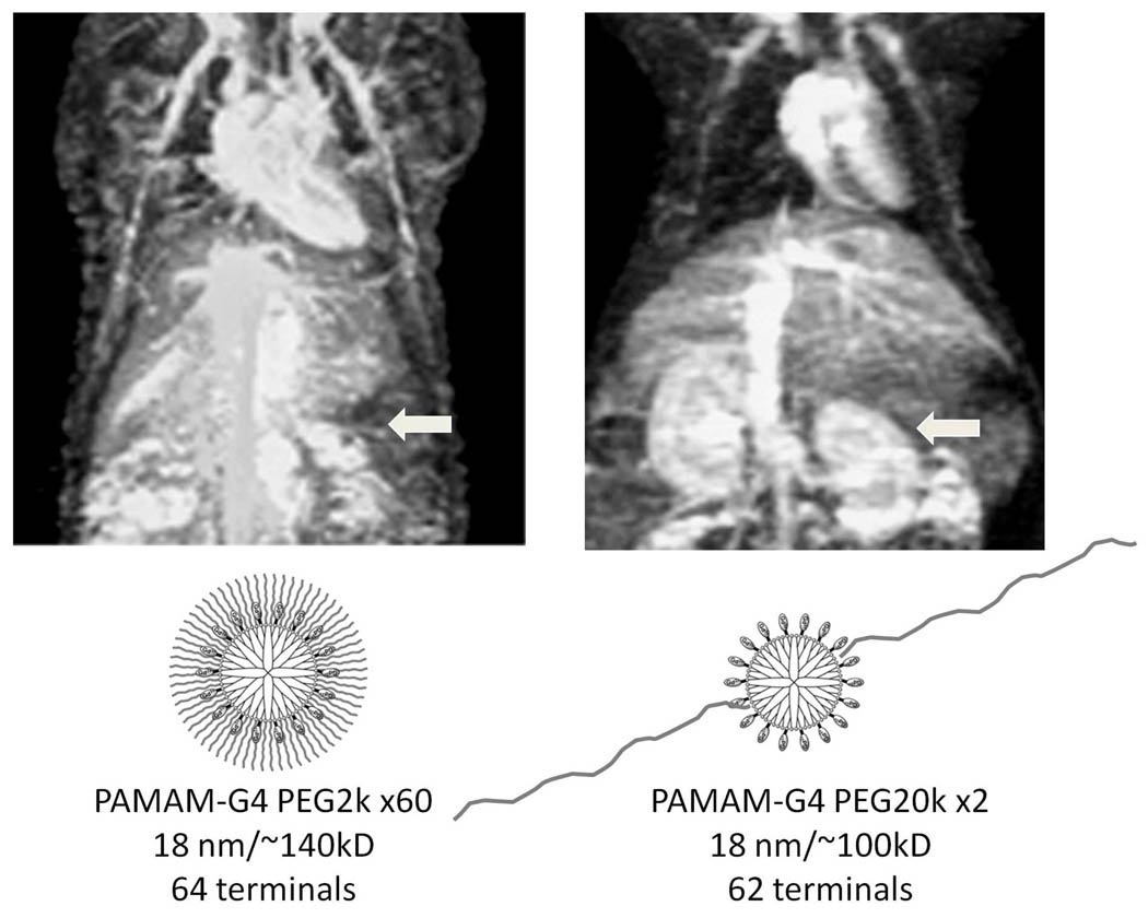
Traditionally, dendrimers were synthesized as spherical nanoparticles. However, recently surface modifications conjugating linear molecules to the surface produce nonspherical shapes with different pharmacokinetics. Despite a large particle size conferred by one or two linear, long PEGylated tails 10 , the particles may still undergo relatively rapid renal clearance. In contrast, the same generation of dendrimers with short PEG tails do not show renal clearance (Figure 1). 25
Figure 1.
Two long linear PEG (20kD) conjugated PAMAM-G4-Gd shows renal excretion. However, Short PEG (2kD) conjugated PAMAM-G4-Gd of similar physical size shows no renal excretion.
Surface modifications to alter biodistribution
To achieve target tissue accumulation, probes must avoid non-specific uptake. Of the strategies to alter in vivo behavior, addition of poly(ethylene glycol)(PEG) is among the most widely utilized approaches. One consistent limitation to the use of polymeric nanoparticles in vivo is their premature elimination from the circulatory system by the mononuclear phagocyte system. 7, 26 This process is initiated by serum opsonins attaching to the surface of nanoparticles in circulation. Following opsonization, macrophages recognize, phagocytose, and then sequester the particles in the liver, spleen, and/or bone marrow. PEGylation is one method to camouflage nano-size molecules and particles to prevent adhesion of opsonins so that the particles remain in circulation and evade the reticuloendothelial system, a process referred to as the “stealth effect.” 7 It is proposed that PEG reduces opsonization through steric and hydration effects. 7, 27 Interestingly, although PEGylation increases size, which has been associated with faster uptake by the liver and RES, numerous reports indicate that PEGylation increases circulatory retention times and decreases liver uptake. 28
Flexibility
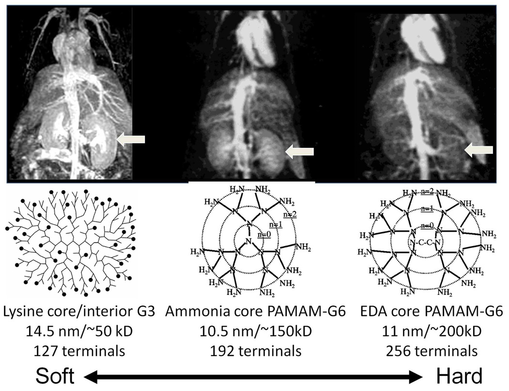
At this time, particle flexibility is not widely considered an important determinant of in vivo nano-sized molecule or particle behavior. However, given the dynamic nature of living systems and the time course over which particles interact with internal tissues, particle flexibility may have an important role in how nano-sized molecules or particles interface with living tissues. Research suggests flexibility is an important determinant of in vivo behavior. Given the highly adaptable nature of these particles, it is possible to achieve high degrees of intramolecular flexibility. Dendrimers may represent an ideal particle to study the effects of particle flexibility in living systems as well as an ideal platform to then harness the insights for optimizing particle platforms. In one example, a large particle conferred by one or two linear, long PEGylated tails 17 or the use of more flexible interiors including lysine dendrimers 29 or ammonia core PAMAM dendrimer 30 , the more flexibile particle demonstrated increased renal clearance (Figure 2). In contrast, less flexible EDA-core dendrimers 17 or dendrimers grafted with short PEG tails did not show the renal clearance. 25 Interestingly, flexible dendrimer based MR contrast agents exhibited improved renal clearance but retained similar relaxivity compared with “hard” dendrimers, which were not excreted by the kidneys. 19
Figure 2.
Despite of small size, hard interior (high density) EDA-core PAMAM-G6 does not show renal excretion. Two other “softer” interior dendrimers show renal excretion.
Biological application of physical characteristics
The in vivo affinity of nanoparticles for specific anatomic locations and their behavior at specific biological interfaces plays an important role in probe characteristics and toxicity. Specific interfaces include the glomerular basement membrane, the blood-brain barrier, vascular and lymphatic endothelial linings, and hepatic sinusoids. The ability or inability of nanoparticles to cross these interfaces will in large part determine their biodistribution and hence their utility.
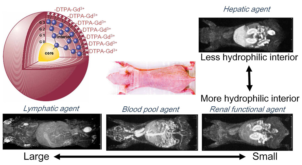
Dendrimers have shown to be useful imaging agents of various anatomic and physiologic processes. Preclinical studies demonstrate PAMAM-based contrast agents are effective for MR liver imaging 31, 32 , renal functional imaging 33, 34 , and angiography 35–37 , and lymphography. 20, 38 (Figure 3) Macromolecular MR contrast agents prepared from dendrimers have the advantage of uniform molecular weight distribution, relatively controlled structure, high relaxivities, and high loading of gadolinium chelates on the surface. 39, 40 Dendrimers are particularly well suited for lymphatic imaging 41–43 as this requires a modality with high spatial resolution and high sensitivity for contrast agents, since the lymphatics themselves are so small. 19, 41–52 Dendrimers are retained within the lymphatics, especially when their surface charge is modified with an appropriate coating or when conjugated to specific fluorophores 35, 40, 53–55 . Smaller molecules (< 6 nm) are taken up by the lymphatics, however, do not remain in the lymphatic vessels, whereas large molecules (> 20 nm) are taken up very slowly but are retained. 41 Dendrimer-based contrast agents between 6–10nm are retained within the lymphatics but are cleared from the sentinel lymph nodes within a day, which enables repeated injections. Additionally, these particles can be labeled with organic fluorophores (for optical imaging), radioisotopes (for scintigraphy) and paramagnetic lanthanide ions (for MRI). 40 Pre-clinical MR lymphangiography demonstrates that intradermally injected dendrimers are effective for imaging lymphatics in small and large animal models. 41, 43 This may eventually play a vital role in identifying otherwise life threatening thoracic duct injuries that occur during some pediatric cardiovascular surgeries.
Figure 3.
Strategic use of dendrimer size to achieve organ-specific imaging. This schema depits a generation 3 dendrimer, with core chemistry, shape, and surface modifications functionalized for use as an MRI contrast agent. Images of mice demonstrate that stragtegic selection of dendrimer size enables target organ-specific imaging. For example, PAMAM-G8 shows the lymphatic system; PAMAM-G6 shows the blood pool; PAMAM-G4 depicts renal function; and PPI/DAM-G4 depicts liver parenchyma.
Dendrimers are also useful nanoparticles for urinary tract imaging as well as for studying nano-sized molecular pharmacokinetics. PAMAM-based Gd(III) complexes have shown size-dependent pharmacokinetics and renal filtration. 19 For instance, particles 2 nm in size are handled very similarly to low molecular weight contrast agents, yet chemically similar but larger (6 nm) probes undergo comparatively slow renal clearance and particles that are 11 nm or larger do not undergo renal clearance at all. 56 Research suggests that the generation-4 (G4) dendrimer, which is approximately 6 nm in diameter, is ideal for renal imaging. 15, 57 When administered in a normally functioning kidney, the G4 dendrimer accumulates in the proximal tubules, which is seen on imaging as enhancement of the outer stripe of the medulla. 56 This phenomenon enables the depiction of acute tubular injury (seen as loss of the medullary stripe), which correlates with the extent of renal impairment. 16 Advancements in dendrimer-based contrast agents include the advent of biodegradable agents, which typically rely on endogenous enzymes to cleave the probe, creating smaller, more easily cleared bi-products. Recently, a biodegradable macromolecular dendrimer-based agent, nanoglobule-G4-cystamin-(Gd-DO3A) conjugate was created as an extracellular degradable nano-sized contrast agent for dynamic contrast enhanced MRI. A disulfide spacer was introduced to accelerate the renal excretion of gadolinium chelates by reducing the spacer with free endogenous thiols and homocysteine in the plasma such that released chelates underwent rapid renal filtration with subsequent urinary tract accumulation sufficient for MR urography. The nanoglobular dendrimer demonstrated lower cytotoxicity, relatively rapid renal filtration, and low liver uptake 58 and in vivo studies demonstrated fast elimination kinetics that were ideal for evaluation of the kidneys. 59
Effect of targeting on biodistribution
Conjugation of targeting ligands directly and indirectly alters biodistribution and clearance of nanoparticle based imaging agents. Numerous strategies have been developed to manipulate in vivo behavior of nanoparticles by modifying the surface chemistry. Among the various nanoparticles, dendrimers present an ideal platform for conjugation of various targeting ligands, such as folic acid, avidin-biotin complex, RGD peptides, and carbohydrate molecules. Attachment of such ligands alters particle shape, flexibility, and size. Yang et al conjugated anti-EGF antibody to boronated PAMAM-G4 dendrimers for targeting to EGF receptor expressing tumor cells and found that at 24 h post-injection, the conjugates were highly localized in EGFR-positive glioma with a tumor to blood ratio substantially higher than mice bearing tumors without EGFR receptors or mice injected with non-conjugated PAMAM dendrimers. 60 Dendrimers have also been targeted with smaller molecules such as folate. 61–67 Folate-modified PAMAM dendrimers showed significantly high probe accumulation, however, hepatic and renal accumulation was also observed, likely due to the presence of folate receptors in hepatic macrophages and renal proximal tubules.68
Another targeting molecule is the avidin-biotin complex system, which selectively targets various types of tumors. Wilbur et al showed that 125I–labeled iodobenzoate-biotinylated PAMAM dendrimers (G0–4) were quickly cleared, giving low blood levels and high kidney and liver accumulation compared to unmodified counter parts. 68, 69 G4 dendrimers complexed with olig-DNA and 111In demonstrated lower hepatic uptake and higher kidney and spleen uptake that similar constructs without a dendrimer carrier. A similar construct containing oligo-DNA and avidin-biotin conjugated to a G4 dendrimer demonstrated very high uptake in the lung, which was thought to be due to trapping of large molecular weight complexes within the lung. 70 Avidin-biotin targeting has been extended for MRI, Gd-neutron capture therapy, and gene delivery. 71, 72 After intraperitoneal administration, avidin-conjugated PAMAM-G6-Gd conjugates exhibited specific accumulation in intraperitoneally disseminated SHIN3 ovarian cancer tumors with 366- and 3.4 fold greater values than Gd-DTPA and unconjugated PAMAM-Gd respectively. 71
Baker et al have used PAMAM dendrimers as a vehicle to achieve cancer-targeted drug delivery by functionalizing the particles with riboflavin, methotrexate, and other tumor-cell binding moieties. 73–75 By utilizing generation-5 PAMAM dendrimers, it is possible to deliver sizable chemotherapeutic payloads and the particles can be synthesized as an appropriately monodispersed product that demonstrates optimal pharmacokinetics.
Conclusion
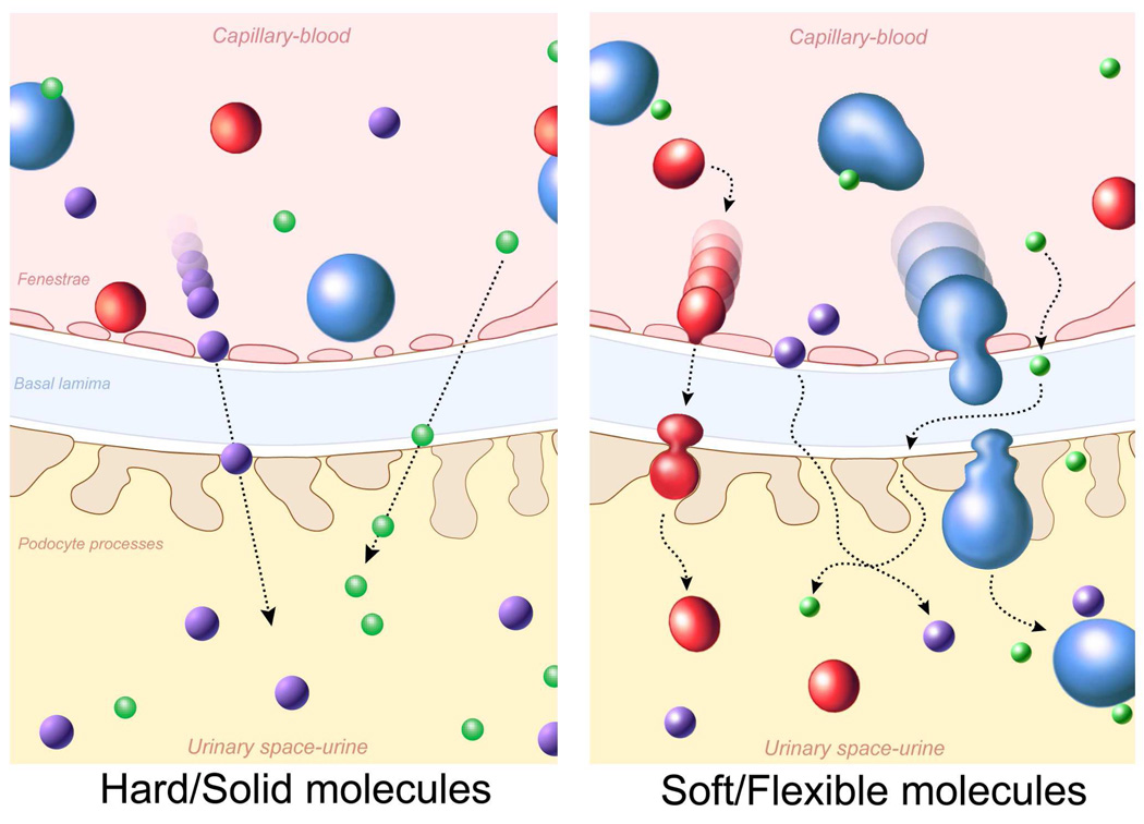
To effectively move nano-sized molecules or particles from the bench to human applications will require gaining a better understanding of their in vivo behavior. At this time, our understanding is rather limited as studies have primarily focused on the pharmacokinetics of relatively spherical molecules and size has been the primary parameter evaluated. To improve upon this, we need to consider other molecular parameters such as shape and flexibility as well as expand biodistribution studies beyond hard and spherical particles (Figure 4). As more information becomes available regarding the effects of these other parameters and particle types, we may move towards categorization systems that reflect in vivo behavior. Such a step forward will facilitate efficient and effective design and chemical synthesis and may also expedite the approval process for use in humans.
Figure 4.
A schema for glomerular filtration of hard and soft nano-sized molecules or particles.
Acknowledgments
This research was supported by the Intramural Research Program of the National Institutes of Health, National Cancer Institute, Center for Cancer Research.
References
- 1.Alexis F, Pridgen E, Molnar LK, Farokhzad OC. Factors affecting the clearance and biodistribution of polymeric nanoparticles. Mol Pharm. 2008;5:505–515. doi: 10.1021/mp800051m. [DOI] [PMC free article] [PubMed] [Google Scholar]
- 2.Bartneck M, Keul HA, Zwadlo-Klarwasser G, Groll J. Phagocytosis independent extracellular nanoparticle clearance by human immune cells. Nano Lett. 2010;10:59–63. doi: 10.1021/nl902830x. [DOI] [PubMed] [Google Scholar]
- 3.Briley-Saebo KC, Johansson LO, Hustvedt SO, Haldorsen AG, Bjørnerud A, Fayad ZA, Ahlstrom HK. Clearance of iron oxide particles in rat liver: effect of hydrated particle size and coating material on liver metabolism. Invest Radiol. 2006;41:560–571. doi: 10.1097/01.rli.0000221321.90261.09. [DOI] [PubMed] [Google Scholar]
- 4.Bulte JW, Kraitchman DL. Iron oxide MR contrast agents for molecular and cellular imaging. NMR Biomed. 2004;17:484–499. doi: 10.1002/nbm.924. [DOI] [PubMed] [Google Scholar]
- 5.Choi HS, Liu W, Misra P, Tanaka E, Zimmer JP, Itty Ipe B, Bawendi MG, Frangioni JV. Renal clearance of quantum dots. Nat Biotechnol. 2007;25:1165–1170. doi: 10.1038/nbt1340. [DOI] [PMC free article] [PubMed] [Google Scholar]
- 6.Choi HS, Liu W, Liu F, Nasr K, Misra P, Bawendi MG, Frangioni JV. Design considerations for tumour-targeted nanoparticles. Nat Nanotechnol. 2010;5:42–47. doi: 10.1038/nnano.2009.314. [DOI] [PMC free article] [PubMed] [Google Scholar]
- 7.Gonçalves C, Torrado E, Martins T, Pereira P, Pedrosa J, Gama M. Dextrin nanoparticles: studies on the interaction with murine macrophages and blood clearance. Colloids Surf B Biointerfaces. 2010;75:483–489. doi: 10.1016/j.colsurfb.2009.09.024. [DOI] [PubMed] [Google Scholar]
- 8.He X, Nie H, Wang K, Tan W, Wu X, Zhang P. In vivo study of biodistribution and urinary excretion of surface-modified silica nanoparticles. Anal Chem. 2008;80:9597–9603. doi: 10.1021/ac801882g. [DOI] [PubMed] [Google Scholar]
- 9.Jain TK, Reddy MK, Morales MA, Leslie-Pelecky DL, Labhasetwar V. Biodistribution, clearance, and biocompatibility of iron oxide magnetic nanoparticles in rats. Mol Pharm. 2008;5:316–327. doi: 10.1021/mp7001285. [DOI] [PubMed] [Google Scholar]
- 10.Kobayashi H, Sato N, Kawamoto S, Saga T, Hiraga A, Ishimori T, Konishi J, Togashi K, Brechbiel MW. Novel intravascular macromolecular MRI contrast agent with generation-4 polyamidoamine dendrimer core: accelerated renal excretion with coinjection of lysine. Magn Reson Med. 2001;46:457–464. doi: 10.1002/mrm.1214. [DOI] [PubMed] [Google Scholar]
- 11.Longmire M, Choyke PL, Kobayashi H. Clearance properties of nano-sized particles and molecules as imaging agents: considerations and caveats. Nanomedicine (Lond) 2008;3:703–717. doi: 10.2217/17435889.3.5.703. [DOI] [PMC free article] [PubMed] [Google Scholar]
- 12.Schipper ML, Iyer G, Koh AL, Cheng Z, Ebenstein Y, Aharoni A, Keren S, Bentolila LA, Li J, Rao J, Chen X, Banin U, Wu AM, Sinclair R, Weiss S, Gambhir SS. Particle size, surface coating, and PEGylation influence the biodistribution of quantum dots in living mice. Small. 2009;5:126–134. doi: 10.1002/smll.200800003. [DOI] [PMC free article] [PubMed] [Google Scholar]
- 13.Sigovan M, Boussel L, Sulaiman A, Sappey-Marinier D, Alsaid H, Desbleds-Mansard C, Ibarrola D, Gamondès D, Corot C, Lancelot E, Raynaud JS, Vives V, Laclédère C, Violas X, Douek PC, Canet-Soulas E. Rapid-clearance iron nanoparticles for inflammation imaging of atherosclerotic plaque: initial experience in animal model. Radiology. 2009;252:401–409. doi: 10.1148/radiol.2522081484. [DOI] [PubMed] [Google Scholar]
- 14.Kobayashi H, Kawamoto S, Jo SK, Bryant HL, Brechbiel MW, Star RA. Macromolecular MRI contrast agents with small dendrimers: pharmacokinetic differences between sizes and cores. Bioconjug Chem. 2003;14:388–394. doi: 10.1021/bc025633c. [DOI] [PubMed] [Google Scholar]
- 15.Kobayashi H, Brechbiel MW. Dendrimer-based nanosized MRI contrast agents. Curr Pharm Biotechnol. 2004;5:539–549. doi: 10.2174/1389201043376571. [DOI] [PubMed] [Google Scholar]
- 16.Kobayashi H, Jo SK, Kawamoto S, Yasuda H, Hu X, Knopp MV, Brechbiel MW, Choyke PL, Star RA. Polyamine dendrimer-based MRI contrast agents for functional kidney imaging to diagnose acute renal failure. J Magn Reson Imaging. 2004;20:512–518. doi: 10.1002/jmri.20147. [DOI] [PubMed] [Google Scholar]
- 17.Kobayashi H, Kawamoto S, Saga T, Sato N, Hiraga A, Ishimori T, Konishi J, Togashi K, Brechbiel MW. Positive effects of polyethylene glycol conjugation to generation-4 polyamidoamine dendrimers as macromolecular MR contrast agents. Magn Reson Med. 2001;46:781–788. doi: 10.1002/mrm.1257. [DOI] [PubMed] [Google Scholar]
- 18.Tomalia DA. In quest of a systematic framework for unifying and defining nanoscience. J Nanopart Res. 2009;11:1251–1310. doi: 10.1007/s11051-009-9632-z. [DOI] [PMC free article] [PubMed] [Google Scholar]
- 19.Kobayashi H, Brechbiel MW. Nano-sized MRI contrast agents with dendrimer cores. Adv Drug Deliv Rev. 2005;57:2271–2286. doi: 10.1016/j.addr.2005.09.016. [DOI] [PubMed] [Google Scholar]
- 20.Sato N, Kobayashi H, Hiraga A, Saga T, Togashi K, Konishi J, Brechbiel MW. Pharmacokinetics and enhancement patterns of macromolecular MR contrast agents with various sizes of polyamidoamine dendrimer cores. Magn Reson Med. 2001;46:1169–1173. doi: 10.1002/mrm.1314. [DOI] [PubMed] [Google Scholar]
- 21.Canelas DA, Herlihy KP, DeSimone JM. Top-down particle fabrication: control of size and shape for diagnostic imaging and drug delivery. Wiley Interdiscip Rev Nanomed Nanobiotechnol. 2009;1:391–404. doi: 10.1002/wnan.40. [DOI] [PMC free article] [PubMed] [Google Scholar]
- 22.Sadekar S, Ray A, Janàt-Amsbury M, Peterson CM, Ghandehari H. Comparative biodistribution of PAMAM dendrimers and HPMA copolymers in ovarian-tumor-bearing mice. Biomacromolecules. 2011;12:88–96. doi: 10.1021/bm101046d. [DOI] [PMC free article] [PubMed] [Google Scholar]
- 23.Geng Y, Dalhaimer P, Cai S, Tsai R, Tewari M, Minko T, Discher DE. Shape effects of filaments versus spherical particles in flow and drug delivery. Nat Nanotechnol. 2007;2:249–255. doi: 10.1038/nnano.2007.70. [DOI] [PMC free article] [PubMed] [Google Scholar]
- 24.Arnida Janát-Amsbury MM, Ray A, Peterson CM, Ghandehari H. Geometry and surface characteristics of gold nanoparticles influence their biodistribution and uptake by macrophages. Eur J Pharm Biopharm. 2010 doi: 10.1016/j.ejpb.2010.11.010. [DOI] [PMC free article] [PubMed] [Google Scholar]
- 25.Kojima C, Regino C, Umeda Y, Kobayashi H, Kono K. Influence of dendrimer generation and polyethylene glycol length on the biodistribution of PEGylated dendrimers. Int J Pharm. 2010;383:293–296. doi: 10.1016/j.ijpharm.2009.09.015. [DOI] [PubMed] [Google Scholar]
- 26.Owens DE, Peppas NA. Opsonization, biodistribution, and pharmacokinetics of polymeric nanoparticles. Int J Pharm. 2006;307:93–102. doi: 10.1016/j.ijpharm.2005.10.010. [DOI] [PubMed] [Google Scholar]
- 27.Sheng Y, Yuan Y, Liu C, Tao X, Shan X, Xu F. In vitro macrophage uptake and in vivo biodistribution of PLA-PEG nanoparticles loaded with hemoglobin as blood substitutes: effect of PEG content. J Mater Sci Mater Med. 2009;20:1881–1891. doi: 10.1007/s10856-009-3746-9. [DOI] [PubMed] [Google Scholar]
- 28.Gaumet M, Vargas A, Gurny R, Delie F. Nanoparticles for drug delivery: the need for precision in reporting particle size parameters. Eur J Pharm Biopharm. 2008;69:1–9. doi: 10.1016/j.ejpb.2007.08.001. [DOI] [PubMed] [Google Scholar]
- 29.Ogawa M, Regino CA, Marcelino B, Williams M, Kosaka N, Bryant LH, Jr, Choyke PL, Kobayashi H. New nanosized biocompatible MR contrast agents based on lysine-dendri-graft macromolecules. Bioconjug Chem. 2010;21:955–960. doi: 10.1021/bc9005442. [DOI] [PMC free article] [PubMed] [Google Scholar]
- 30.Kobayashi H, Sato N, Kawamoto S, Saga T, Hiraga A, Haque TL, Ishimori T, Konishi J, Togashi K, Brechbiel MW. Comparison of the macromolecular MR contrast agents with ethylenediamine-core versus ammonia-core generation-6 polyamidoamine dendrimer. Bioconjug Chem. 2001;12:100–107. doi: 10.1021/bc000075s. [DOI] [PubMed] [Google Scholar]
- 31.Kobayashi H, Saga T, Kawamoto S, Sato N, Hiraga A, Ishimori T, Konishi J, Togashi K, Brechbiel MW. Dynamic micro-magnetic resonance imaging of liver micrometastasis in mice with a novel liver macromolecular magnetic resonance contrast agent DAB-Am64-(1B4M–Gd)(64) Cancer Res. 2001;61:4966–4970. [PubMed] [Google Scholar]
- 32.Kobayashi H, Kawamoto S, Saga T, Sato N, Hiraga A, Ishimori T, Akita Y, Mamede MH, Konishi J, Togashi K, Brechbiel MW. Novel liver macromolecular MR contrast agent with a polypropylenimine diaminobutyl dendrimer core: comparison to the vascular MR contrast agent with the polyamidoamine dendrimer core. Magn Reson Med. 2001;46:795–802. doi: 10.1002/mrm.1259. [DOI] [PubMed] [Google Scholar]
- 33.Dear JW, Kobayashi H, Jo SK, Holly MK, Hu X, Yuen PS, Brechbiel MW, Star RA. Dendrimer-enhanced MRI as a diagnostic and prognostic biomarker of sepsis-induced acute renal failure in aged mice. Kidney Int. 2005;67:2159–2167. doi: 10.1111/j.1523-1755.2005.00321.x. [DOI] [PubMed] [Google Scholar]
- 34.Kobayashi H, Kawamoto S, Jo S, Sato N, Saga T, Hiraga A, Konishi J, Hu S, Togashi K, Brechbiel MW, Star RA. Renal tubular damage detected by dynamic micro-MRI with a dendrimer-based MR contrast agent. Kidney Int. 2002;61:1980–1985. doi: 10.1046/j.1523-1755.2002.00364.x. [DOI] [PubMed] [Google Scholar]
- 35.Kobayashi H, Sato N, Hiraga A, Saga T, Nakamoto Y, Ueda H, Konishi J, Togashi K, Brechbiel MW. 3D–micro-MR angiography of mice using macromolecular MR contrast agents with polyamidoamine dendrimer core with reference to their pharmacokinetic properties. Magn Reson Med. 2001;45:454–460. doi: 10.1002/1522-2594(200103)45:3<454::aid-mrm1060>3.0.co;2-m. [DOI] [PubMed] [Google Scholar]
- 36.Kobayashi H, Kawamoto S, Saga T, Sato N, Hiraga A, Konishi J, Togashi K, Brechbiel MW. Micro-MR angiography of normal and intratumoral vessels in mice using dedicated intravascular MR contrast agents with high generation of polyamidoamine dendrimer core: reference to pharmacokinetic properties of dendrimer-based MR contrast agents. J Magn Reson Imaging. 2001;14:705–713. doi: 10.1002/jmri.10025. [DOI] [PubMed] [Google Scholar]
- 37.Kobayashi H, Sato N, Kawamoto S, Saga T, Hiraga A, Ishimori T, Konishi J, Togashi K, Brechbiel MW. 3D MR angiography of intratumoral vasculature using a novel macromolecular MR contrast agent. Magn Reson Med. 2001;46:579–585. doi: 10.1002/mrm.1230. [DOI] [PubMed] [Google Scholar]
- 38.Xu R, Wang Y, Wang X, Jeong EK, Parker DL, Lu ZR. In Vivo evaluation of a PAMAM-cystamine-(Gd-DO3A) conjugate as a biodegradable macromolecular MRI contrast agent. Exp Biol Med (Maywood) 2007;232:1081–1089. doi: 10.3181/0702-RM-33. [DOI] [PubMed] [Google Scholar]
- 39.Kobayashi H, Brechbiel MW. Dendrimer-based macromolecular MRI contrast agents: characteristics and application. Mol Imaging. 2003;2:1–10. doi: 10.1162/15353500200303100. [DOI] [PubMed] [Google Scholar]
- 40.Kobayashi H, Ogawa M, Kosaka N, Choyke PL, Urano Y. Multicolor imaging of lymphatic function with two nanomaterials: quantum dot-labeled cancer cells and dendrimer-based optical agents. Nanomedicine (Lond) 2009;4:411–419. doi: 10.2217/nnm.09.15. [DOI] [PMC free article] [PubMed] [Google Scholar]
- 41.Kobayashi H, Kawamoto S, Choyke PL, Sato N, Knopp MV, Star RA, Waldmann TA, Tagaya Y, Brechbiel MW. Comparison of dendrimer-based macromolecular contrast agents for dynamic micro-magnetic resonance lymphangiography. Magn Reson Med. 2003;50:758–766. doi: 10.1002/mrm.10583. [DOI] [PubMed] [Google Scholar]
- 42.Koyama Y, Talanov VS, Bernardo M, Hama Y, Regino CA, Brechbiel MW, Choyke PL, Kobayashi H. A dendrimer-based nanosized contrast agent dual-labeled for magnetic resonance and optical fluorescence imaging to localize the sentinel lymph node in mice. J Magn Reson Imaging. 2007;25:866–871. doi: 10.1002/jmri.20852. [DOI] [PubMed] [Google Scholar]
- 43.Sena LM, Fishman SJ, Jenkins KJ, Xu H, Brechbiel MW, Regino CA, Kosaka N, Bernardo M, Choyke PL, Kobayashi H. Magnetic resonance lymphangiography with a nano-sized gadolinium-labeled dendrimer in small and large animal models. Nanomedicine (Lond) 2010;5:1183–1191. doi: 10.2217/nnm.10.70. [DOI] [PMC free article] [PubMed] [Google Scholar]
- 44.Hama Y, Koyama Y, Urano Y, Choyke PL, Kobayashi H. Two-color lymphatic mapping using Ig-conjugated near infrared optical probes. J Invest Dermatol. 2007;127:2351–2356. doi: 10.1038/sj.jid.5700892. [DOI] [PubMed] [Google Scholar]
- 45.Kim S, Lim YT, Soltesz EG, De Grand AM, Lee J, Nakayama A, Parker JA, Mihaljevic T, Laurence RG, Dor DM, Cohn LH, Bawendi MG, Frangioni JV. Near-infrared fluorescent type II quantum dots for sentinel lymph node mapping. Nat Biotechnol. 2004;22:93–97. doi: 10.1038/nbt920. [DOI] [PMC free article] [PubMed] [Google Scholar]
- 46.Kobayashi H, Kawamoto S, Bernardo M, Brechbiel MW, Knopp MV, Choyke PL. Delivery of gadolinium-labeled nanoparticles to the sentinel lymph node: comparison of the sentinel node visualization and estimations of intra-nodal gadolinium concentration by the magnetic resonance imaging. J Control Release. 2006;111:343–351. doi: 10.1016/j.jconrel.2005.12.019. [DOI] [PubMed] [Google Scholar]
- 47.Kobayashi H, Kawamoto S, Sakai Y, Choyke PL, Star RA, Brechbiel MW, Sato N, Tagaya Y, Morris JC, Waldmann TA. Lymphatic drainage imaging of breast cancer in mice by micro-magnetic resonance lymphangiography using a nano-size paramagnetic contrast agent. J Natl Cancer Inst. 2004;96:703–708. doi: 10.1093/jnci/djh124. [DOI] [PubMed] [Google Scholar]
- 48.Kobayashi H, Kawamoto S, Star RA, Waldmann TA, Tagaya Y, Brechbiel MW. Micro-magnetic resonance lymphangiography in mice using a novel dendrimer-based magnetic resonance imaging contrast agent. Cancer Res. 2003;63:271–276. [PubMed] [Google Scholar]
- 49.Kobayashi H, Kawamoto S, Brechbiel MW, Bernardo M, Sato N, Waldmann TA, Tagaya Y, Choyke PL. Detection of lymph node involvement in hematologic malignancies using micromagnetic resonance lymphangiography with a gadolinum-labeled dendrimer nanoparticle. Neoplasia. 2005;7:984–991. doi: 10.1593/neo.05454. [DOI] [PMC free article] [PubMed] [Google Scholar]
- 50.Kobayashi H, Hama Y, Koyama Y, Barrett T, Regino CA, Urano Y, Choyke PL. Simultaneous multicolor imaging of five different lymphatic basins using quantum dots. Nano Lett. 2007;7:1711–1716. doi: 10.1021/nl0707003. [DOI] [PubMed] [Google Scholar]
- 51.Kobayashi H, Koyama Y, Barrett T, Hama Y, Regino CA, Shin IS, Jang BS, Le N, Paik CH, Choyke PL, Urano Y. Multimodal nanoprobes for radionuclide and five-color near-infrared optical lymphatic imaging. ACS Nano. 2007;1:258–264. doi: 10.1021/nn700062z. [DOI] [PMC free article] [PubMed] [Google Scholar]
- 52.Mounzer R, Shkarin P, Papademetris X, Constable T, Ruddle NH, Fahmy TM. Dynamic imaging of lymphatic vessels and lymph nodes using a bimodal nanoparticulate contrast agent. Lymphat Res Biol. 2007;5:151–158. doi: 10.1089/lrb.2007.5302. [DOI] [PubMed] [Google Scholar]
- 53.Kono K, Kojima C, Hayashi N, Nishisaka E, Kiura K, Watarai S, Harada A. Preparation and cytotoxic activity of poly(ethylene glycol)-modified poly(amidoamine) dendrimers bearing adriamycin. Biomaterials. 2008;29:1664–1675. doi: 10.1016/j.biomaterials.2007.12.017. [DOI] [PubMed] [Google Scholar]
- 54.Kojima C, Kono K, Maruyama K, Takagishi T. Synthesis of polyamidoamine dendrimers having poly(ethylene glycol) grafts and their ability to encapsulate anticancer drugs. Bioconjug Chem. 2000;11:910–917. doi: 10.1021/bc0000583. [DOI] [PubMed] [Google Scholar]
- 55.Sato N, Kobayashi H, Saga T, Nakamoto Y, Ishimori T, Togashi K, Fujibayashi Y, Konishi J, Brechbiel MW. Tumor targeting and imaging of intraperitoneal tumors by use of antisense oligo-DNA complexed with dendrimers and/or avidin in mice. Clin Cancer Res. 2001;7:3606–3612. [PubMed] [Google Scholar]
- 56.Choyke PL, Kobayashi H. Functional magnetic resonance imaging of the kidney using macromolecular contrast agents. Abdom Imaging. 2006;31:224–231. doi: 10.1007/s00261-005-0390-9. [DOI] [PubMed] [Google Scholar]
- 57.Kobayashi H, Reijnders K, English S, Yordanov AT, Milenic DE, Sowers AL, Citrin D, Krishna MC, Waldmann TA, Mitchell JB, Brechbiel MW. Application of a macromolecular contrast agent for detection of alterations of tumor vessel permeability induced by radiation. Clin Cancer Res. 2004;10:7712–7720. doi: 10.1158/1078-0432.CCR-04-1175. [DOI] [PubMed] [Google Scholar]
- 58.Kaneshiro TL, Jeong EK, Morrell G, Parker DL, Lu ZR. Synthesis and evaluation of globular Gd-DOTA-monoamide conjugates with precisely controlled nanosizes for magnetic resonance angiography. Biomacromolecules. 2008;9:2742–2748. doi: 10.1021/bm800486c. [DOI] [PMC free article] [PubMed] [Google Scholar]
- 59.Xu R, Kaneshiro TL, Jeong EK, Parker DL, Lu ZR. Synthesis and evaluation of nanoglobule-cystamine-(Gd-DO3A), a biodegradable nanosized magnetic resonance contrast agent for dynamic contrast-enhanced magnetic resonance urography. Int J Nanomedicine. 2010;5:707–713. doi: 10.2147/ijn.s12224. [DOI] [PMC free article] [PubMed] [Google Scholar]
- 60.Yang W, Barth RF, Adams DM, Soloway AH. Intratumoral delivery of boronated epidermal growth factor for neutron capture therapy of brain tumors. Cancer Res. 1997;57:4333–4339. [PubMed] [Google Scholar]
- 61.Kono K, Liu M, Frechet JM. Design of dendritic macromolecules containing folate or methotrexate residues. Bioconjug Chem. 1999;10:1115–1121. doi: 10.1021/bc990082k. [DOI] [PubMed] [Google Scholar]
- 62.Sideratou Z, Kontoyianni C, Drossopoulou GI, Paleos CM. Synthesis of a folate functionalized PEGylated poly(propylene imine) dendrimer as prospective targeted drug delivery system. Bioorg Med Chem Lett. 2010;20:6513–6517. doi: 10.1016/j.bmcl.2010.09.058. [DOI] [PubMed] [Google Scholar]
- 63.Wiener EC, Konda S, Shadron A, Brechbiel M, Gansow O. Targeting dendrimer-chelates to tumors and tumor cells expressing the high-affinity folate receptor. Invest Radiol. 1997;32:748–754. doi: 10.1097/00004424-199712000-00005. [DOI] [PubMed] [Google Scholar]
- 64.Zhang Y, Sun Y, Xu X, Zhang X, Zhu H, Huang L, Qi Y, Shen YM. Synthesis, biodistribution, and microsingle photon emission computed tomography (SPECT) imaging study of technetium-99m labeled PEGylated dendrimer poly(amidoamine) (PAMAM)-folic acid conjugates. J Med Chem. 2010;53:3262–3272. doi: 10.1021/jm901910j. [DOI] [PubMed] [Google Scholar]
- 65.Cao W, Zhou J, Wang Y, Zhu L. Synthesis and in vitro cancer cell targeting of folate-functionalized biodegradable amphiphilic dendrimer-like star polymers. Biomacromolecules. 2010;11:3680–3687. doi: 10.1021/bm101154r. [DOI] [PubMed] [Google Scholar]
- 66.Glickson JD, Lund-Katz S, Zhou R, Choi H, Chen IW, Li H, Corbin I, Popov AV, Cao W, Song L, Qi C, Marotta D, Nelson DS, Chen J, Chance B, Zheng G. Lipoprotein nanoplatform for targeted delivery of diagnostic and therapeutic agents. Mol Imaging. 2008;7:101–110. [PubMed] [Google Scholar]
- 67.Konda SD, Aref M, Wang S, Brechbiel M, Wiener EC. Specific targeting of folate-dendrimer MRI contrast agents to the high affinity folate receptor expressed in ovarian tumor xenografts. MAGMA. 2001;12:104–113. doi: 10.1007/BF02668091. [DOI] [PubMed] [Google Scholar]
- 68.Wijagkanalan W, Kawakami S, Hashida M. Designing Dendrimers for Drug Delivery and Imaging: Pharmacokinetic Considerations. Pharm Res. 2010 doi: 10.1007/s11095-010-0339-8. [DOI] [PubMed] [Google Scholar]
- 69.Wilbur DS, Pathare PM, Hamlin DK, Buhler KR, Vessella RL. Biotin reagents for antibody pretargeting. 3. Synthesis, radioiodination, and evaluation of biotinylated starburst dendrimers. Bioconjug Chem. 1998;9:813–825. doi: 10.1021/bc980055e. [DOI] [PubMed] [Google Scholar]
- 70.Mamede M, Saga T, Ishimori T, Higashi T, Sato N, Kobayashi H, Brechbiel MW, Konishi J. Hepatocyte targeting of 111In-labeled oligo-DNA with avidin or avidin-dendrimer complex. J Control Release. 2004;95:133–141. doi: 10.1016/j.jconrel.2003.11.015. [DOI] [PubMed] [Google Scholar]
- 71.Kobayashi H, Kawamoto S, Saga T, Sato N, Ishimori T, Konishi J, Ono K, Togashi K, Brechbiel MW. Avidin-dendrimer-(1B4M–Gd)(254): a tumor-targeting therapeutic agent for gadolinium neutron capture therapy of intraperitoneal disseminated tumor which can be monitored by MRI. Bioconjug Chem. 2001;12:587–593. doi: 10.1021/bc010002o. [DOI] [PubMed] [Google Scholar]
- 72.Xu H, Regino CA, Koyama Y, Hama Y, Gunn AJ, Bernardo M, Kobayashi H, Choyke PL, Brechbiel MW. Preparation and preliminary evaluation of a biotin-targeted, lectin-targeted dendrimer-based probe for dual-modality magnetic resonance and fluorescence imaging. Bioconjug Chem. 2007;18:1474–1482. doi: 10.1021/bc0701085. [DOI] [PubMed] [Google Scholar]
- 73.Baker JR., Jr Dendrimer-based nanoparticles for cancer therapy. Hematology Am Soc Hematol Educ Program. 2009:708–719. doi: 10.1182/asheducation-2009.1.708. [DOI] [PubMed] [Google Scholar]
- 74.Majoros IJ, Williams CR, Baker JR., Jr Current dendrimer applications in cancer diagnosis and therapy. Curr Top Med Chem. 2008;8:1165–1179. doi: 10.2174/156802608785849049. [DOI] [PubMed] [Google Scholar]
- 75.Quintana A, Raczka E, Piehler L, Lee I, Myc A, Majoros I, Patri AK, Thomas T, Mule J, Baker JR., Jr Design and function of a dendrimer-based therapeutic nanodevice targeted to tumor cells through the folate receptor. Pharm Res. 2002;19:1310–1316. doi: 10.1023/a:1020398624602. [DOI] [PubMed] [Google Scholar]


