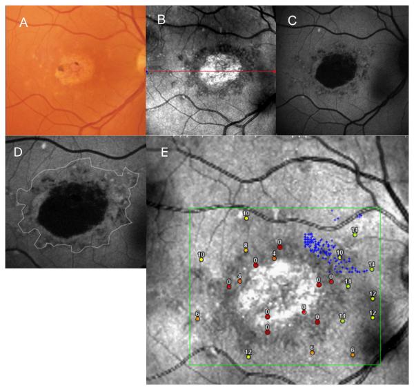
FIGURE 1.
(A, B, C) Composite of a color fundus photograph (gray scale), and SLO infrared and FAF images from patient 5 with a geographic, atrophic-appearing macular lesion.
(D) An outline of the atrophic macular lesion on SLO infrared imaging of patient 5, superimposed on the FAF image of this patient.
(E) Microperimetry responses superimposed on the SLO infrared image of patient 5.

