Abstract
The MEF2 factors regulate transcription during cardiac and skeletal myogenesis. MEF2 factors establish skeletal muscle commitment by amplifying and synergizing with MyoD. While phosphorylation is known to regulate MEF2 function, lineage-specific regulation is unknown. Here, we show that phosphorylation of MEF2C on T80 by skeletal myosin light chain kinase (skMLCK) enhances skeletal and not cardiac myogenesis. A phosphorylation-deficient MEF2C mutant (MEFT80A) enhanced cardiac, but not skeletal myogenesis in P19 stem cells. Further, MEFT80A was deficient in recruitment of p300 to skeletal but not cardiac muscle promoters. In gain-of-function studies, skMLCK upregulated myogenic regulatory factor (MRF) expression, leading to enhanced skeletal myogenesis in P19 cells and more efficient myogenic conversion. In loss-of-function studies, MLCK was essential for efficient MRF expression and subsequent myogenesis in embryonic stem (ES) and P19 cells as well as for proper activation of quiescent satellite cells. Thus, skMLCK regulates MRF expression by controlling the MEF2C-dependent recruitment of histone acetyltransferases to skeletal muscle promoters. This work identifies the first kinase that regulates MyoD and Myf5 expression in ES or satellite cells.
Keywords: MEF2, MRFs, skeletal myogenesis, skMLCK, stem cells
Introduction
Cellular differentiation is controlled by cascades of regulatory genes comprising combinations of widely expressed and cell type-restricted transcription factors. The MEF2 proteins (MEF2A-D) consist of a family of transcription factors that have a central role during the development of several tissues, including cardiac and skeletal muscle (Potthoff and Olson, 2007). While MEF2 can synergize with tissue-specific transcription factors in the various lineages (Molkentin et al, 1995; Naidu et al, 1995; Black et al, 1996; Morin et al, 2000), the signalling pathways that regulate MEF2 function in a lineage-specific manner remain to be determined.
P19 embryonal carcinoma (EC) cells are a well-established pluripotent embryonic stem (ES) cell model that has shed light on unique aspects of molecular mechanisms regulating cardiac and skeletal muscle development (Skerjanc, 1999; van der Heyden and Defize, 2003). Results in P19 cells have been confirmed in animal models and/or ES cells (Pandur et al, 2002; Karamboulas et al, 2006a; Kennedy et al, 2009). Following 4 days of cellular aggregation in the presence of dimethylsulfoxide (DMSO) to induce differentiation, P19-derived cardiomyocytes first appear on day 6, while skeletal muscle first appears on day 9. Similarly, mouse ES cells differentiate into cardiac muscle by day 6 and skeletal muscle by day 15, with a profile of gene expression analogous to P19 cells (van der Heyden and Defize, 2003; Kennedy et al, 2009; Gessert and Kuhl, 2010). Previously, we have shown that MEF2C can induce skeletal and cardiac myogenesis as well as neurogenesis in aggregated P19 stem cells (Skerjanc et al, 1998; Ridgeway et al, 2000; Skerjanc and Wilton, 2000), providing a unique tissue culture system in which to examine the cell type-specific regulation of MEF2C.
The activity and stability of MEF2 transcription factors are controlled by phosphorylation. The transcriptional activity of MEF2 family members can be enhanced upon phosphorylation by several kinases, including p38 MAPK, ERK5/BMK1, protein kinase C, and casein kinase-II (Molkentin et al, 1996b; Han et al, 1997; Kato et al, 1997; Yang et al, 1998; Ornatsky et al, 1999; Zhao et al, 1999; Cox et al, 2003; Barsyte-Lovejoy et al, 2004). Phosphorylation of MEF2A by ERK family members can target it for degradation, suggesting that ERK kinases have a dual function during myogenesis (Cox et al, 2003). In contrast, MEF2D transactivation properties are potently abolished upon phosphorylation by PKA at Ser-121 and Ser-190 (Du et al, 2008). Thus, it is clear that the MEF2 family is regulated by posttranslational modification, although the regulation of MEF2 by kinases in P19 EC or ES cells is not understood.
MLCK is important in regulating muscle contraction, cell motility, membrane events, and cell morphology (Gallagher and Stull, 1997; Kamm and Stull, 2001). In vertebrates, three genes code for MLCK, including smooth muscle (sm), skeletal muscle (sk), and cardiac muscle (c) MLCK (Kamm and Stull, 1986; Kennelly et al, 1987; Seguchi et al, 2007; Chan et al, 2008). Unlike, skeletal myosin light chain kinase (skMLCK) and cMLCK, which are specifically expressed in skeletal and cardiac muscles, respectively, smMLCK is expressed ubiquitously in a wide range of tissues. MLCK has a serine/threonine-kinase catalytic core and a regulatory segment containing autoinhibitory and calmodulin-binding domains (Herring et al, 1990; Takashima, 2009). Myosin II regulatory light chain is the only known substrate for MLCK (Kamm and Stull, 2001; Takashima, 2009).
To discover proteins that may regulate MEF2C function in a tissue-specific manner, we used the tandem affinity purification strategy (Cox et al, 2003) and identified skMLCK as a MEF2C-interacting protein during P19 cell differentiation. We set out to determine if skMLCK regulates MEF2C and if this regulation was lineage specific. We identified a novel role for skMLCK in regulating skeletal muscle commitment by controlling MEF2C-mediated recruitment of p300 to specific promoters.
Results
skMLCK binds and phosphorylates MEF2C
In order to identify proteins that regulate MEF2C, P19 stem cells expressing a TAP-tagged MEF2C were generated and characterized, confirming that MEF2C-TAP is functional and can enhance both cardiomyogenesis and skeletal myogenesis (Supplementary Figure S1A–C), as shown previously for untagged MEF2C (Skerjanc et al, 1998; Ridgeway et al, 2000). Purification and mass spectrometric analysis of proteins interacting with tagged MEF2C on day 5 of differentiation led to the identification of skMLCK as a novel MEF2C-interacting protein (Supplementary Figure S1D). Western blot analysis with anti-skMLCK antibodies validated the mass spectrometric findings (Supplementary Figure S1D) and demonstrated that skMLCK is present at low levels in C2C12 myoblasts and upregulated during differentiation (Supplementary Figure S1F). SkMLCK can be localized to the nucleus and the cytoplasm (Pujol et al, 1993) (Supplementary Figure S1E), indicating that it can be located in the same subcellular compartment as the transcription factor MEF2C.
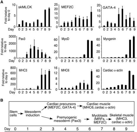
Identification of the interaction of MEF2C and skMLCK led us to study the expression profile of skMLCK during P19 EC myogenesis. Quantitative real-time PCR (Q–PCR) analysis showed that the expression of skMLCK transcripts increased during differentiation, almost 10-fold by day 9, compared with day 0 (Figure 1A). The transcripts of MEF2C increased on days 2–5 and 9, representing cardiac and skeletal muscle induction, respectively. GATA-4, found in cardiac muscle precursors (Grepin et al, 1997), showed increased levels starting at day 2 and peaking at days 5–6 (Figure 1A). Cardiomyocytes were observed by day 6 of differentiation (Figure 1A; Supplementary Figure S3). The expression of the skeletal premyogenic mesoderm gene, Pax3, peaked from days 4 to 6 and the myogenic regulatory factors (MRFs) increased starting on day 7, indicating commitment into the skeletal muscle lineage (Figure 1A). Skeletal myocytes were observed by day 9 of differentiation (Figure 1; Supplementary Figure S3). Expression of structural proteins such as myosin heavy chain 3 (MHC3), MHC6, and cardiac α-actin showed increased levels from days 5–6 and 8–9, indicating the waves of cardiac and skeletal muscle formation, respectively (Figure 1A). The difficulty of discerning skeletal versus cardiac muscle-specific structural protein markers is evident, although MHC3 appears to be predominantly skeletal muscle specific. The time line for the various stages of skeletal and cardiac muscle, along with the genes expressed, is outlined in Figure 1B.
Figure 1.
skMLCK is upregulated during skeletal myogenesis in P19 cells. (A) Q–PCR was performed for the indicated genes in a time course of P19 cell differentiation with DMSO. Results were normalized to β-actin, and expressed relative to day 0. Data are shown as mean±s.e.m. (n=3). (B) A schematic outline of the cardiac and skeletal muscle differentiation programmes that occur simultaneously during P19 cell differentiation.
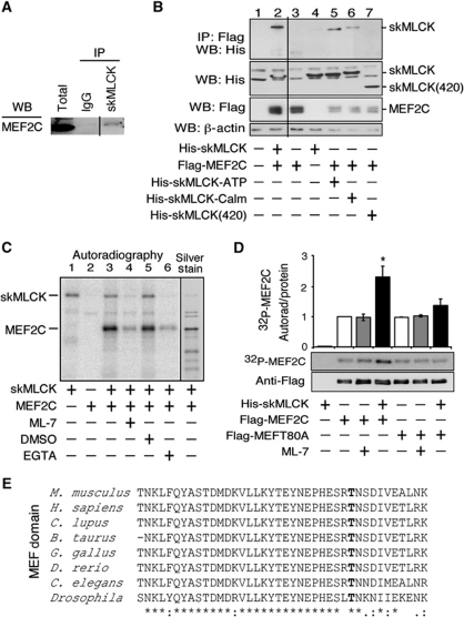
The novel interaction between TAP-tagged MEF2C and skMLCK was validated using co-immunoprecipitation assays. Endogenous MEF2C was co-immunoprecipitated with endogenous skMLCK in extracts from C2C12 myocytes using skMLCK-specific antibodies (Figure 2A). Further, co-transfection of HEK-293 cells with Flag-MEF2C with constructs containing wild-type skMLCK, revealed that the full-length skMLCK protein physically interacted with MEF2C (Figure 2B, lane 2). To determine which domain of skMLCK interacted with MEF2C, several skMCLK mutants were examined. MEF2C-interacting mutants included His-skMLCK-ATP and His-skMLCK-Calm, defective in the putative ATP and calmodulin-binding domains, respectively (Figure 2B, lanes 5 and 6). In contrast, a C-terminally truncated skMLCK mutant (skMLCK420) was inefficiently co-immunoprecipitated with MEF2C, indicating that the interaction required the C-terminal domain of skMLCK (Figure 2B, lane 7).
Figure 2.
skMLCK physically interacts with and phosphorylates MEF2C in vivo and in vitro. (A) The in vivo interaction between MEF2C and skMLCK was observed in C2C12 myoblasts that were differentiated under serum starvation conditions. Co-immunoprecipitation (IP) was performed using anti-skMLCK antibodies conjugated to magnetic beads followed by western blot with antibodies against MEF2C. Anti-IgG antibodies were used in a control IP. Intervening lanes have been removed for clarity, marked by a black line. (B) Flag-tagged MEF2C and His-tagged skMLCK or its mutants were co-transfected into HEK-293 cells. Co-immunoprecipitation (IP) using anti-Flag-agarose resin was followed by western blot analysis (WB) with anti-His antibodies. WB for His-, Flag-, and β-actin show expression prior to IP. Intervening lanes have been removed for clarity, marked by a black line. (C) In vitro kinase assays were performed with recombinant His-MEF2C incubated with purified skMLCK as indicated and visualized by silver stain or autoradiography. (D) In vivo kinase assays were performed in HEK-293 cells co-transfected as indicated. After immunopurification with an anti-Flag resin, western blot analysis and autoradiography were performed. An Image J program was used to measure band intensities and the intensity of each 32P-radiolabelled band was normalized to the corresponding level of MEF2C protein. The data are shown as the normalized average 32P intensity±s.e.m. (n=3). (E) The MEF-domain protein sequence alignment from different species, showing conservation of T80. *P<0.05.
To determine if skMLCK could phosphorylate MEF2C, an in vitro kinase assay was performed. Purified His-tagged MEF2C protein was phosphorylated by a commercially available skMLCK protein preparation in the presence of Ca2+, Mg2+, and [32P]-γ-ATP as a phosphate donor and the absence of calmodulin (Figure 2C, lane 3). Phosphorylation of MEF2C was lost in the absence of skMLCK and reduced in the presence of EGTA or the skMLCK inhibitor ML-7 (Figure 2C, lanes 4 and 6). Phosphorylation of MEF2C also occurred in the presence of calmodulin, which is required for optimal myosin light chain phosphorylation, but the autophosphorylation of skMLCK was more intense (Gao et al, 1992) (Supplementary Figure S2). Mass spectrometric analysis of the skMLCK preparation did not reveal any contaminating kinases. Silver staining showed an equal loading of the blot (Supplementary Figure S2). Thus, skMLCK phosphorylates MEF2C in vitro.
To identify the sites in MEF2C that are phosphorylated by skMLCK, in vitro kinase assays were performed followed by LC–MS/MS analysis (Abu-Farha et al, 2008). Phosphorylation of MEF2C was observed on a tryptic peptide corresponding to amino acids 80–89 of MEF2C, identifying a single phosphorylation in this peptide at Threonine-80 (T80). Notably, the peptide was mapped to the MEF2 domain, which is essential for homodimerization and heterodimerization of MEF2 family members, binding to DNA, and interaction with the inhibitor HDAC4 (Lu et al, 2000). Alignment of the MEF2 domain shows that T80 is conserved in MEF2C proteins from different species (Figure 2E).
To investigate whether skMLCK phosphorylates MEF2C in cells, in vivo phosphorylation assays were conducted. After incubation of transfected HEK-293 cells with [32P]-orthophosphate, Flag-MEF2C was purified on an anti-Flag-agarose resin and examined by western blot analysis to determine Flag-MEF2C expression and by autoradiography to identify the level of phosphorylation. Quantification revealed a statistically significant 2.5-fold increase in the intensity of Flag-MEF2C phosphorylation in the presence of skMLCK (Figure 2D). Mutation of T80 to Alanine, creating MEFT80A, resulted in a lack of enhancement of phosphorylation in the presence of skMLCK, suggesting that T80 is the major site of MEF2C phosphorylation by skMLCK (Figure 2D). HEK-293 cells did not contain endogenous MLCK activity, since treatment with ML-7 did not change the baseline MEF2C phosphorylation levels (Figure 2D) and skMLCK protein was not detected by western blotting (Supplementary Figure S1F). Immunoblotting with anti-flag antibodies indicated an equal loading of the purified Flag-MEF2C protein (Figure 2D). Therefore, skMLCK binds and phosphorylates MEF2C on T80.
Phosphorylation-deficient MEF2C cannot enhance skeletal myogenesis
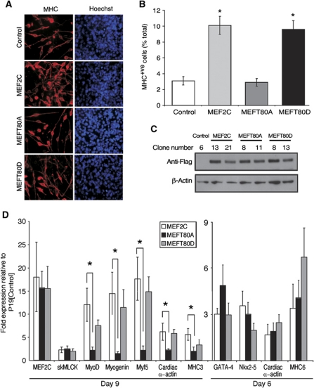
To explore the functional relevance of MEF2C phosphorylation at T80 on endogenous promoters during myogenesis, stable P19 cells expressing wild-type Flag-MEF2C, Flag-MEFT80A, or Flag-MEFT80D mutants were isolated and aggregated to induce differentiation. While MEFT80A is phosphorylation deficient, MEFT80D should be a phosphomimetic mutant. On day 9 of differentiation, immunofluorescence using antibodies against the muscle-specific marker MHC showed that both P19[MEF2C] and P19[MEFT80D] cultures, but not P19[MEFT80A], enhanced skeletal muscle generation three-fold compared with P19[Control] cultures (Figure 3A and B). Notably, all three cell lines induced comparable numbers of cardiomyocytes expressing MHC (MHC+ve), indicated by their rounded morphology (data not shown), and contained comparable levels of Flag-tagged wild-type or mutant MEF2C protein (Figure 3C). Taken together, these data indicate that phosphorylation of T80 in MEF2C is required for MEF2C-enhanced skeletal myogenesis.
Figure 3.
MEFT80D, but not MEFT80A enhances skeletal myogenesis. (A) P19[Control], P19[MEF2C], P19[MEFT80A], and P19[MEFT80D] stable cell lines were differentiated and examined by immunofluorescence with an anti-MHC antibody, MF20, to detect skeletal muscle, and counter stained with Hoechst dye to visualize the nuclei (× 400). (B) Skeletal myocytes and total nuclei for 10 random fields from different clones were counted and shown as a percentage of total cells, ±s.e.m. (n=3). (C) Western blot analysis of total protein extracts with anti-flag antibodies, showed similar levels of exogenous wild-type and mutant MEF2C protein. (D) Q–PCR analysis from RNA harvested on day 6 or 9 was performed with the genes indicated. Results were normalized to β-actin, and expressed as normalized fold-change relative to P19[Control] cells (n=15). *P<0.05.
Q–PCR analyses supported the results obtained by immunofluorescence. While P19[MEF2C] and P19[MEFT80D] cultures showed a statistically significant enhancement of the skeletal myoblast/myocyte-specific markers MyoD, myogenin and Myf5, compared with P19[Control] cells, P19[MEFT80A] cultures did not (Figure 3D). A similar pattern was observed for mRNA transcripts of the muscle structural proteins cardiac α-actin and MHC3 on day 9, indicating a lack of upregulation of skeletal myogenesis in P19[MEFT80A] cultures. Note that under the conditions used, MEF2C enhanced skeletal myogenesis more efficiently than cardiomyogenesis (Figure 3D). Interestingly, the cardiac muscle markers GATA-4 and Nkx2-5 had similar increases in transcript levels in the wild-type MEF2C and its mutant cell cultures (Figure 3D). Furthermore, cardiac α-actin and MHC6 transcript levels on day 6 of differentiation, representative of cardiac muscle, were similar in all cell lines examined (Figure 3D), indicating a similar extent of cardiomyogenesis. The reduced ability to generate skeletal muscle by the MEFT80A mutant was not due to lower levels of MEF2C or skMLCK expression, since the transcript levels of wild-type and mutant MEF2C and skMLCK on day 9 were similar in all three cell lines (Figure 3D). Taken together, our results show that the phosphorylation of T80 in the MADS/MEF2 domain has a critical effect on the ability of MEF2C to enhance skeletal but not cardiac myogenesis.
skMLCK is necessary and sufficient for enhanced skeletal myogenesis
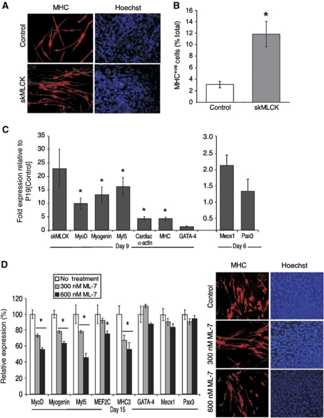
The importance of the T80 phosphorylation and the interaction of MEF2C with skMLCK prompted us to study the effect of skMLCK stable overexpression during P19 cell myogenesis. Cells were examined by immunofluorescence with MF20 on day 9 of differentiation and the number of MHC+ve skeletal myocytes was counted and found to be increased four-fold in P19[skMLCK] cultures compared with P19[Control] cells (Figure 4A and B). Q–PCR analysis of MRF expression revealed a 10–15-fold induction over control of MyoD, Myogenin, and Myf5 transcripts, indicating enhanced commitment into skeletal muscle (Figure 4C). Further, a five-fold increase was observed in mRNA levels of the structural genes cardiac α-actin and MHC in P19[skMLCK] compared with P19[Control] cultures (Figure 4C). In contrast, the cardiac muscle marker, GATA-4, and the skeletal muscle progenitor markers Pax3 and Meox1, were not significantly upregulated on day 6. These results suggest that skMLCK exerts its effect at the commitment stage of P19 skeletal myogenesis, and not cardiac myogenesis, by amplifying the expression of the MRFs and not the premyogenic mesoderm markers.
Figure 4.
skMLCK is necessary and sufficient for efficient myogenesis. (A) Immunofluorescence with MF20 antibodies, showing enhanced myogenesis in day 9 differentiated P19[skMLCK] cells compared with P19[Control] cells. Cells were counter stained with Hoechst dye (× 400). (B) MHC+ve skeletal myocytes and total nuclei were counted and the percentage of total cells was calculated (n=3). (C) Q–PCR analysis of RNA harvested on days 6 and 9 of differentiation from P19[Control] and P19[skMLCK] cells (n=10). (D) Skeletal myogenesis was inhibited in mouse ES cells differentiated in the presence of ML-7 for 15 days. Q–PCR analysis was performed and the results were normalized to GAPDH, with levels expressed as normalized fold-change over undifferentiated cells and as a percentage of the control untreated cells (n=3). Immunofluorescence was performed with MF20 antibodies and stained with Hoechst dye (× 400). *P<0.05.
To investigate the effect of skMLCK inhibition on skeletal myogenesis in mES and P19 cells, ML-7, a cell-permeable, potent, and selective competitive inhibitor for the ATP-binding site of MLCK was used (Kuhlmann et al, 2007). mES cells were differentiated in the presence of ML-7 (0, 300, 600 nM) and examined by immunofluorescence for MHC on day 15. ML-7 treatment resulted in a decrease of skeletal, but not cardiac, muscle formation (Figure 4D). The inhibition of skeletal myogenesis was due to a significant reduction (35–55%) in MyoD, myogenin, Myf5, and MHC3 mRNA transcript levels (Figure 4D). In contrast, GATA-4 transcripts were unaffected (Figure 4D), indicating no loss of cardiomyogenesis. Furthermore, the skeletal premyogenic mesoderm markers, Meox1 and Pax3 showed no significant changes in their mRNA levels (Figure 4D). Inhibition of skeletal, but not cardiac, myogenesis at the stage of muscle commitment was also observed in P19 cells treated with ML-7 (Supplementary Figure S3). Therefore, gain-and-loss-of-function studies identify skMLCK as a key regulatory kinase during the commitment stage of skeletal, but not cardiac, muscle formation in differentiating embryonic and P19 stem cells.
Inhibition of skMLCK reduced the activation and subsequent differentiation of quiescent satellite cells
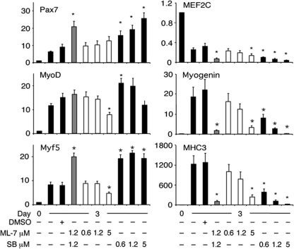
Since previous studies have shown that MEF2C is highly expressed in quiescent satellite cells (Pallafacchina et al, 2010), we isolated adult mouse satellite cells and examined their activation and differentiation status when freshly isolated (day 0) and after 3 days culture, with or without ML-7. Q–PCR analysis of satellite cells cultured for 3 days showed upregulated levels of Pax7, MyoD, and Myf5, indicative of satellite cell activation, and upregulated levels of myogenin and MHC3, indicative of early myoblast differentiation (Figure 5). In the presence of ML-7, MyoD, Myf5, myogenin, and MHC3 were significantly downregulated but Pax7 levels remained relatively unchanged (Figure 5). MEF2C was expressed at high levels in the freshly isolated satellite cells, in agreement with previous findings (Pallafacchina et al, 2010), and was still present but at reduced levels after 3 days of culture. Thus, MEF2C appears to be expressed at all stages of satellite cell myogenesis. Notably, treatment with ML-7 reduced MEF2C expression significantly by day 3. Taken together, our data suggest that skMLCK is important for the activation of satellite cells and their subsequent differentiation.
Figure 5.
Inhibition of skMLCK reduced the activation of adult satellite cells. Isolated mouse satellite cells were cultured in the presence or absence of increasing concentrations of ML-7, SB203580, separately or together, as indicated. On day 3, RNA was harvested and Q–PCR analysis was performed. Results were normalized to β-actin, and expressed as fold-change relative to non-treated cells (n=5; *P<0.05).
Recent work has shown that Pax7 expression in satellite cells is regulated by the p38α kinase-dependent repression by PRC2 (Palacios et al, 2010). We compared inhibition of skMLCK, by ML-7, with inhibition of p38 kinase by SB203580 (SB). In agreement with the previous study (Palacios et al, 2010), SB markedly enhanced Pax7 expression and efficiently inhibited expression of MEF2C, myogenin, and MHC (Figure 5). Notably, both MyoD and Myf5 were upregulated by SB, although MyoD was upregulated only at the lowest concentration. Thus, in accordance with what has been previously reported (Palacios et al, 2010), SB expanded the population of activated satellite cells and inhibited their subsequent differentiation.
In comparing the two pathways, SB was a better inhibitor of differentiation than ML-7 and the latter was a superior inhibitor of satellite cell activation than SB. To determine how the two pathways would interact, we treated satellite cells with both SB and ML-7. The observed outcome was more similar to that of SB inhibition, with high Pax7 and Myf5 expression and greatly reduced MEF2C, myogenin, and MHC3 (Figure 5). Thus, the two pathways did not synergize but the SB treatment overrode the inhibition of MyoD and Myf5 by ML-7. In summary, skMLCK regulates the expression of MyoD and Myf5 during satellite cell activation.
skMLCK phosphorylation of MEF2C regulates MyoD function
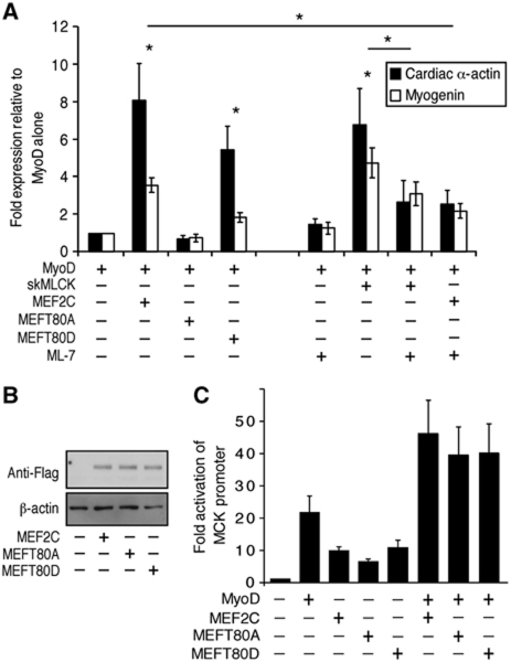
In light of the ability of skMLCK to phosphorylate MEF2C and enhance skeletal muscle commitment in P19 cells, the effect of mutant MEF2C and skMLCK was examined on the myogenic conversion of C3H10T1/2 embryonic fibroblasts by MyoD. Myogenic conversion was evaluated by Q–PCR analysis of cardiac α-actin and myogenin transcripts, in cells transiently transfected with MyoD and/or skMLCK, MEF2C or its T80 mutants. Co-transfection of MEF2C or MEFT80D with MyoD significantly increased the transcript levels of cardiac α-actin and myogenin (Figure 6A). In contrast, MEFT80A did not increase myogenin or cardiac α-actin transcript levels over MyoD alone (Figure 6A). Co-transfection with skMLCK significantly enhanced MyoD-directed myogenic conversion and this enhancement was reduced by the skMLCK inhibitor, ML-7. Finally, ML-7 reduced the observed synergy between MyoD and MEF2C (Figure 6A). Similar levels of wild-type and mutant MEF2C proteins were detected in these experiments, as shown by a western blot analysis with an anti-Flag antibody (Figure 6B). SkMLCK transcripts were present at low levels in 10T1/2 fibroblasts and were upregulated by MyoD and MEF2C expression (data not shown), similar to the upregulation of skMLCK in myogenesis of C2C12 (Supplementary Figure S1F) and P19 cells (Figure 1). These results indicate that the synergy between MyoD and MEF2C activity on endogenous promoters is regulated, at least in part, by the phosphorylation of MEF2C-T80 by skMLCK.
Figure 6.
MEFT80A cannot synergize with MyoD on endogenous skeletal muscle-specific promoters. (A) Myogenic conversion assays were performed in C3H10T1/2 fibroblasts, transiently transfected with plasmids as indicated, with or without ML-7 treatment. Q–PCR analysis of cardiac α-actin and myogenin transcript levels were normalized to transfected GFP transcripts and expressed relative to cells transfected with MyoD alone (n=3). (B) Western blot analysis with anti-flag antibodies, showing similar protein expression of wild-type and mutant MEF2C in transfected C3H10T1/2 cells. (C) A reporter assay with the muscle reporter MCK-luciferase shows similar levels of synergy for MEF2C or its mutants with MyoD on an exogenous promoter. Luciferase activity was measured and normalized against Renilla (n=4). *P<0.05.
Promoter analysis was performed to determine if the phosphorylation-deficient (MEFT80A) or phosphomimetic (MEFT80D) mutants would modulate the transcriptional activity of MEF2C. Transient expression of wild-type or mutant MEF2C resulted in similar levels of enhancement of luciferase activity, driven by a MyoD- and MEF2-responsive promoter, in the presence or absence of MyoD (Figure 6C). Thus, in contrast to the above findings analysing endogenous gene expression, the phosphorylation of T80 is not essential for the transcriptional activity of MEF2C, or its synergistic activation with MyoD, on non-chromatinized, exogenous promoters.
skMLCK phosphorylation of MEF2C regulates recruitment of histone acetyltransferases to skeletal muscle promoters
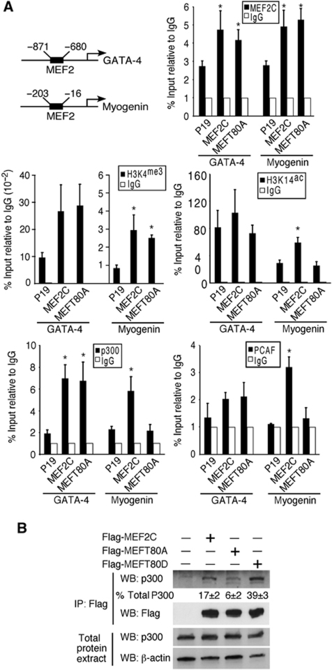
Since MEFT80A could activate exogenous (Figure 6C) but not endogenous (Figures 3 and 6A) promoters, we examined the ability of MEFT80A to bind to endogenous MEF2 DNA-binding sites, using chromatin immunoprecipitation (ChIP) followed by Q–PCR. Both MEF2C and MEFT80A bound to the myogenin and GATA-4 promoters (Figure 7A), specifically in the region of a conserved MEF2 DNA-binding site. Thus, the MEFT80A mutant appeared to bind MEF2 sites in chromatin as efficiently as wild-type MEF2C protein.
Figure 7.
MEFT80A cannot recruit p300/PCAF to endogenous skeletal muscle-specific promoters. (A) ChIP was performed using the indicated antibodies and analysed by Q–PCR with primers flanking the MEF2C site in the myogenin or GATA-4 promoters. Graphs represent Q–PCR analysis from day 7 of differentiation for P19, P19[MEF2C], or P19[MEFT80A] cultures. Relative enrichment was calculated as the percent chromatin input normalized to IgG (n=4). (B) HEK-293 cells were transfected with Flag-MEF2C, -MEFT80A, or -MEFT80D. Co-immunoprecipitation (IP) using anti-Flag-agarose resin was followed by western blot analysis (WB) with antibodies against endogenous p300. Western blots were prepared from total extracts, reacted with antibodies as indicated, and quantified using the Image J program. The p300 Co-IP band intensities were normalized to the intensity of their corresponding control β-actin bands and then to total p300 for each sample (n=3). *P<0.05.
To determine if histone methylation/acetylation levels were changed, ChIP was performed with antibodies against trimethylated-K4 on Histone 3 (H3K4me3) and against acetylated H3K14 (H3K14ac), indicative of actively transcribed chromatin. The levels of H3K4me3 on the myogenin promoter were comparable in P19[MEF2C] and P19[MEFT80A] cell lines, indicating similar levels of H3K4 trimethylation in these cell lines (Figure 7A). However, ChIP performed with antibodies against acetylated H3K14 indicated that the levels of H3K14ac were significantly lower at the myogenin promoter, but not the GATA-4 promoter, in P19[MEFT80A] cells as compared with P19[MEF2C] cells (Figure 7A).
To determine if the loss of acetylation was due to deficient recruitment of a histone acetyltransferase (HAT), we performed ChIP with antibodies against p300 and PCAF, which have been shown to bind MEF2C (Sartorelli et al, 1997). While wild-type MEF2C could recruit both p300 and PCAF to the myogenin promoter, the MEFT80A mutant could not (Figure 7A). In contrast, p300 was recruited efficiently by both MEF2C and MEFT80A to the GATA-4 promoter.
To determine if the decrease of p300 recruitment was due to inefficient binding to MEFT80A, co-immunoprecipitation studies were performed in HEK-293 cells transfected with wild-type MEF2C, MEFT80A, or MEFT80D. A decrease in the interaction of p300 with MEFT80A was observed, compared with the interaction with MEF2C and MEFT80D (Figure 7B). Therefore, the MEFT80A mutant appears defective in enhancing H3K14 acetylation on skeletal, but not cardiac, promoters, likely due to a loss of recruitment of p300 and/or PCAF.
Discussion
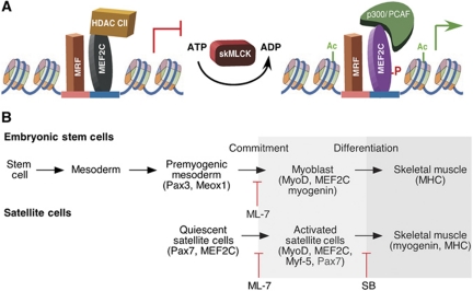
Our data support a model whereby the ability of MEF2C to establish commitment to the skeletal muscle lineage is regulated, at least in part, by skMLCK phosphorylation of MEF2C on T80. In the absence of phosphorylation, MEF2C can bind to endogenous skeletal muscle promoters but cannot recruit p300/PCAF, leading to a lack of histone acetylation (Figure 8A). As a consequence, the synergy between MRFs and MEF2C is disrupted, resulting in minimal MRF upregulation and a deficit in the formation of committed skeletal myoblasts (Figures 3, 4, and 6) or in the activation of quiescent satellite cells (Figure 5). Thus, skMLCK and, by implication, regulators of skMLCK (Kamm and Stull, 2001), control the transition from skeletal muscle progenitors or quiescent satellite cells, which can repopulate the satellite cell niche (Montarras et al, 2005; Kuang et al, 2008), to skeletal myoblasts or activated satellite cells, respectively (Figure 8B).
Figure 8.
Working model for the mechanism by which skMLCK can enhance myogenesis. (A) Previous studies have shown that MEF2C is inhibited by class II HDACs (HDAC CII), synergizes with MRFs, and recruits p300 to promoters (Sartorelli et al, 1997; Lu et al, 2000; Potthoff and Olson, 2007). Here, we show that skMLCK directly phosphorylates MEF2C, leading to p300/PCAF recruitment, increased acetylation of skeletal muscle-specific genes, and enhanced skeletal myogenesis. (B) Comparison of the stages of myogenesis in embryonic stem cells and satellite cells. ML-7 reduced the efficient upregulation of MRFs in the ES-derived premyogenic mesoderm or during the activation of quiescent satellite cells. In agreement with other studies (Palacios et al, 2010), SB inhibited the loss of Pax7, required for myoblast formation and subsequent differentiation.
SkMLCK belongs to a family of Ca2+-dependent protein kinases and the phosphorylation of MEF2C by skMLCK was found to require Ca2+ (Figure 2). Sufficient intracellular Ca2+ levels are vital for C2C12 differentiation into skeletal muscle (Porter et al, 2002). Further, Ca2+-activated signalling has a crucial role in regulating myogenesis by a variety of mechanisms, including the promotion of E protein-MyoD heterodimerization (Hauser et al, 2008), the loss of HDAC4/5 repression of MEF2 by Ca2+/CaM-dependent kinase (Lu et al, 2000; McKinsey et al, 2000), and the activation of calcineurin (Delling et al, 2000; Friday et al, 2000). Thus, our results are consistent with the importance of Ca2+ in regulating skeletal muscle development and reveal a novel mechanism of transcriptional regulation by Ca2+.
The regulation of myogenesis by skMLCK was both lineage and stage specific, having little effect on the formation of either cardiac muscle or skeletal muscle progenitors. Cardiac muscle genes, such as GATA-4 and Nkx2-5, were still upregulated by the MEFT80A mutant or after treatment with ML-7, and were not upregulated by skMLCK overexpression. Similarly, skeletal muscle progenitor genes, such as Pax3/7 and Meox1, were unaffected by changes in skMLCK activity. In contrast, expression of the MRFs along with muscle structural genes was dependent on skMLCK activity. Finally, although MEF2C binds and recruits p300 to muscle promoters (Sartorelli et al, 1997), the mutant MEFT80A appeared deficient in p300 recruitment. It is likely that other transcription factors present in MEF2C complexes in the cardiac muscle lineage can compensate for the loss of p300 recruitment by MEFT80A. Thus, specificity of regulation by skMLCK appears to be mediated by the ability of MEF2C to recruit p300 to skeletal versus cardiac muscle promoters at the stage of muscle commitment.
Interestingly, the phosphorylation-deficient MEF2C mutant could still enhance H3K4 trimethylation on skeletal muscle promoters, indicating a specific role for skMLCK in regulating histone acetylation, as opposed to p38, which can regulate histone trimethylation via MEF2D, but not p300 recruitment in myoblasts (Rampalli et al, 2007; Serra et al, 2007; Guasconi and Puri, 2009). However, other effects of skMLCK on chromatin cannot be ruled out. Overall, our data suggest that, in contrast to the cardiac muscle lineage, the complex of transcription factors bound to MEF2C in the skeletal muscle pathway requires MEF2C-T80 phosphorylation for efficient p300 recruitment.
Previous studies have analysed T80 in the context of mutating ESRT77−80 to VNQA and found that this mutant could still homodimerize and heterodimerize and bind to HDAC4 and DNA (Molkentin et al, 1996a; Lu et al, 2000). These studies agree with our finding that MEFT80A could bind and activate exogenous promoters as efficiently as wild-type MEF2C.
The finding that MEF2C is highly expressed in quiescent satellite cells (Pallafacchina et al, 2010) (Figure 6) suggests that the recruitment of class II HDACs by MEF2C (Lu et al, 2000; McKinsey et al, 2000) may have an important role in satellite cell formation or maintenance. From gene profiling analysis, HDAC2, 4, and 11 transcripts are present at high levels in quiescent satellite cells (Pallafacchina et al, 2010). Release of HDAC repression by CamK or PKD1 signalling (McKinsey et al, 2000; Kim et al, 2008) may then allow for the phosphorylation of MEF2C by skMLCK and the subsequent upregulation of MyoD and Myf5, creating an activated satellite cell (Figure 8). This transition appears crucial in that MRF expression in activated satellite cells is detrimental towards their ability to reconstitute the satellite cell niche after transplantation (Montarras et al, 2005; Kuang et al, 2007).
The recent finding that p38α signalling regulates Pax7 expression and expansion of satellite cells (Palacios et al, 2010) led us to compare the inhibition of skMLCK with that of p38. As expected, SB treatment blocked the downregulation of Pax7, and appeared to enhance activated satellite cell proliferation by upregulating Myf5 expression. Treatment with both drugs resulted in a blockade of ML-7 inhibition, consistent with SB functioning downstream to prevent a reduction in Pax7 expression. Higher concentrations of ML-7 may provide a more extensive downregulation of MyoD and Myf5. The efficient inhibition of differentiation by SB may be due in part to a reduction in MEF2 activity, since p38 kinase phosphorylation of MEF2 is important for its activity during muscle differentiation (Wu et al, 2000; Penn et al, 2004). Future experiments will determine whether ML-7 treatment is beneficial towards the use of satellite cells for muscle therapy.
Mice lacking skMLCK showed no obvious phenotype, including no change in body mass or viability, although the efficiency of satellite cell formation was not examined in these mice (Zhi et al, 2005). Since smMLCK is also expressed in skeletal muscle, it is possible that it could compensate for the loss of skMLCK during development, similar to the previously shown compensation of MyoD by MRF4 and Myf5 (Kassar-Duchossoy et al, 2004). Similarly, mice lacking MEF2C in skeletal or cardiac muscle lineages do not display early differentiation defects (Lin et al, 1997; Potthoff et al, 2007). This is likely due to compensation by other MEF2 family members, since disruption of MEF2 function with dominant negative approaches results in a loss of differentiation into cardiac or skeletal muscle in transgenic mice or C2C12 cells, respectively (Ornatsky et al, 1997; Karamboulas et al, 2006a). ML-7, which inhibited MyoD and Myf5 upregulation in mouse ES, P19, and satellite cells, inhibits all forms of MLCK. Thus, skMLCK may not be the only MLCK that can regulate skeletal muscle commitment.
In addition to muscle contraction, MLCK regulates a variety of other processes, including cell morphology, cell motility, and membrane events (Kamm and Stull, 2001). These events may be mediated in non-muscle cells by MLCK phosphorylation of myosin II. Since the MEF2 family is expressed in a wide variety of tissues and regulates a broad range of proteins (Sandmann et al, 2006; Potthoff et al, 2007), our results implicate the regulation of MEF2 as a mechanism for MLCK function in both muscle and non-muscle cells. Thus, it is possible that MEF2 factors may be regulated by MLCK in a variety of biological processes.
In summary, we have identified a novel phosphorylation of MEF2C by skMLCK that regulates the commitment of cells to the skeletal muscle lineage at least in part by regulating the ability of MEF2C to recruit p300 to skeletal muscle-specific promoters. Inhibition of MLCK activity reduced the upregulation of MRFs that occurs during the commitment of mES and P19 cells to myoblasts and during the activation of quiescent satellite cells. Our work supports the use of P19 cells as a model system to identify novel molecular pathways regulating myogenesis and our findings may lead to innovative approaches to muscle replacement therapies.
Materials and methods
P19 cell culture and DNA transfections
P19 EC cells were cultured as described (Wilton and Skerjanc, 1999; Karamboulas et al, 2006a) and stable cell lines were generated following the published procedures (Ridgeway and Skerjanc, 2001; Karamboulas et al, 2006b). Briefly, P19 cells were transfected using the FuGENE-6 transfection system (Roche Diagnostics, USA) with 0.7 μg of PGK-Puro, 1 μg of PGK-LacZ, and 2.5 μg of B17 in the presence of 2.04 μg MEF2C-TAP, Flag-MEF2C, Flag-MEFT80A, Flag-MEFT80D, or His-skMLCK constructs. P19 control cells were transfected with the appropriate vector control. Stable clonal populations were selected in a media containing 2 μg/ml puromycin. Clones with relatively similar expression of the exogenous genes were selected for further studies.
The differentiation protocol was performed as described previously (Karamboulas et al, 2006a, 2006b) with minor modifications. Cells were aggregated at a density of 5 × 10⁵ in Petri dishes in serum-supplemented medium containing 1% DMSO. Four days later, the aggregates were plated in tissue culture plates. At days 6 or 9 following differentiation, cultures were fixed or RNA/protein were harvested, as described (Skerjanc et al, 1998; Skerjanc and Wilton, 2000). Suboptimal serum conditions were used to minimize the extent of skeletal muscle development in the control cultures (Skerjanc, 1999; Wilton and Skerjanc, 1999).
Mouse ES differentiation and treatment with ML-7
D3 mouse ES cells were grown and differentiated as described previously (Kennedy et al, 2009). Briefly, differentiation was induced by aggregating the cells at a density of 4 × 10⁴ cells/ml, for 2 days in hanging drops and 5 days in NTC plates. On day 7, the embryoid bodies were transferred to TC plates or 0.1% gelatin-coated glass coverslips. On day 10, the cells were cultured in low serum medium containing N2 supplement and allowed to grow for a further 5 days, at which time skeletal myocytes were observed. The cells were treated with ML-7 (0, 300, or 600 nM) from days 3 to 15 of differentiation. These concentrations are far below the concentrations at which ML-7 inhibits other kinases. According to the manufacturer (http://www.emdchemicals.com/life-science-research/ml-7-hydrochloride/EMD_BIO-475880/p_uuid?ProductID=PwGb.s1OzZcAAAEiLTxCeVC_), the primary target for ML-7 is MLCK with Ki=300 nM, the secondary targets are PKA (Ki=21 μM) and PKC (Ki=42 μM).
Isolation of mouse satellite cells
The hind limb muscles were isolated from 3-week-old C57BL/6 mice (five animals treated individually). The muscles were digested with 10 mg/ml collagenase in DMEM for 1 h at 37°C. Satellite cells were isolated by three cycles (1 h each) of incubation in tissue culture plates to remove fibroblasts. The satellite cells were placed on Matrigel-coated (Invitrogen) plates in growth media containing 10% fetal-calf serum, 100 U/ml penicillin/streptomycin, and 292 ng/ml L-glutamine in DMEM for 3 days. Under these conditions, both the activation of satellite cells, as shown by MyoD and Myf5 upregulation, and their early differentiation, as shown by myogenin and MHC3 upregulation, were observed. However, the cultures appeared to be just beginning to differentiate and multinucleated myotubes were not yet observed. On a daily basis, cells were treated with ML-7 or SB203580 (Calbiochem) at final concentrations of 0.6, 1.2, or 5 μM, or both drugs at 1.2 μM.
DNA constructs and mutagenesis
Human MEF2C cDNA was subcloned into the appropriate restriction sites of the eukaryotic expression plasmids pCMV-Taq2 and pCTAP (Stratagene, USA). MEF2C mutants, MEFT80A and MEFT80D, were generated by a Site-Directed Mutagenesis Kit (Stratagene, USA) using the primers in Supplementary Table S1 and following the protocol recommended by the manufacturer. Human skMLCK was cloned from cDNA generated from human skeletal RNA (Clontech, USA) by PCR using Advanced-GC cDNA PCR kit (Clontech, USA) and skMLCK-specific primers (Supplementary Table S1). The skMLCK mutants resulting in the loss of the ATP-binding site, or the calmodulin-binding site were generated using a Site-Directed Mutagenesis Kit (Stratagene, USA; Supplementary Table S1). The skMLCK C-terminal deletion mutant was generated by an insertion of a stop codon after amino-acid 420, resulting in a deletion of the C-terminal 300 aa containing the putative kinase active site and the calmodulin domains.
Western blot, immunoprecipitation, and silver stain
Total cellular proteins were prepared by lysing cells in modified RIPA buffer and western blots were performed as described (Al-Madhoun et al, 2004; Abu-Farha et al, 2008). Western blots were probed with antibodies against the calmodulin-binding peptide, MEF2C, skMLCK (Santa Cruz Biotechnology, USA), anti-His tag (Invitrogen, USA), monoclonal anti-FLAG M2-peroxidase (HRP) or β-actin (Sigma-Aldrich, USA). Co-immunoprecipitation and silver staining was performed as described previously (Al-Madhoun et al, 2007; Abu-Farha et al, 2008).
Immunofluorescence and luciferase activity
For immunofluorescent labelling, cells were fixed in cold methanol and incubated first with mouse anti-MHC monoclonal antibody supernatant MF20 (Bader et al, 1982), followed by goat anti-mouse IgG(H+L) Cy3-linked antibody (Jackson ImmunoResearch Laboratories), as described previously (Gianakopoulos and Skerjanc, 2005). Luciferase activity was measured using the Dual luciferase assay system (Promega, USA) as previously described (Al-Madhoun et al, 2004).
RNA extraction, cDNA synthesis, and quantitative Q–PCR reactions
Total RNA was extracted from cells using the RNeasy Kit (Qiagen Inc., Canada) following the protocol described by the manufacturers. First-strand cDNA was synthesized from 1 μg RNA by reverse transcription using QuantiTect Reverse Transcription Kit (Qiagen Inc., Canada). Q–PCR reactions were performed as described (Savage et al, 2009). Primer pairs (Supplementary Table S2) were selected from the PrimerBank (Wang and Seed, 2003) at website (http://pga.mgh.harvard.edu/primerbank). The reactions and data analysis were performed on the ABI 7300 system (Applied Biosystems, Canada) using SDS software. Relative gene expression was calculated using the comparative Ct method as previously described (Savage et al, 2009). Unless indicated otherwise, results were normalized to β-actin, and averages±s.e.m. are shown expressed as fold-change relative to P19[Control] cells.
Expression and purification of recombinant proteins
MEF2C cDNA was constructed into PET30a bacterial expression vector and was expressed in Escherichia coli, BL 21 (DH3) pLys host cells, which were induced by 0.5 mM IPTG (Al-Madhoun et al, 2002). The protein was purified on Ni-NTA His-binding resins column (Invitrogen, USA) (Al-Madhoun et al, 2002). Purified human skMLCK was obtained from a commercially available source (ProQinase Tools and Tests, USA).
Statistical analysis and sequence alignment
Statistical significance was estimated with a one-tailed Student's t-test assuming equal variance and error is s.e.m. (* indicates P<0.05). Sequences were extracted from PubMed website and alignments were performed using Clustal W program (http://www.ebi.ac.uk/Tools/clustalw2). The accession numbers for MEF2C were H. sapiens NP_002388; M. musculus NP_079558.1; C. lupus XP_536298.1; B. taurus NP_001039578.1; G. gallus XP_001231662.1; D. rerio AAC05226.1; C. elegans NM_060040.4; Drosophila NP_477018.1.
In vitro and in vivo kinase assays
The skMLCK phosphorylation assay was carried out as described (Kennelly et al, 1990) with minor modifications. Purified His-MEF2C and skMLCK were incubated at 30°C for 30 min in a buffer system containing 20 mM Tris–HCl, pH 7.5, 10 mM MgCl2, 0.5 mM NaCl, 5 mM DTT, 1 μM ATP, 0.3 μM [γ-32P]-ATP (NEN Life Science Products, USA), with and without 0.5 mM CaCl2, 25 mM EGTA, or 5 μM ML-7. The enzyme activity was terminated by heat inactivation in the presence of 1 × SDS-loading buffer.
The in vivo kinase assay was performed as described previously (Huang and Chen, 2005) with some modifications. HEK-293 were co-transfected with Flag-MEF2C or Flag-MEFT80A constructs with/without His-skMLCK in the presence or absence of ML-7. Eighteen hours later, the cells were incubated in phosphate-free Dulbecco's modified Eagle's medium for 5 h. Fresh phosphate-free DMEM media containing 0.05 mCi/ml [32P]-orthophosphate (NEN Life Science Products, USA) was then added to the cells for another 4 h. The labelled cells were washed with Tris-buffered saline and lysed in 1 ml of lysis buffer containing 50 mM Tris–HCl, pH 7.5, 300 mM NaCl, and 1 × Mini Complete protease-inhibitor cocktail and 1 × phosphatase-inhibitor cocktail, PhosSTOP (Roche, Laval, Canada). Labelled Flag-MEF2C was purified from cell extracts on anti-Flag-agarose resin as described in the manufacturer's protocol (Sigma-Aldrich, USA), and examined by western blot using monoclonal anti-FLAG M2-peroxidase (HRP; Sigma-Aldrich, USA) antibodies. Labelled proteins were visualized using a phosphorimager (Typhoon TRIO, variable mode imager, GE Healthcare Bio-science).
Myogenic conversion of C3H10T1/2 fibroblasts
10T1/2 fibroblasts were cultured in 10% 1:1 cosmic calf-fetal bovine serum in α-minimum Eagle's medium. One day before transfection, cells were seeded in a six-well plate at a density of 1 × 10⁵ cells/well. The cells were then transfected using the FuGENE-6 transfection reagent following the procedure recommended by the manufacturer (Roche Diagnostics, USA). The myogenic conversion was driven by 1 μg Flag-MyoD in the absence or presence of 1 μg His-skMLCK, Flag-MEF2C, Flag-MEFT80A, or Flag-MEF2T80D constructs and 0.2 μg pEGFP-N1 vector (Clontech Laboratories, Inc., Palo Alto, CA). After 48 h, cells were transferred to differentiation medium, containing 0.5% horse serum, for 4 days with or without daily administration of 600 nM ML-7 (Calbiochem, USA). Transfection efficiency was scored by analysing GFP transcripts and myogenic conversion was quantified by real-time Q–PCR analysis.
Chromatin immunoprecipitation
ChIP assays were performed as described (Savage et al, 2009) with minor modifications. P19, P19[MEF2C], or P19[MEFT80A] clones were aggregated for 4 days in the presence of DMSO, and cells were harvested for ChIP analysis on day 7. Relative enrichment of binding sites compared with the IgG negative control immunoprecipitation was analysed from 50 μg of chromatin using Q–PCR, as described above. Primers sequences for myogenin and GATA-4 are listed in (Supplementary Table S2).
Supplementary Material
Acknowledgments
We thank Valerie Wallace, Alexandre Blais, Robert Screaton, Mary-Ellen Harper, Jean-Francois Couture, Anastassia Voronova, and Diba Ebadi for their helpful comments on the manuscript. We thank Eric Olson and Rhonda Bassel-Duby for their generous gift of the MCK-luciferase construct. We thank Mary-Ellen Harper and Mahmoud Salkhordeh for assistance with these experiments. We thank Alain Stintzi for help with the Q–PCR experiments. We thank Qiao Li for her generous gift of p300 and PCAF antibodies and helpful discussions. ASM was supported by an OWHC/CIHR IGH Fellowship. ISS was funded by a Canadian Institute of Aging Investigator Award. This work was funded by grants to ISS from the CIHR (MOP-84458 and -53277).
Author contributions: ASM and ISS designed the study, collected and analysed the data. ASM performed the experiments except for Supplementary Figure S3, which was carried out by VM. GL isolated satellite cells under the supervision of NWB, who also helped interpret the data for the satellite cells analysis. DF interpreted the data for the mass spectroscopy analysis. ASM and ISS wrote and edited the manuscript. All of the authors discussed the results and commented on the manuscript.
Footnotes
The authors declare that they have no conflict of interest.
References
- Abu-Farha M, Lambert JP, Al-Madhoun AS, Elisma F, Skerjanc IS, Figeys D (2008) The tale of two domains: proteomics and genomics analysis of SMYD2, a new histone methyltransferase. Mol Cell Proteomics 7: 560–572 [DOI] [PubMed] [Google Scholar]
- Al-Madhoun AS, Chen YX, Haidari L, Rayner K, Gerthoffer W, McBride H, O’Brien ER (2007) The interaction and cellular localization of HSP27 and ERbeta are modulated by 17beta-estradiol and HSP27 phosphorylation. Mol Cell Endocrinol 270: 33–42 [DOI] [PubMed] [Google Scholar]
- Al-Madhoun AS, Johnsamuel J, Yan J, Ji W, Wang J, Zhuo JC, Lunato AJ, Woollard JE, Hawk AE, Cosquer GY, Blue TE, Eriksson S, Tjarks W (2002) Synthesis of a small library of 3-(carboranylalkyl)thymidines and their biological evaluation as substrates for human thymidine kinases 1 and 2. J Med Chem 45: 4018–4028 [DOI] [PubMed] [Google Scholar]
- Al-Madhoun AS, Talianidis I, Eriksson S (2004) Transcriptional regulation of the mouse deoxycytidine kinase: identification and functional analysis of nuclear protein binding sites at the proximal promoter. Biochem Pharmacol 68: 2397–2407 [DOI] [PubMed] [Google Scholar]
- Bader D, Masaki T, Fischman DA (1982) Immunochemical analysis of myosin heavy chain during avian myogenesis in vivo and in vitro. J Cell Biol 95: 763–770 [DOI] [PMC free article] [PubMed] [Google Scholar]
- Barsyte-Lovejoy D, Galanis A, Clancy A, Sharrocks AD (2004) ERK5 is targeted to myocyte enhancer factor 2A (MEF2A) through a MAPK docking motif. Biochem J 381(Part 3): 693–699 [DOI] [PMC free article] [PubMed] [Google Scholar]
- Black BL, Ligon KL, Zhang Y, Olson EN (1996) Cooperative transcriptional activation by the neurogenic basic helix-loop-helix protein mash1 and members of the myocyte enhancer factor-2 (Mef2) family. J Biol Chem 271: 26659–26663 [DOI] [PubMed] [Google Scholar]
- Chan JY, Takeda M, Briggs LE, Graham ML, Lu JT, Horikoshi N, Weinberg EO, Aoki H, Sato N, Chien KR, Kasahara H (2008) Identification of cardiac-specific myosin light chain kinase. Circ Res 102: 571–580 [DOI] [PMC free article] [PubMed] [Google Scholar]
- Cox DM, Du M, Marback M, Yang EC, Chan J, Siu KW, McDermott JC (2003) Phosphorylation motifs regulating the stability and function of myocyte enhancer factor 2A. J Biol Chem 278: 15297–15303 [DOI] [PubMed] [Google Scholar]
- Delling U, Tureckova J, Lim HW, De Windt LJ, Rotwein P, Molkentin JD (2000) A calcineurin-NFATc3-dependent pathway regulates skeletal muscle differentiation and slow myosin heavy-chain expression. Mol Cell Biol 20: 6600–6611 [DOI] [PMC free article] [PubMed] [Google Scholar]
- Du M, Perry RL, Nowacki NB, Gordon JW, Salma J, Zhao J, Aziz A, Chan J, Siu KW, McDermott JC (2008) Protein kinase A represses skeletal myogenesis by targeting myocyte enhancer factor 2D. Mol Cell Biol 28: 2952–2970 [DOI] [PMC free article] [PubMed] [Google Scholar]
- Friday BB, Horsley V, Pavlath GK (2000) Calcineurin activity is required for the initiation of skeletal muscle differentiation. J Cell Biol 149: 657–666 [DOI] [PMC free article] [PubMed] [Google Scholar]
- Gallagher PJ, Stull JT (1997) Localization of an actin binding domain in smooth muscle myosin light chain kinase. Mol Cell Biochem 173: 51–57 [DOI] [PubMed] [Google Scholar]
- Gao ZH, Moomaw CR, Hsu J, Slaughter CA, Stull JT (1992) Autophosphorylation of skeletal muscle myosin light chain kinase. Biochemistry 31: 6126–6133 [DOI] [PubMed] [Google Scholar]
- Gessert S, Kuhl M (2010) The multiple phases and faces of wnt signaling during cardiac differentiation and development. Circ Res 107: 186–199 [DOI] [PubMed] [Google Scholar]
- Gianakopoulos PJ, Skerjanc IS (2005) Hedgehog signaling induces cardiomyogenesis in P19 cells. J Biol Chem 280: 21022–21028 [DOI] [PubMed] [Google Scholar]
- Grepin C, Nemer G, Nemer M (1997) Enhanced cardiogenesis in embryonic stem cells overexpressing the GATA-4 transcription factor. Development 124: 2387–2395 [DOI] [PubMed] [Google Scholar]
- Guasconi V, Puri PL (2009) Chromatin: the interface between extrinsic cues and the epigenetic regulation of muscle regeneration. Trends Cell Biol 19: 286–294 [DOI] [PMC free article] [PubMed] [Google Scholar]
- Han J, Jiang Y, Li Z, Kravchenko VV, Ulevitch RJ (1997) Activation of the transcription factor MEF2C by the MAP kinase p38 in inflammation. Nature 386: 296–299 [DOI] [PubMed] [Google Scholar]
- Hauser J, Saarikettu J, Grundstrom T (2008) Calcium regulation of myogenesis by differential calmodulin inhibition of basic helix-loop-helix transcription factors. Mol Biol Cell 19: 2509–2519 [DOI] [PMC free article] [PubMed] [Google Scholar]
- Herring BP, Stull JT, Gallagher PJ (1990) Domain characterization of rabbit skeletal muscle myosin light chain kinase. J Biol Chem 265: 1724–1730 [PMC free article] [PubMed] [Google Scholar]
- Huang WC, Chen CC (2005) Akt phosphorylation of p300 at Ser-1834 is essential for its histone acetyltransferase and transcriptional activity. Mol Cell Biol 25: 6592–6602 [DOI] [PMC free article] [PubMed] [Google Scholar]
- Kamm KE, Stull JT (1986) Activation of smooth muscle contraction: relation between myosin phosphorylation and stiffness. Science 232: 80–82 [DOI] [PubMed] [Google Scholar]
- Kamm KE, Stull JT (2001) Dedicated myosin light chain kinases with diverse cellular functions. J Biol Chem 276: 4527–4530 [DOI] [PubMed] [Google Scholar]
- Karamboulas C, Dakubo GD, Liu J, De Repentigny Y, Yutzey K, Wallace VA, Kothary R, Skerjanc IS (2006a) Disruption of MEF2 activity in cardiomyoblasts inhibits cardiomyogenesis. J Cell Sci 119(Part 20): 4315–4321 [DOI] [PubMed] [Google Scholar]
- Karamboulas C, Swedani A, Ward C, Al-Madhoun AS, Wilton S, Boisvenue S, Ridgeway AG, Skerjanc IS (2006b) HDAC activity regulates entry of mesoderm cells into the cardiac muscle lineage. J Cell Sci 119(Part 20): 4305–4314 [DOI] [PubMed] [Google Scholar]
- Kassar-Duchossoy L, Gayraud-Morel B, Gomes D, Rocancourt D, Buckingham M, Shinin V, Tajbakhsh S (2004) Mrf4 determines skeletal muscle identity in Myf5: Myod double-mutant mice. Nature 431: 466–471 [DOI] [PubMed] [Google Scholar]
- Kato Y, Kravchenko VV, Tapping RI, Han JH, Ulevitch RJ, Lee JD (1997) Bmk1/Erk5 regulates serum-induced early gene expression through transcription factor Mef2c. EMBO J 16: 7054–7066 [DOI] [PMC free article] [PubMed] [Google Scholar]
- Kennedy KA, Porter T, Mehta V, Ryan SD, Price F, Peshdary V, Karamboulas C, Savage J, Drysdale TA, Li SC, Bennett SA, Skerjanc IS (2009) Retinoic acid enhances skeletal muscle progenitor formation and bypasses inhibition by bone morphogenetic protein 4 but not dominant negative beta-catenin. BMC Biol 7: 67. [DOI] [PMC free article] [PubMed] [Google Scholar]
- Kennelly PJ, Edelman AM, Blumenthal DK, Krebs EG (1987) Rabbit skeletal muscle myosin light chain kinase. The calmodulin binding domain as a potential active site-directed inhibitory domain. J Biol Chem 262: 11958–11963 [PubMed] [Google Scholar]
- Kennelly PJ, Starovasnik MA, Edelman AM, Krebs EG (1990) Modulation of the stability of rabbit skeletal muscle myosin light chain kinase through the calmodulin-binding domain. J Biol Chem 265: 1742–1749 [PubMed] [Google Scholar]
- Kim MS, Fielitz J, McAnally J, Shelton JM, Lemon DD, McKinsey TA, Richardson JA, Bassel-Duby R, Olson EN (2008) Protein kinase D1 stimulates MEF2 activity in skeletal muscle and enhances muscle performance. Mol Cell Biol 28: 3600–3609 [DOI] [PMC free article] [PubMed] [Google Scholar]
- Kuang S, Gillespie MA, Rudnicki MA (2008) Niche regulation of muscle satellite cell self-renewal and differentiation. Cell Stem Cell 2: 22–31 [DOI] [PubMed] [Google Scholar]
- Kuang S, Kuroda K, Le Grand F, Rudnicki MA (2007) Asymmetric self-renewal and commitment of satellite stem cells in muscle. Cell 129: 999–1010 [DOI] [PMC free article] [PubMed] [Google Scholar]
- Kuhlmann CR, Tamaki R, Gamerdinger M, Lessmann V, Behl C, Kempski OS, Luhmann HJ (2007) Inhibition of the myosin light chain kinase prevents hypoxia-induced blood-brain barrier disruption. J Neurochem 102: 501–507 [DOI] [PubMed] [Google Scholar]
- Lin Q, Schwarz J, Bucana C, Olson EN (1997) Control of mouse cardiac morphogenesis and myogenesis by transcription factor MEF2C. Science 276: 1404–1407 [DOI] [PMC free article] [PubMed] [Google Scholar]
- Lu J, McKinsey TA, Zhang CL, Olson EN (2000) Regulation of skeletal myogenesis by association of the MEF2 transcription factor with class II histone deacetylases. Mol Cell 6: 233–244 [DOI] [PubMed] [Google Scholar]
- McKinsey TA, Zhang CL, Lu J, Olson EN (2000) Signal-dependent nuclear export of a histone deacetylase regulates muscle differentiation. Nature 408: 106–111 [DOI] [PMC free article] [PubMed] [Google Scholar]
- Molkentin JD, Black BL, Martin JF, Olson EN (1995) Cooperative activation of muscle gene expression by MEF2 and myogenic bHLH proteins. Cell 83: 1125–1136 [DOI] [PubMed] [Google Scholar]
- Molkentin JD, Black BL, Martin JF, Olson EN (1996a) Mutational analysis of the DNA binding, dimerization, and transcriptional activation domains Of Mef2c. Mol Cell Biol 16: 2627–2636 [DOI] [PMC free article] [PubMed] [Google Scholar]
- Molkentin JD, Li L, Olson EN (1996b) Phosphorylation of the MADS-Box transcription factor MEF2C enhances its DNA binding activity. J Biol Chem 271: 17199–17204 [DOI] [PubMed] [Google Scholar]
- Montarras D, Morgan J, Collins C, Relaix F, Zaffran S, Cumano A, Partridge T, Buckingham M (2005) Direct isolation of satellite cells for skeletal muscle regeneration. Science 309: 2064–2067 [DOI] [PubMed] [Google Scholar]
- Morin S, Charron F, Robitaille L, Nemer M (2000) GATA-dependent recruitment of MEF2 proteins to target promoters. EMBO J 19: 2046–2055 [DOI] [PMC free article] [PubMed] [Google Scholar]
- Naidu PS, Ludolph DC, To RQ, Hinterberger TJ, Konieczny SF (1995) Myogenin and MEF2 function synergistically to activate the MRF4 promoter during myogenesis. Mol Cell Biol 15: 2707–2718 [DOI] [PMC free article] [PubMed] [Google Scholar]
- Ornatsky OI, Andreucci JJ, McDermott JC (1997) A dominant-negative form of transcription factor MEF2 inhibits myogenesis. J Biol Chem 272: 33271–33278 [DOI] [PubMed] [Google Scholar]
- Ornatsky OI, Cox DM, Tangirala P, Andreucci JJ, Quinn ZA, Wrana JL, Prywes R, Yu YT, McDermott JC (1999) Post-translational control of the MEF2A transcriptional regulatory protein. Nucleic Acids Res 27: 2646–2654 [DOI] [PMC free article] [PubMed] [Google Scholar]
- Palacios D, Mozzetta C, Consalvi S, Caretti G, Saccone V, Proserpio V, Marquez VE, Valente S, Mai A, Forcales SV, Sartorelli V, Puri PL (2010) TNF/p38alpha/polycomb signaling to Pax7 locus in satellite cells links inflammation to the epigenetic control of muscle regeneration. Cell Stem Cell 7: 455–469 [DOI] [PMC free article] [PubMed] [Google Scholar]
- Pallafacchina G, Francois S, Regnault B, Czarny B, Dive V, Cumano A, Montarras D, Buckingham M (2010) An adult tissue-specific stem cell in its niche: a gene profiling analysis of in vivo quiescent and activated muscle satellite cells. Stem Cell Res 4: 77–91 [DOI] [PubMed] [Google Scholar]
- Pandur P, Lasche M, Eisenberg LM, Kuhl M (2002) Wnt-11 activation of a non-canonical Wnt signalling pathway is required for cardiogenesis. Nature 418: 636–641 [DOI] [PubMed] [Google Scholar]
- Penn BH, Bergstrom DA, Dilworth FJ, Bengal E, Tapscott SJ (2004) A MyoD-generated feed-forward circuit temporally patterns gene expression during skeletal muscle differentiation. Genes Dev 18: 2348–2353 [DOI] [PMC free article] [PubMed] [Google Scholar]
- Porter GA Jr, Makuck RF, Rivkees SA (2002) Reduction in intracellular calcium levels inhibits myoblast differentiation. J Biol Chem 277: 28942–28947 [DOI] [PubMed] [Google Scholar]
- Potthoff MJ, Arnold MA, McAnally J, Richardson JA, Bassel-Duby R, Olson EN (2007) Regulation of skeletal muscle sarcomere integrity and postnatal muscle function by Mef2c. Mol Cell Biol 27: 8143–8151 [DOI] [PMC free article] [PubMed] [Google Scholar]
- Potthoff MJ, Olson EN (2007) MEF2: a central regulator of diverse developmental programs. Development 134: 4131–4140 [DOI] [PubMed] [Google Scholar]
- Pujol MJ, Bosser R, Vendrell M, Serratosa J, Bachs O (1993) Nuclear calmodulin-binding proteins in rat neurons. J Neurochem 60: 1422–1428 [DOI] [PubMed] [Google Scholar]
- Rampalli S, Li L, Mak E, Ge K, Brand M, Tapscott SJ, Dilworth FJ (2007) p38 MAPK signaling regulates recruitment of Ash2L-containing methyltransferase complexes to specific genes during differentiation. Nat Struct Mol Biol 14: 1150–1156 [DOI] [PMC free article] [PubMed] [Google Scholar]
- Ridgeway AG, Skerjanc IS (2001) Pax3 is essential for skeletal myogenesis and the expression of Six1 and Eya2. J Biol Chem 276: 19033–19039 [DOI] [PubMed] [Google Scholar]
- Ridgeway AG, Wilton S, Skerjanc IS (2000) Myocyte enhancer factor 2C and myogenin up-regulate each other's expression and induce the development of skeletal muscle in P19 cells [in process citation]. J Biol Chem 275: 41–46 [DOI] [PubMed] [Google Scholar]
- Sandmann T, Jensen LJ, Jakobsen JS, Karzynski MM, Eichenlaub MP, Bork P, Furlong EE (2006) A temporal map of transcription factor activity: mef2 directly regulates target genes at all stages of muscle development. Dev Cell 10: 797–807 [DOI] [PubMed] [Google Scholar]
- Sartorelli V, Huang J, Hamamori Y, Kedes L (1997) Molecular mechanisms of myogenic coactivation By P300—direct interaction with the activation domain of myod and with the mads box of Mef2c. Mol Cell Biol 17: 1010–1026 [DOI] [PMC free article] [PubMed] [Google Scholar]
- Savage J, Conley AJ, Blais A, Skerjanc IS (2009) SOX15 and SOX7 differentially regulate the myogenic program in P19 cells. Stem Cell 27: 1231–1243 [DOI] [PubMed] [Google Scholar]
- Seguchi O, Takashima S, Yamazaki S, Asakura M, Asano Y, Shintani Y, Wakeno M, Minamino T, Kondo H, Furukawa H, Nakamaru K, Naito A, Takahashi T, Ohtsuka T, Kawakami K, Isomura T, Kitamura S, Tomoike H, Mochizuki N, Kitakaze M (2007) A cardiac myosin light chain kinase regulates sarcomere assembly in the vertebrate heart. J Clin Invest 117: 2812–2824 [DOI] [PMC free article] [PubMed] [Google Scholar]
- Serra C, Palacios D, Mozzetta C, Forcales SV, Morantte I, Ripani M, Jones DR, Du K, Jhala US, Simone C, Puri PL (2007) Functional interdependence at the chromatin level between the MKK6/p38 and IGF1/PI3K/AKT pathways during muscle differentiation. Mol Cell 28: 200–213 [DOI] [PMC free article] [PubMed] [Google Scholar]
- Skerjanc IS (1999) Cardiac and skeletal muscle development in P19 embryonal carcinoma cells. Trends Cardiovasc Med 9: 139–143 [DOI] [PubMed] [Google Scholar]
- Skerjanc IS, Petropoulos H, Ridgeway AG, Wilton S (1998) Myocyte enhancer factor 2C and Nkx2-5 up-regulate each other's expression and initiate cardiomyogenesis in P19 cells. J Biol Chem 273: 34904–34910 [DOI] [PubMed] [Google Scholar]
- Skerjanc IS, Wilton S (2000) Myocyte enhancer factor 2C upregulates MASH-1 expression and induces neurogenesis in P19 cells. FEBS Lett 472: 53–56 [DOI] [PubMed] [Google Scholar]
- Takashima S (2009) Phosphorylation of myosin regulatory light chain by myosin light chain kinase, and muscle contraction. Circ J 73: 208–213 [DOI] [PubMed] [Google Scholar]
- van der Heyden MA, Defize LH (2003) Twenty one years of P19 cells: what an embryonal carcinoma cell line taught us about cardiomyocyte differentiation. Cardiovasc Res 58: 292–302 [DOI] [PubMed] [Google Scholar]
- Wang X, Seed B (2003) A PCR primer bank for quantitative gene expression analysis. Nucleic Acids Res 31: e154. [DOI] [PMC free article] [PubMed] [Google Scholar]
- Wilton S, Skerjanc IS (1999) Factors in serum regulate muscle development in P19 cells. In Vitro Cell Dev Biol Animal 35: 175–177 [DOI] [PubMed] [Google Scholar]
- Wu Z, Woodring PJ, Bhakta KS, Tamura K, Wen F, Feramisco JR, Karin M, Wang JY, Puri PL (2000) p38 and extracellular signal-regulated kinases regulate the myogenic program at multiple steps. Mol Cell Biol 20: 3951–3964 [DOI] [PMC free article] [PubMed] [Google Scholar]
- Yang CC, Ornatsky OI, McDermott JC, Cruz TF, Prody CA (1998) Interaction of myocyte enhancer factor 2 (MEF2) with a mitogen- activated protein kinase, ERK5/BMK1. Nucleic Acids Res 26: 4771–4777 [DOI] [PMC free article] [PubMed] [Google Scholar]
- Zhao M, New L, Kravchenko VV, Kato Y, Gram H, di Padova F, Olson EN, Ulevitch RJ, Han J (1999) Regulation of the MEF2 family of transcription factors by p38. Mol Cell Biol 19: 21–30 [DOI] [PMC free article] [PubMed] [Google Scholar]
- Zhi G, Ryder JW, Huang J, Ding P, Chen Y, Zhao Y, Kamm KE, Stull JT (2005) Myosin light chain kinase and myosin phosphorylation effect frequency-dependent potentiation of skeletal muscle contraction. Proc Natl Acad Sci USA 102: 17519–17524 [DOI] [PMC free article] [PubMed] [Google Scholar]
Associated Data
This section collects any data citations, data availability statements, or supplementary materials included in this article.


