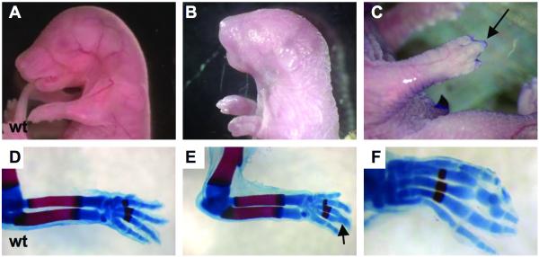
Figure 5. Clft1 skeletal defects.
A clft1 mutant at P0.5 (B) with apparently shortened forelimbs as compared to E18.5 wild-type (A). (C) An E18.5 clft1 embryo with syndactyly. (D-F) Skeletal preparations to highlight bone and cartilage. Note the fusion of phalanges on an otherwise normal clft1 limb (E,F).

