Abstract
Recent studies indicate that systemic administration of tumor necrosis factor (TNF)-α induces increases in corticotrophin releasing hormone (CRH) and CRH type 1 receptors in the hypothalamic paraventricular nucleus (PVN). In this study, we explored the hypothesis that CRH in the PVN contributes to sympathoexcitation via interaction with neurotransmitters in heart failure (HF). Sprague–Dawley rats with HF or sham-operated controls (SHAM) were treated for 4 weeks with a continuous bilateral PVN infusion of the selective CRH-R1 antagonist NBI-27914 or vehicle. Rats with HF had higher levels of glutamate, norepinephrine (NE) and tyrosine hydroxylase (TH), and lower levels of gamma-aminobutyric acid (GABA) and the 67-kDa isoform of glutamate decarboxylase (GAD67) in the PVN when compared to SHAM rats. Plasma levels of cytokines, NE, ACTH and renal sympathetic nerve activity (RSNA) were increased in HF rats. Bilateral PVN infusions of NBI-27914 attenuated the decreases in PVN GABA and GAD67, and the increases in RSNA, ACTH and PVN glutamate, NE and TH observed in HF rats. These findings suggest that CRH in the PVN modulates neurotransmitters and contributes to sympathoexcitation in rats with ischemia-induced HF.
Keywords: Corticotrophin releasing hormone, Neurotransmitters, Hypothalamic paraventricular nucleus, Sympathetic nervous system, Heart failure
Introduction
Stress is an important risk factor in the development and progression of cardiovascular disease. Acute myocardial infarction can be induced by stress in susceptible patients or animals. One well-studied stress system is the hypothalamo–pituitary–adrenal (HPA) axis. In rats with HF, the HPA axis is activated, likely by increased circulating and/or brain proinflammatory cytokines (PICs). The physiological marker of HPA axis activation is increased corticotrophin releasing hormone (CRH) in the hypothalamic paraventricular nucleus (PVN). The cell bodies of CRH-producing neurons are located in the PVN. The CRH is the principal hormone involved in the HPA axis activation, with CRH receptors (CRH-R) being the primary site for HPA axis induction. Previous studies demonstrated that centrally administered CRH elicits cardiovascular and autonomic responses [53]. Recent studies indicate that circulating PICs act upon the CRH neurons in the PVN [9, 12, 13, 38]. However, the mechanisms by which these PICs activate the sympathetic nervous system are not clear. Most of the CRH neurons in the PVN are involved in neuroendocrine functions [41]. Even though preautonomic and neuroendocrine CRH neurons co-mingle in the PVN, they might be differentially regulated. CRH acts upon CRH type 1 receptors (CRH-R1) and can induce autonomic responses in reaction to the initial stimuli. Under normal physiological conditions, CRH-R1 have only a scant presence in the PVN [32]. The levels of CRH-R1 in the PVN are upregulated under conditions of stress or in the presence of increased CRH [20, 29, 39].
Either HPA axis activation or CRH injection into the forebrain can increase both peripheral sympathetic nerve activity and circulating epinephrine and norepinephrine (NE). A number of excitatory and inhibitory neurotransmitters converge in the PVN to influence its neuronal activity. Among these neurotransmitters are glutamate, NE, and gamma-aminobutyric acid (GABA). Increased PICs in the PVN cause an imbalance in PVN neurotransmitters and contribute to sympathoexcitation in heart failure [21]. Despite the abundant evidence that cytokines respond to and drive the HPA axis, very few studies have examined the role of HPA axis activation in HF. Recent work indicates that myocardial infarction increases CRH in the PVN of HF rats [24, 25], and that blockade of PICs decreases sympathetic activity and downregulates the activation of CRH neurons in the PVN of HF rats [24, 25]. We recently found that central blockade of PICs restores of the balance between excitatory and inhibitory neurotransmitters in the PVN of HF rats [21]. The aim of this study was to determine the role of PIC-driven HPA and CRH activity in inducing sympathoexcitation via interaction with neurotransmitters in the PVN of HF rats.
Methods
Animals
All experimental procedures were conducted using adult male Sprague-Dawley rats (275–300 g). Rats were housed in light- and temperature-controlled (12 h light/dark cycle, 23 ± 2°C, respectively) animal quarters and were fed rat chow and tap water ad libitum. All protocols were approved by the Institutional Animal Care and Use Committees of Xi’an Jiaotong University and Louisiana State University. All procedures requiring use of animals were in compliance with the “National Institutes of Health Guide for the Care and Use of Laboratory Animals” published by the US National Institutes of Health (NIH Publication No. 85-23, revised 1996).
Coronary ligation and cannula implantation
Rats underwent sterile surgery under anesthesia [90 mg/kg ketamine + 7.5 mg/kg xylazine, intraperitoneally (IP)] for induction of HF by ligation of the left anterior descending coronary artery, or the same surgery without vessel ligation (SHAM), as previously described [21, 22, 24, 25]. While still under anesthesia, each rat had two cannulae implanted, using stereotaxic coordinates [36], to facilitate bilateral infusions into the PVN [15]. Animals received buprenorphine (0.01 mg/kg, SC) immediately following surgery and 12 h post-operation.
Echocardiographic assessment of left ventricular function
Echocardiography was performed under ketamine (25 mg/kg, IP) sedation for assessment of left ventricular (LV) function as previously described [22, 25]. Ischemic zone (IZ) was estimated by planimetry of the region of the LV endocardial silhouette which demonstrated akinesis or dyskinesis, and expressed as a percentage of the whole (%IZ). From these measurements, LV ejection fraction (LVEF), and LV end-diastolic volume (LVEDV) were also determined.
Drug infusion
Within 24 h of coronary ligation or sham operation, each rat was anesthetized (60 mg/kg ketamine + 5 mg/kg xylazine, IP) and underwent subcutaneous implantation of osmotic mini-pumps (Alzet Model #1004). Mini-pumps were connected to the bilateral PVN cannulae for continuous infusion (0.11 μl/h/side) of the selective CRH-R1 antagonist NBI-27914 (5-chloro-4-[N-(cyclo-propyl)methyl-N-propylamino]-2-methyl-6-(2,4,6-trichlor-ophenyl)amino-pyridine, Sigma) at a total dose of 10 μg/h, or vehicle, over a 4-week treatment period. Another set of HF and SHAM rats were treated with IP infusion of a similar dose of NBI-27914 or vehicle over a 4-week treatment period. NBI-27914 was dissolved in dimethyl sulfoxide (DMSO) and further diluted with artificial cerebrospinal fluid for PVN infusion or saline for IP infusion to the desired concentration.
Electrophysiological recordings and anatomical measurements
Arterial pressure (AP), heart rate (HR) and renal sympathetic nerve activity (RSNA) were measured as described previously [21, 23]. Maximum RSNA was measured using an intravenous bolus administration of sodium nitroprusside (SNP, 10 μg) [21, 35] at the end of the experiment, the background noise, defined as the signal-recorded postmortem, was subtracted from actual RSNA and expressed as percent of maximum (in response to SNP) [17]. The left ventricular end-diastolic pressure (LVEDP), the right ventricle (RV)/body weight (BW) ratio and lung/BW ratio were measured as described previously [21, 22, 25].
The survival rate between the first and second echo-cardiograms for each group was calculated by dividing the number of rats at the second echocardiography assessment by the number of rats at the first echocardiography assessment.
ELISA studies
Plasma and tissue cytokine (TNF-α, IL-1β and IL-6) levels were measured using ELISA (Biosource International Inc.) techniques, as previously described [21–23]. Plasma ACTH was measured using an ELISA kit (MD Biosciences) according to manufacturer instructions.
HPLC measurements of tissue neurotransmitter levels and circulating catecholamine levels
Tissue concentrations of glutamate and GABA were measured using HPLC with electrochemical detection (ECD-300, Eicom Corporation, Japan) and tissue NE concentration was measured using HPLC with electrochemical detection (HTEC-500, Eicom Corporation, Japan) as previously described [21]. Plasma NE and epinephrine were measured using HPLC as previously described [17, 18].
Tissue microdissection
Microdissection procedure was used to isolate the PVN, as previously described [21]. The PVN was punched with the help of a stereotaxic atlas [36]. The samples were stored at −70°C until analyzed for cytokines using ELISA and neurotransmitters using high performance liquid chromatography (HPLC).
Immunohistochemical studies
Transverse sections from brains were obtained from the region approximately 1.80 mm from the bregma. Immunohistochemical labeling was performed in floating sections as described previously [24, 25] to identify Fra-like protein (Fra-LI, a marker of chronic neuronal activation; sc-253, Santa Cruz Biotechnology), and CRH (Phoenix Pharmaceuticals). For each rat, the neurons positive for Fra-LI or CRH within the bilateral borders of the PVN were manually counted in three consecutive sections and an average value was reported. Neurons positive for Fra-LI or CRH within a window superimposed over the dorsal parvocellular (dpPVN), ventrolateral parvocellular (vlpPVN), and magnocellular (mPVN) subregions of the PVN and were counted similarly for data analysis.
Western blot
Measurement of tissue protein was performed as previously described [22–25, 30, 43]. Briefly, protein extracted from the PVN was used for measurements of tyrosine hydroxylase (TH, Abcam) and the 67-kDa isoform of glutamate decarboxylase (GAD67, Abcam) expression by western blot. Equal protein loading was determined by probing all blots with β-actin antibody (Santa Cruz Biotechnology) and normalizing their protein intensities to that of β-actin. The bands were analyzed using NIH Image J software.
Statistical analysis
All data are expressed as mean ± SEM. Data were analyzed by two-way ANOVA. Multiple testing was corrected for by using Tukey’s test. The echocardiography data were analyzed with repeated measures ANOVA. A probability value of P < 0.05 was considered statistically significant.
Results
Echocardiography
Echocardiography performed within 24 h of coronary artery ligation revealed that HF rats had a lower LVEF, a higher LVEDV, and a higher LVEDV/M ratio than SHAM rats (Table 1). The %IZ, LVEF, LVEDV, and LVEDV/M ratio were equivalent among rats assigned to vehicle versus drug treatment. At 4 weeks, LVEF was higher in the HF rats that received NBI-27914 when compared with the HF rats that received vehicle (Table 1). However, there were no significant differences in LVEDV, LVEDV/mass ratio or %IZ between NBI-27914 and VEH-treated HF rats.
Table 1.
Echocardiographic measurements (n = 14)
| HF + NBI-27914 | HF + vehicle | SHAM + NBI-27914 | SHAM + vehicle | |
|---|---|---|---|---|
| Measurements at baseline | ||||
| LVEDV (ml) | 0.72 ± 0.08* | 0.71 ± 0.07* | 0.36 ± 0.05 | 0.39 ± 0.05 |
| LVEDV/Mass | 1.05 ± 0.08* | 1.08 ± 0.09* | 0.54 ± 0.05 | 0.59 ± 0.06 |
| LVEF | 0.35 ± 0.04* | 0.37 ± 0.05* | 0.82 ± 0.05 | 0.84 ± 0.07 |
| IZ (%) | 48 ± 4 | 46 ± 4 | – | – |
| Measurements at 4 weeks | ||||
| LVEDV (ml) | 1.35 ± 0.07*,‡ | 1.40 ± 0.08*,‡ | 0.32 ± 0.03 | 0.35 ± 0.04 |
| LVEDV/Mass | 1.61 ± 0.12*,‡ | 1.70 ± 0.15*,‡ | 0.56 ± 0.05 | 0.61 ± 0.06 |
| LVEF | 0.34 ± 0.03*,† | 0.24 ± 0.03*,‡ | 0.83 ± 0.07 | 0.80 ± 0.06 |
| IZ (%) | 46 ± 4 | 50 ± 5 | – | – |
Values are mean ± SEM
SHAM sham-operated control, HF heart failure, LVEDV left ventricular end-diastolic volume, LVEF left ventricular ejection fraction, IZ% percent ischemic zone
P < 0.05 versus SHAM + NBI-27914 or SHAM + vehicle;
P < 0.05 HF + NBI-27914 versus HF + vehicle;
P < 0.05, 4 weeks versus 24 h value
Functional/anatomical indicators of heart failure
Compared with SHAM rats, HF rats had higher LVEDP, RV/BW and lung/BW ratio. The NBI-27914-treated HF rats had significantly lower LVEDP and lung/BW ratios than vehicle-treated HF rats (Table 2). IP treatment with the same doses of NBI-27914 did not affect LVEDP, RV/BW or lung/BW ratio (Table 2).
Table 2.
Hemodynamic and anatomical measurements (n = 7)
| Measurements at 4 weeks | RV/BW (mg/g) | Lung/BW (mg/g) | HR (beats/min) | MAP (mmHg) | PP (mmHg) | LVEDP (mmHg) |
|---|---|---|---|---|---|---|
| HF + NBI-27914 | 0.71 ± 0.09† | 5.1 ± 0.6† | 328 ± 14 | 93 ± 8 | 33 ± 4 | 7.61 ± 1.57† |
| HF + vehicle | 1.10 ± 0.12* | 10.2 ± 0.7* | 334 ± 14 | 91 ± 7 | 35 ± 5 | 20.26 ± 1.90* |
| SHAM + NBI-27914 | 0.56 ± 0.06 | 4.3 ± 0.4 | 323 ± 12 | 101 ± 8 | 36 ± 4 | 6.56 ± 1.42 |
| SHAM + vehicle | 0.64 ± 0.07 | 4.8 ± 0.5 | 326 ± 13 | 103 ± 9 | 38 ± 5 | 7.01 ± 1.46 |
Values are mean ± SEM
SHAM sham-operated control, HF heart failure, BW body weight, RV right ventricular, HR heart rate, MAP mean arterial pressure, PP pulse pressure, LVEDP left ventricular end-diastolic pressure
P < 0.05 versus SHAM + NBI-27914 or SHAM + vehicle;
P < 0.05 HF + NBI-27914 versus HF + vehicle
NBI-27914 treatment improved the survival (P < 0.05) (HF + NBI-27914, 82.4%; HF + VEH, 70.0%) over the 4-week interval between the first and second echocardiograms.
Humoral indicators of heart failure
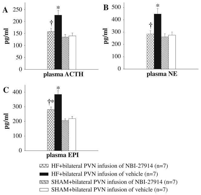
Humoral indicators of heart failure paralleled the PVN findings. Plasma levels of NE, EPI, ACTH, TNF-α, IL-1β and IL-6 were all higher in HF rats than in SHAM rats. Bilateral PVN infusions of NBI-27914 attenuated the increases in plasma levels of these factors in HF rats (Table 3, and Fig. 1). However, the plasma levels were unaffected by IP treatment with the same dose of NBI-27914.
Table 3.
Proinflammatory cytokines in the PVN and plasma (n = 7)
| Measurements at 4 weeks | PVN (pg/mg protein)
|
Plasma (pg/ml)
|
||||
|---|---|---|---|---|---|---|
| TNF-α | IL-1β | IL-6 | TNF-α | IL-1β | IL-6 | |
| HF + NBI-27914 | 3.9 ± 0.4† | 20.2 ± 2.7† | 25.2 ± 2.4† | 14.4 ± 1.3† | 63.4 ± 6.1† | 41.5 ± 4.3† |
| HF + vehicle | 7.2 ± 0.6* | 50.2 ± 4.9* | 63.2 ± 6.0* | 34.1 ± 3.3* | 112.5 ± 10.2* | 94.5 ± 9.2* |
| SHAM + NBI-27914 | 3.2 ± 0.4 | 17.9 ± 1.6 | 20.3 ± 1.9 | 11.2 ± 1.2 | 54.3 ± 4.7 | 34.2 ± 3.8 |
| SHAM + vehicle | 3.4 ± 0.4 | 19.1 ± 2.4 | 23.4 ± 2.0 | 12.5 ± 1.2 | 58.4 ± 5.2 | 38.3 ± 4.1 |
P < 0.05 versus SHAM + NBI-27914 or SHAM + vehicle;
P < 0.05 HF + NBI-27914 versus HF + vehicle
Fig. 1.
Plasma ACTH, NE and epinephrine (EPI) were higher in HF rats than in SHAM rats. Bilateral PVN infusions of NBI-27914 attenuated the increases in plasma ACTH, NE and EPI of HF rats. *P < 0.05 versus SHAM + NBI-27914 or SHAM + vehicle; †P < 0.05 HF + NBI-27914 versus HF + vehicle
CRH in the PVN
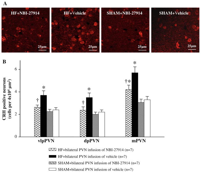
Compared with SHAM rats, HF rats had higher levels of CRH expression in the PVN as revealed by immunohistochemistry (Fig. 2). HF rats treated with NBI-27914 had fewer CRH-positive PVN neurons than vehicle-treated HF rats (Fig. 2).
Fig. 2.
Immunohistochemistry for CRH expression in the PVN of hypothalamus. CRH expression in the PVN was lower in the NBI-27914-treated HF rats than in vehicle-treated HF rats. *P < 0.05 versus SHAM + NBI-27914 or SHAM + vehicle; †P < 0.05 HF + NBI-27914 versus HF + vehicle
PVN neurotransmitters
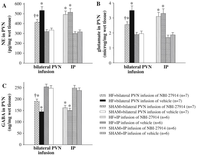
HF rats had higher levels of NE and glutamate, and lower levels of GABA in the PVN. Four-week bilateral infusions of NBI-27914 into the PVN prevented the decrease in PVN GABA and the increases in PVN glutamate and NE in HF rats (Fig. 3). However, IP treatment with the same dose of NBI-27914 did not alter NE, glutamate, or GABA in the PVN of HF rats.
Fig. 3.
PVN levels of norepinephrine (NE), glutamate and GABA in heart failure (HF) and sham operated (SHAM) rats treated for 4 weeks with bilateral PVN infusion of NBI-27914 or vehicle. Bilateral PVN infusions of the selective CRH-R1 antagonist NBI-27914 prevented, the decrease in PVN GABA, and the increases in PVN glutamate and NE observed in HF rats. IP treatment with the same dose of NBI-27914 did not alter NE, glutamate, and GABA in the PVN of HF rats. *P < 0.05 versus SHAM + NBI-27914 or SHAM + vehicle; †P < 0.05 HF + NBI-27914 versus HF + vehicle
TH and GAD67 protein expression in the PVN
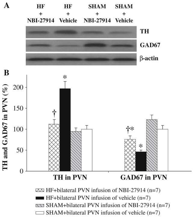
Western blot showed that HF rats had higher TH levels and lower GAD67 levels in the PVN when compared with SHAM rats (Fig. 4). Bilateral PVN infusion of NBI-27914 for 4 weeks prevented the decrease in PVN GAD67, and the increases in TH in the PVN of HF rats (Fig. 4).
Fig. 4.
Western blot of TH and GAD67 in the PVN showed that HF rats had higher levels of TH and lower levels of GAD67 when compared with SHAM rats. Bilateral PVN infusions of NBI-27914 decreased expression of TH, and increased GAD67 expression in the PVN of HF rats. *P < 0.05 versus SHAM + NBI-27914 or SHAM + vehicle; †P < 0.05 HF + NBI-27914 versus HF + vehicle
Fra-LI activity, an indicator of chronic neuronal activation, in the PVN
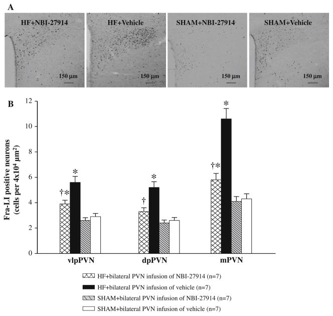
Compared with SHAM rats, Fra-LI activity was higher in the PVN of HF rats. Bilateral PVN infusions of NBI-27914 prevented the increases in Fra-LI of HF rats (Fig. 5). However, IP treatment with the same dose of NBI-27914 had no effect on the number of Fra-LI in the PVN.
Fig. 5.
The effects of bilateral PVN infusion of NBI-27914 on neuronal activity in the PVN of rats with ischemia-induced HF. Fra-LI activity (black dots), an indicator of chronic neuronal excitation, increased in the PVN of HF rats when compared with SHAM rats. Fra-LI activity was lower in the NBI-27914-treated HF rats than in vehicle-treated HF rats. *P < 0.05 versus SHAM + NBI-27914 or SHAM + vehicle; †P < 0.05 HF + NBI-27914 versus HF + vehicle
PVN levels of proinflammatory cytokines
PVN levels of TNF-α, IL-1β and IL-6 were higher in HF than in SHAM rats. PVN levels of TNF-α, IL-1β and IL-6 were lower in HF rats that received bilateral PVN infusions of NBI-27914 (Table 3). IP treatment with the same dose of NBI-27914 had no effect on PIC levels in the PVN of HF rats.
Renal sympathetic nerve activity (RSNA)
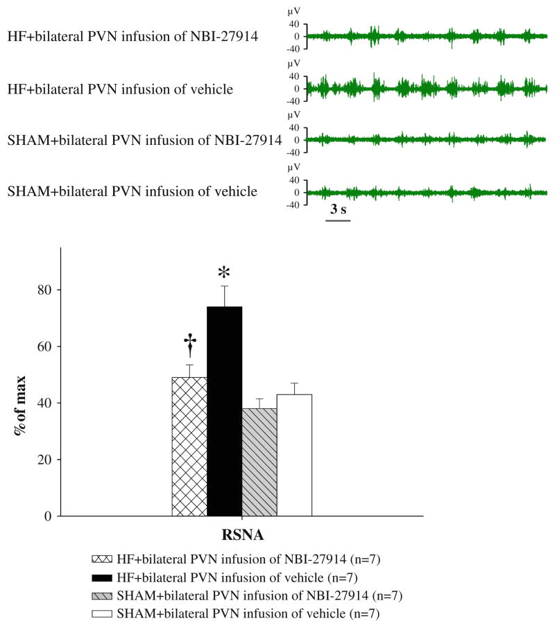
At the conclusion of the study, HF rats exhibited higher renal sympathetic nerve activity (RSNA, % of max) when compared to SHAM rats. Bilateral PVN infusions of NBI-27914 inhibited RSNA in HF rats (Fig. 6). IP treatment with the same dose of NBI-27914 did not affect RSNA.
Fig. 6.
Renal sympathetic nerve activity (RSNA) was increased in HF rats compared with SHAM rats. RSNA of HF rats treated with bilateral PVN infusion of NBI-27914 was lower than vehicle-treated HF rats. *P < 0.05 versus SHAM + NBI-27914 or SHAM + vehicle; †P < 0.05 HF + NBI-27914 versus HF + vehicle
Discussion
The novel finding of this study is that CRH, possibly through a PIC activation mechanism, induces an imbalance between excitatory and inhibitory neurotransmitters in the PVN of HF rats, which contributes to sympathoexcitation. Treatment with PVN infusion of a CRH-R1 antagonist attenuated this imbalance and sympathoexcitation in HF rats. Similar doses of these blockers given peripherally did not restore neurotransmitter levels in the PVN of HF rats, suggesting that central nervous system cytokines modulate neurotransmitters, especially NE, in the PVN, and contribute to sympathoexcitation in heart failure post-MI.
One of the pathophysiological characteristics of HF is elevated sympathetic drive, which is a major factor contributing to the morbidity and mortality of HF patients. Recent evidence points to a central nervous system mechanism that contributes to the sympathetic abnormality typically observed in HF. The excitatory and inhibitory neurotransmitters converge in the PVN to influence its neuronal activity [46]. In the brain, the PVN is an important central integration site for sympathetic nerve activity [48, 49], as well as an important region for cardiovascular control and homeostasis [40, 44]. The primary controlling neurotransmitters include glutamate, NE, and GABA. It has been reported that functional glutamate receptors are expressed in the PVN [5, 34] and are involved in cardiovascular reflexes [1, 3]. It has also been shown that sympathetic hyperactivity in rats with HF is associated with increased NE in the resting PVN [47, 50], and it is well known that NE plays a critical role in the pathophysiologic process of HF [37, 48]. Furthermore, a large body of evidence suggests that GABA plays an important role in central sympathetic and cardiovascular regulation [7, 8, 45] and that it is the dominant inhibitory neurotransmitter within the PVN. Considerable evidence suggests that the PVN is one of the sites in which the cardiovascular effects of GABA are elicited. Previously, work from Patel’s [52] laboratory demonstrated that inhibitory mechanisms of sympathetic regulation within the PVN via GABA were reduced in HF rats. Through a western blot approach, we also identified the neurons expressing GAD67, a marker used to identify GABAergic neurons in the PVN. Our results show that the expression of GAD67 in PVN neurons of HF rats was lower when compared with SHAM rats, and that this reduction was normalized in HF rats treated with bilateral PVN infusion of the selective CRH-R1 antagonist. Elevated excitatory neurotransmitters and decreased inhibitory neurotransmitters in the PVN are shown to contribute to sympathetic dysregulation in HF [2, 11].
Proinflammatory cytokines, including TNF-α, IL-1β and IL-6 [6, 8, 21, 27, 28], are released into the circulation post-MI [26, 51]. In HF rats, TNF-α, which appears quickly in the cytokine cascade [9], increases in the blood, brain and heart within minutes after an acute MI and continues to rise over the ensuing weeks [14]. IL-1β has a similar pattern of early appearance post-MI [16]. The PVN is particularly sensitive to the influences of inflammatory stress and peripheral cytokine production. Blood-borne and brain cytokines are shown to stimulate COX-2 for the eventual generation of prostaglandin E2 (PGE2), which acts centrally to increase sympathetic drive [24, 25] and to induce expression of CRH [31] in the PVN neurons that mediate the HPA axis’s stress response. Within the PVN, the parvocellular CRH neurons are specifically activated by peripheral administration of IL-1β [4]. Central nervous system signaling by blood-borne cytokines activates CRH-producing neurons in the PVN. In rats, deafferentation of the hypothalamus and lesions of the PVN blocked the plasma ACTH response to IL-1β [12]. These findings suggest that PICs contribute to HPA axis activation, such that the early stage PICs act to increase CRH, which then causes an imbalance between excitatory and inhibitory neurotransmitters and initiating sympathoexcitation in these HF rats. This is shown by our finding that a decrease in plasma ACTH after blockade of CRH and this was accompanied by modulation of neurotransmitters within the PVN, thereby affecting the typical negative feedback system at several levels of the HPA axis activation contributing to the attenuated sympathoexcitation in heart failure.
The endocrine (i.e., glucocorticoid) response of the HPA axis to an acute cytokine challenge depends upon the noradrenergic activation of CRH-containing neurons in the PVN. CRH can excite PVN neurons and elicit a sympathoexcitatory response. ICV infusion of CRH caused significant increases in PVN neuronal activity and RSNA [53]. Our results in this study suggest that sympathoexcitatory effects of CRH in PVN may be a primary mechanism of HF progression after MI. Due to the different distributions and functions of CRH in the brain [10, 19, 42], it is difficult to observe identical responses to CRH antagonists. However, this study has clearly demonstrated that CRH is involved in the regulation of sympathetic activity; this is based upon the effect of bilateral PVN infusion of the CRH antagonist NBI-27914 on blocking the RSNA response to HF. We have also previously demonstrated that PICs were increased in the PVN in HF rats [21, 22], and elevated PICs in the PVN can cause an imbalance in PVN neurotransmitters and contribute to sympathoexcitation in HF [21]. Moreover, high levels of circulating cytokines post-MI have additional effects on the brain that may promote the development of HF. Central nervous system signaling by blood-borne cytokines activates CRH producing neurons in the PVN, where TNF-α, IL-1β, and IL-6 all share a common property of activating the HPA axis [9, 12, 33, 49] and increasing sympathetic nerve activity [54]. The present study suggests that a HF-induced increase in CRH in the PVN causes an imbalance in PVN neurotransmitters and contributes to sympathoexcitation in HF rats.
In summary, the results of the present study indicate that elevated brain PICs in heart failure increase CRH neuronal activity and CRH-R1 expression in the PVN. This increased CRH then causes an imbalance between excitatory and inhibitory neurotransmitters in the PVN neuronal tissue, thereby contributing to sympathoexcitation in HF rats. Central blockade of CRH restored these alterations in the PVN of HF rats. Though further investigations are needed to determine the mechanisms by which these interactions occur, as well as an extended follow-up period to assess the potential long-term effect of CRH modulation on HF mortality, these findings outline a possible therapeutic approach whereby central inhibition of CRH and a restoration of neurotransmitter imbalance may be beneficial for the treatment of heart failure.
Acknowledgments
Funding: Supported by National Natural Science Foundation of China (No. 81070199), US National Institutes of Health (NIH) Grant RO1-HL-080544-01, and Fundamental Research Funds for the Central Universities of China (No. 08142001).
Abbreviations
- AP
Arterial pressure
- ANOVA
Analysis of variance
- BW
Body weight
- CRH
Corticotrophin releasing hormone
- dpPVN
Dorsal parvocellular PVN
- ECD
Electrochemical detection
- EPI
Epinephrine
- ELISA
Enzyme-linked immunosorbentassay
- GABA
Gamma-aminobutyric acid
- GAD67
67-kDa isoform of glutamate decarboxylase
- GLU
Glutamate
- HF
Heart failure
- HPA
Hypothalamo–pituitary–adrenal axis
- HPLC
High performance liquid chromatography
- HR
Heart rate
- IZ
Ischemic zone
- IP
Intraperitoneal injection
- ICV
Intracerebroventricular injection
- LV
Left ventricle
- LVEF
LV ejection fraction
- LVEDV
LV end-diastolic volume
- LVEDP
Left ventricular end-diastolic pressure
- mPVN
Magnocellular PVN
- MI
Myocardial infarction
- NE
Norepinephrine
- PVN
Hypothalamic paraventricular nucleus
- PICs
Pro-inflammatory cytokines
- PGE2
Prostaglandin E2
- RSNA
Renal sympathetic nerve activity
- RV
Right ventricle
- SHAM
Sham-operated control
- SNP
Sodium nitroprusside
- TNF
Tumour necrosis factor
- TH
Tyrosine hydroxylase
- vlpPVN
Ventrolateral parvocellular PVN
Contributor Information
Yu-Ming Kang, Email: ykang@mail.xjtu.edu.cn, Department of Physiology and Pathophysiology, Xi’an Jiaotong University School of Medicine, Xi’an 710061, China.
Ai-Qun Zhang, Institute of Hepatobiliary Surgery, General Hospital of Chinese People’s Liberation Army, Beijing, China.
Xiu-Fang Zhao, Department of Internal Medicine, General Hospital of Chinese People’s Armed Police Forces, Beijing, China.
Jeffrey P. Cardinale, Comparative Biomedical Sciences, School of Veterinary Medicine, Louisiana State University, Baton Rouge, LA 70803, USA
Carrie Elks, Comparative Biomedical Sciences, School of Veterinary Medicine, Louisiana State University, Baton Rouge, LA 70803, USA.
Xi-Mei Cao, Department of Physiology, Shanxi Medical University, Taiyuan, China.
Zhen-Wen Zhang, Department of Internal Medicine, General Hospital of Chinese People’s Armed Police Forces, Beijing, China.
Joseph Francis, Email: jfrancis@lsu.edu, Comparative Biomedical Sciences, School of Veterinary Medicine, Louisiana State University, Baton Rouge, LA 70803, USA.
References
- 1.Antonaccio MJ, Kerwin L, Taylor DG. Reductions in blood pressure, heart rate and renal sympathetic nerve discharge in cats after the central administration of muscimol, a GABA agonist. Neuropharmacology. 1978;17:783–791. doi: 10.1016/0028-3908(78)90065-5. [DOI] [PubMed] [Google Scholar]
- 2.Borkowski KR, Finch L. Cardiovascular changes in anaesthetised rats after the intra-hypothalamic administration of adrenaline. Clin Exp Hypertens. 1978;1:279–291. doi: 10.3109/1064196 7809068609. [DOI] [PubMed] [Google Scholar]
- 3.Brennan TJ, Haywood JR, Ticku MK. GABA receptor binding and hemodynamic responses to ICV GABA in adult spontaneously hypertensive rats. Life Sci. 1983;33:701–709. doi: 10. 1016/0024-3205(83)90774-9. [DOI] [PubMed] [Google Scholar]
- 4.Buller K, Xu Y, Dayas C, Day T. Dorsal and ventral medullary catecholamine cell groups contribute differentially to systemic interleukin-1beta-induced hypothalamic pituitary adrenal axis responses. Neuroendocrinology. 2001;73:129–138. doi: 10. 1159/000054629. [DOI] [PubMed] [Google Scholar]
- 5.Butcher KS, Cethetto DF. Receptors in lateral hypothalamic area involved in insular cortex sympathetic responses. Am J Physiol. 1998;275:H689–H696. doi: 10.1152/ajpheart.1998.275.2.H689. 0363-6135/98. [DOI] [PubMed] [Google Scholar]
- 6.Chappell D, Hofmann-Kiefer K, Jacob M, Rehm M, Briegel J, Welsch U, Conzen P, Becker BF. TNF-alpha induced shedding of the endothelial glycocalyx is prevented by hydrocortisone and antithrombin. Basic Res Cardiol. 2009;104:79–89. doi: 10.1007/s00395-008-0749-5. [DOI] [PubMed] [Google Scholar]
- 7.Chen QH, Haywood JR, Toney GM. Sympathoexcitation by PVN injected bicuculline requires activation of excitatory amino acid receptors. Hypertension. 2003;42:725–731. doi: 10.1161/01.HYP.0000085197.20043.44. [DOI] [PubMed] [Google Scholar]
- 8.Chorianopoulos E, Heger T, Lutz M, Frank D, Bea F, Katus HA, Frey N. FGF-inducible 14-kDa protein (Fn14) is regulated via the RhoA/ROCK kinase pathway in cardiomyocytes and mediates nuclear factor-kappaB activation by TWEAK. Basic Res Cardiol. 2010;105:301–313. doi: 10.1007/s00395-009-0046-y. [DOI] [PubMed] [Google Scholar]
- 9.Chrousos GP. The stress response and immune function: clinical implications. The 1999 Novera H. Spector Lecture. Ann NY Acad Sci. 2000;917:38–67. doi: 10.1111/j.1749-6632.2000.tb05371.x. [DOI] [PubMed] [Google Scholar]
- 10.Cole RL, Sawchenko PE. Neurotransmitter regulation of cellular activation and neuropeptide gene expression in the paraventricular nucleus of the hypothalamus. J Neurosci. 2002;22:959–969. doi: 10.1523/JNEUROSCI.22-03-00959.2002. 0270-6474/02/220959-11. [DOI] [PMC free article] [PubMed] [Google Scholar]
- 11.Dampney RAL. Functional organization of central pathways regulating the cardiovascular system. Physiol Rev. 1994;74:323–364. doi: 10.1152/physrev.1994.74.2.323. 0031-9333/94. [DOI] [PubMed] [Google Scholar]
- 12.Dunn AJ. Cytokine activation of the HPA axis. Ann NY Acad Sci. 2000;917:608–617. doi: 10.1111/j.1749-6632.2000.tb05426.x. [DOI] [PubMed] [Google Scholar]
- 13.Elenkov IJ, Wilder RL, Chrousos GP, Vizi ES. The sympathetic nerve—an integrative interface between two supersystems: the brain and the immune system. Pharmacol Rev. 2000;52:595–638. 0031-6997/00/5204-0595. [PubMed] [Google Scholar]
- 14.Francis J, Beltz T, Johnson AK, Felder RB. Mineralocorticoids act centrally to regulate blood-borne tumor necrosis factor-α in normal rats. Am J Physiol Regul Integr Comp Physiol. 2003;285:R1402–R1409. doi: 10.1152/ajpregu.00027.2003. [DOI] [PubMed] [Google Scholar]
- 15.Francis J, Mohan Kumar SM, Mohan Kumar PS. Correlations of norepinephrine release in the paraventricular nucleus with plasma corticosterone and leptin after systemic lipopoly-saccharide: blockade by soluble IL-1 receptor. Brain Res. 2000;867:180–187. doi: 10.1016/S0006-8993(00)02311-8. [DOI] [PubMed] [Google Scholar]
- 16.Francis J, Chu Yi, Johnson KA, Weiss RM, Felder RB. Acute myocardial infarction induces hypothalamic cytokine synthesis. Am J Physiol Heart Circ Physiol. 2004;286:H2264–H2271. doi: 10.1152/ajpheart.01072.2003. [DOI] [PubMed] [Google Scholar]
- 17.Guggilam A, Haque M, Kerut EK, McIlwain E, Lucchesi P, Seghal I, Francis J. TNF-alpha blockade decreases oxidative stress in the paraventricular nucleus and attenuates sympathoexcitation in heart failure rats. Am J Physiol Heart Circ Physiol. 2007;293:H599–H609. doi: 10.1152/ajpheart.00286.2007. [DOI] [PubMed] [Google Scholar]
- 18.Guggilam A, Patel KP, Haque M, Ebenezer PJ, Kapusta DR, Francis J. Cytokine blockade attenuates sympathoexcitation in heart failure: cross-talk between nNOS, AT-1R and cytokines in the hypothalamic paraventricular nucleus. Eur J Heart Fail. 2008;10:625–634. doi: 10.1016/j.ejheart.2008.05.004. [DOI] [PMC free article] [PubMed] [Google Scholar]
- 19.Guillaume V, Conte-Devolx B, Szafarczyk A, Malaval F, Pares-Herbute N, Grino M, Alonso G, Assenmacher I, Oliver C. The corticotropin-releasing factor release in rat hypophysial portal blood is mediated by brain catecholamines. Neuroendocrinology. 1987;46:143–146. doi: 10.1159/000124811. [DOI] [PubMed] [Google Scholar]
- 20.Imaki T, Naruse M, Harada S, Chikada N, Imaki J, Onodera H, Demura H, Vale W. Corticotropin-releasing factor up-regulates its own receptor mRNA in the paraventricular nucleus of the hypothalamus. Brain Res Mol Brain Res. 1996;38:166–170. doi: 10.1016/0169-328X(96)00011-3. [DOI] [PubMed] [Google Scholar]
- 21.Kang YM, He RL, Yang LM, Qin DN, Guggilam A, Elks C, Yan N, Guo Z, Francis J. Brain tumour necrosis factor-alpha modulates neurotransmitters in hypothalamic paraventricular nucleus in heart failure. Cardiovasc Res. 2009;83:737–746. doi: 10.1093/cvr/cvp160. [DOI] [PMC free article] [PubMed] [Google Scholar]
- 22.Kang YM, Ma Y, Elks C, Zheng JP, Yang ZM, Francis J. Cross-talk between cytokines and renin-angiotensin in hypothalamic paraventricular nucleus in heart failure: role of nuclear factor-κB. Cardiovasc Res. 2008;79:671–678. doi: 10. 1093/cvr/cvn119. [DOI] [PMC free article] [PubMed] [Google Scholar]
- 23.Kang YM, Ma Y, Zheng JP, Elks C, Sriramula S, Yang ZM, Francis J. Brain nuclear factor-kappa B activation contributes to neurohumoral excitation in angiotensin II-induced hypertension. Cardiovasc Res. 2009;82:503–512. doi: 10.1093/cvr/cvp073. [DOI] [PMC free article] [PubMed] [Google Scholar]
- 24.Kang YM, Zhang ZH, Johnson RF, Yu Y, Beltz T, Johnson AK, Weiss RM, Felder RB. Novel effect of mineralocorticoid receptor antagonism to reduce proinflammatory cytokines and hypothalamic activation in rats with ischemia-induced heart failure. Circ Res. 2006;99:758–766. doi: 10.1161/01.RES.00002440 92.95152.86. [DOI] [PubMed] [Google Scholar]
- 25.Kang YM, Zhang ZH, Xue B, Weiss RM, Felder RB. Inhibition of brain proinflammatory cytokine synthesis reduces hypothalamic excitation in rats with ischemia-induced heart failure. Am J Physiol Heart Circ Physiol. 2008;295:H227–H236. doi: 10.1152/ajpheart.01157.2007. [DOI] [PMC free article] [PubMed] [Google Scholar]
- 26.Kleinbongard P, Heusch G, Schulz R. TNF-alpha in atherosclerosis, myocardial ischemia/reperfusion and heart failure. Pharmacol Ther. 2010;127:295–314. doi: 10.1016/j.pharmthera. 2010.05.002. [DOI] [PubMed] [Google Scholar]
- 27.Lacerda L, McCarthy J, Mungly SF, Lynn EG, Sack MN, Opie LH, Lecour S. TNFα protects cardiac mitochondria independently of its cell surface receptors. Basic Res Cardiol. 2010;105:751–762. doi: 10.1007/s00395-010-0113-4. [DOI] [PMC free article] [PubMed] [Google Scholar]
- 28.Li S, Zhong S, Zeng K, Luo Y, Zhang F, Sun X, Chen L. Blockade of NF-kappaB by pyrrolidine dithiocarbamate attenuates myocardial inflammatory response and ventricular dysfunction following coronary microembolization induced by homologous microthrombi in rats. Basic Res Cardiol. 2010;105:139–150. doi: 10.1007/s00395-009-0067-6. [DOI] [PubMed] [Google Scholar]
- 29.Luo X, Kiss A, Makara G, Lolait SJ, Aguilera G. Stress-specific regulation of corticotropin releasing hormone receptor expression in the paraventricular and supraoptic nuclei of the hypothalamus in the rat. J Neuroendocrinol. 1994;6:689–696. doi: 10.1111/j.1365-2826.1994.tb00636.x. [DOI] [PubMed] [Google Scholar]
- 30.Lupia E, Spatola T, Cuccurullo A, Bosco O, Mariano F, Pucci A, Ramella R, Alloatti G, Montrucchio G. Thrombopoietin modulates cardiac contractility in vitro and contributes to myocardial depressing activity of septic shock serum. Basic Res Cardiol. 2010;105:609–620. doi: 10.1007/s00395-010-0103-6. [DOI] [PubMed] [Google Scholar]
- 31.Mann DL. Stress activated cytokines and the heart. Cytokine Growth Factor Rev. 1996;7:341–354. doi: 10.1016/S1359-6101(96)00043-3. [DOI] [PubMed] [Google Scholar]
- 32.Mansi JA, Rivest S, Drolet G. Regulation of corticotropin-releasing factor type 1 (CRF1) receptor messenger ribonucleic acid in the paraventricular nucleus of rat hypothalamus by exogenous CRF. Endocrinology. 1996;137:4619–4629. doi: 10.1210/en.137.11.4619. [DOI] [PubMed] [Google Scholar]
- 33.McCann SM, Kimura M, Karanth S, Yu WH, Mastronardi CA, Rettori V. The mechanism of action of cytokines to control the release of hypothalamic and pituitary hormones in infection. Ann N Y Acad Sci. 2000;917:4–18. doi: 10.1111/j.1749-6632.2000. tb05368.x. [DOI] [PubMed] [Google Scholar]
- 34.Miyawaki T, Minson J, Arnolda L, Chalmers J. Role of excitatory amino acid receptors in cardiorespiratory coupling in ventrolateral medulla. Am J Physiol. 1996;271:R1221–R1230. doi: 10.1152/ajpregu.1996.271.5.R1221. 0363-6119/96. [DOI] [PubMed] [Google Scholar]
- 35.Nagura S, Sakagami T, Kakiichi A, Yoshimoto M, Miki K. Acute shifts in baroreflex control of renal sympathetic nerve activity induced by REM sleep and grooming in rats. J Physiol. 2004;558:975–983. doi: 10.1113/jphysiol.2004.064527. [DOI] [PMC free article] [PubMed] [Google Scholar]
- 36.Paxinos G, Watson C. The rat brain in stereotaxic coordinates. 2. Academic; San Diego: 1987. [DOI] [PubMed] [Google Scholar]
- 37.Plotsky PM, Cunningham ET, Jr, Widmaier EP. Cate-cholaminergic modulation of corticotropin-releasing factor and adrenocorticotropin secretion. Endocr Rev. 1989;10:437–458. doi: 10.1210/edrv-10-4-437. [DOI] [PubMed] [Google Scholar]
- 38.Rivest S, Lacroix S, Vallieres L, Nadeau S, Zhang J, Laflamme N. How the blood talks to the brain parenchyma and the paraventricular nucleus of the hypothalamus during systemic inflammatory and infectious stimuli. Proc Soc Exp Biol Med. 2000;223:22–38. doi: 10.1111/j.1525-1373.2000.22304.x. [DOI] [PubMed] [Google Scholar]
- 39.Rivest S, Laflamme N, Nappi RE. Immune challenge and immobilization stress induce transcription of the gene encoding the CRH receptor in selective nuclei of the rat hypothalamus. J Neurosci. 1995;15:2680–2695. doi: 10.1523/JNEUROSCI.15-04-02680.1995. 0270-6474/95/152680-16. [DOI] [PMC free article] [PubMed] [Google Scholar]
- 40.Saper CB, Loewy AD, Swanson LW, Cowan WM. Direct hypothalamo-autonomic connections. Brain Res. 1976;117:305–312. doi: 10.1016/0006-8993(76)90738-1. [DOI] [PubMed] [Google Scholar]
- 41.Sawchenko PE, Imaki T, Potter E, Kovacs K, Imaki J, Vale W. The functional neuroanatomy of corticotropin-releasing factor. Ciba Found Symp. 1993;172:5–21. doi: 10.1002/97804705 14368.ch2. [DOI] [PubMed] [Google Scholar]
- 42.Smagin GN, Swiergiel AH, Dunn AJ. Peripheral administration of interleukin-1 increases extracellular concentrations of norepinephrine in rat hypothalamus: comparison with plasma corticosterone. Psychoneuroendocrinology. 1996;21:83–93. doi: 10.1016/0306-4530(95)00019-4. [DOI] [PubMed] [Google Scholar]
- 43.Sriramula S, Haque M, Majid DS, Francis J. Involvement of tumor necrosis factor-alpha in angiotensin II-mediated effects on salt appetite, hypertension, and cardiac hypertrophy. Hypertension. 2008;51:1345–1351. doi: 10.1161/HYPERTENSIONAHA.107.102152. [DOI] [PMC free article] [PubMed] [Google Scholar]
- 44.Strack AM, Sawyer WB, Platt KB, Loewy AD. CNS cell groups regulating the sympathetic outflow to the adrenal gland as revealed by transneuronal cell body labeling with pseudorabies virus. Brain Res. 1989;491:274–296. doi: 10.1016/0006-8993 (89)90063-2. [DOI] [PubMed] [Google Scholar]
- 45.Sun AY, Li DX. Cardiovascular responses to intracerebroventricular injection of GABA in renovascular hypertensive rats. Zhongguo Yao Li Xue Bao. 1994;15:136–138. cnki:ISSN: 0253-9756.0.1994-02-009. [PubMed] [Google Scholar]
- 46.Swanson LW, Sawchenko PE. Paraventricular nucleus: a site for the integration of neuroendocrine and autonomic mechanisms. Neuroendocrinology. 1980;31:410–417. doi: 10.1159/000 123111. [DOI] [PubMed] [Google Scholar]
- 47.Swedberg K, Bergh CH, Dickstein K, McNay J, Steinberg M. The effects of moxonidine, a novel imidazoline, on plasma norepinephrine in patients with congestive heart failure. Moxonidine Investigators. J Am Coll Cardiol. 2000;35:398–404. doi: 10.1016/S0735-1097(99)00565-3. [DOI] [PubMed] [Google Scholar]
- 48.Szafarczyk A, Malaval F, Laurent A, Gibaud R, Assenmacher I. Further evidence for a central stimulatory action of catecholamines on adrenocorticotropin release in the rat. Endocrinology. 1987;121:883–892. doi: 10.1210/endo-121-3-883. [DOI] [PubMed] [Google Scholar]
- 49.Turnbull AV, Rivier CL. Regulation of the hypothalamic-pituitary-adrenal axis by cytokines: actions and mechanisms of action. Physiol Rev. 1999;79:1–71. doi: 10.1152/physrev.1999.79.1.1. 0031-9333/99. [DOI] [PubMed] [Google Scholar]
- 50.Van Kerckhoven R, van Veen TAB, Boomsma F, Saxena PR, Schoemaker RG. Chronic administration of moxonidine suppresses sympathetic activation in a rat heart failure model. Eur J Pharmacol. 2000;397:113–120. doi: 10.1016/S0014-2999(00)00232-6. [DOI] [PubMed] [Google Scholar]
- 51.Zhang C, Wu J, Xu X, Potter BJ, Gao X. Direct relationship between levels of TNF-alpha expression and endothelial dysfunction in reperfusion injury. Basic Res Cardiol. 2010;105:453–464. doi: 10.1007/s00395-010-0083-6. [DOI] [PMC free article] [PubMed] [Google Scholar]
- 52.Zhang K, Li YF, Patel KP. Reduced endogenous GABA-mediated inhibition in the PVN on renal nerve discharge in rats with heart failure. Am J Physiol Regul Lntegr Comp Physiol. 2002;282:R1006–R1015. doi: 10.1152/ajpregu.00241.2001. [DOI] [PubMed] [Google Scholar]
- 53.Zhang ZH, Felder RB. Hypothalamic corticotrophin-releasing factor and norepinephrine mediate sympathetic and cardiovascular responses to acute intracarotid injection of tumour necrosis factor-alpha in the rat. J Neuroendocrinol. 2008;20:978–987. doi: 10.1111/j.1365-2826.2008.01750.x. [DOI] [PMC free article] [PubMed] [Google Scholar]
- 54.Zhang ZH, Wei SG, Francis J, Felder RB. Cardiovascular and renal sympathetic activation by blood-borne TNF-alpha in rat: the role of central prostaglandins. Am J Physiol Regul Integr Comp Physiol. 2003;284:R916–R927. doi: 10.1152/ajpregu.00406.2002. [DOI] [PubMed] [Google Scholar]


