

Abstract
Background
Elevated plantar loading has been implicated in the etiology of plantar ulceration in individuals with diabetes mellitus and peripheral neuropathy. Total contact casts and cast walker boots are common off-loading strategies to facilitate ulcer healing and prevent re-ulceration. The purpose of this study was to compare off-loading capabilities of these strategies with respect to plantar loading during barefoot walking.
Methods
Twenty-three individuals with diabetes, peripheral neuropathy, and plantar ulceration were randomly assigned to total contact cast (N=11) or removable cast walker boot (N=12). Each subject underwent plantar loading assessment walking barefoot and wearing the off-loading device. Analysis of covariance was used to compare loading patterns in the off-loading devices for the whole foot, hindfoot, midfoot, and forefoot while accounting for walking speed and barefoot loading.
Findings
For the foot as a whole, there were no differences in off-loading between the two techniques. Subjects wearing cast walker boots had greater reductions in forefoot peak pressure, pressure-time integral, maximum force, and force-time integral with respect to barefoot walking. Healing times were similar between groups, but a greater proportion of ulcers healed in total contact casting compared to cast walker boots.
Interpretation
In subjects with diabetes, peripheral neuropathy, and plantar ulceration, cast walker boots provided greater load reduction in the forefoot, the most frequent site of diabetic ulceration, though a greater proportion of subjects wearing total contact casts experienced ulcer healing. Taken together, the less effective ulcer healing in cast walker boots despite superior forefoot off-loading suggests an important role for patient compliance in ulcer healing.
1. Introduction
In individuals with diabetes mellitus (DM), lower extremity amputation is most frequently preceded by development of peripheral neuropathy (PN) and minor foot trauma caused by repetitive elevated plantar loading [Pecoraro et al. 1990, Margolis et al. 2005]. It has been estimated that up to 85% of lower-extremity amputations in individuals with DM are preceded by plantar ulceration [Pecoraro et al. 1990].
Neuropathic foot ulcers, the most common form of foot trauma leading to amputation, have been linked to excessive plantar pressures [Armstrong et al. 1998a, Boulton et al. 1983, Stokes et al. 1975; Veves et al. 1992]. Stokes and colleagues were first to present quantitative evidence linking elevated plantar pressure to diabetic foot ulcers by demonstrating that diabetic patients with ulcers had higher peak pressures than subjects without diabetes, and that in all patients with diabetes the maximum pressures were located at the site of ulceration [Stokes et al. 1975]. Subsequent evidence suggested that DM, PN, and previous ulceration are all linked to excessive plantar loading: abnormally high plantar pressures (>108 kPa) were present in a higher proportion of individuals with DM, PN, and prior ulceration (100%) compared to DM + PN without prior ulceration (31%), DM without PN (17%), and non-DM control subjects (7%) [Boulton et al 1983]. The link between excessive plantar pressures and ulceration has since been established prospectively [Veves et al. 1992, Lavery et al. 2003], with all incident plantar ulcers occurring in subjects with diabetes who exhibited abnormally high plantar pressures at baseline.
Although Armstrong et al. concluded that there was no clear plantar pressure threshold to screen patients at risk for skin breakdown and plantar ulceration, the authors did conclude that peak plantar pressures were higher in patients with active or recently healed ulcers compared to diabetic controls with no history of ulceration, and higher peak pressures were associated with a higher risk of ulceration [Armstrong et al. 1998a]. Barefoot peak plantar pressures also influence ulcer healing time and risk of re-ulceration: in a group of 25 subjects with diabetes and plantar ulcers who were treated with TCC, those whose barefoot peak plantar pressure exceeded 99 N/cm2 (990 kPa) required significantly more time to heal (roughly 60% longer) than those with peak plantar pressures less than 99 N/cm2 [Armstrong et al. 1998b].
To facilitate ulcer healing and prevent re-ulceration after healing is achieved, the major goal of treatments for diabetic foot ulcers is to off-load the plantar surface of the foot. Numerous off-loading strategies have proved effective in reducing peak plantar pressure at the site of an ulcer, including therapeutic footwear, athletic shoes, total contact casts, and removable cast walkers [Lavery et al. 1996b, Lavery et al. 1997a]. For example, peak pressures were compared in therapeutic extra-depth shoes, total contact cast (TCC), and four types of removable cast walker to a control condition of canvas shoes in a group of subjects with an existing or recently healed neuropathic forefoot plantar ulcer [Lavery et al. 1996b]. All of the off-loading devices reduced peak pressure compared to the canvas shoe condition, with the greatest reductions in the TCC and the DH Pressure Relief Walker brand of removable cast walker boot.
As its name suggests, the TCC reduces plantar pressure by increasing contact area on the plantar surface of the foot. In addition to pressure reduction [Lavery et al. 1996b], the TCC has been proven as an effective tool for healing plantar ulcers [Mueller et al. 1989, Armstrong et al., 1998b, Armstrong et al. 2004]. Still, for many clinicians, commercially-available removable cast walker boots (CWB) have many advantages over TCC. Fitting and preparing a TCC requires considerable time and expertise, and some clinicians may be hesitant to place a TCC on a neuropathic individual who could be prone to infection, swelling, or abrasion from the cast interior [Lavery et al. 1996b]. Moreover, it has been hypothesized that the TCC’s capacity for pressure reduction and healing is due to its tendency to slow patients’ walking speed [Lavery et al. 1997b], an assertion that is in agreement with findings which suggest that speed affects the magnitude and relative distribution of loading variables [Kernozek et al. 1996, Rosenbaum et al. 1994].
The purpose of this study was to compare ulcer healing and off-loading capabilities of TCC and a removable CWB while accounting for variability due to walking speed. We elected not to test all subjects in both devices because we were concerned that subjects would prefer the non-assigned device and possibly even drop out of the study. Instead of testing all subjects in both devices, we elected to use a common baseline condition across all subjects to allow comparative assessment of off-loading. Rather than use minimalist footwear such as a simple canvas shoe as the baseline condition [Lavery et al 1996b, Lavery et al 1997a], which would have been an unfamiliar footwear condition for many subjects, we chose barefoot walking for its simplicity, cost-effectiveness, and familiarity among subjects. In addition, barefoot walking is a more appropriate baseline condition because it generally represents the highest loads encountered in the diabetic foot, and neuropathic individuals often ignore clinical advice and walk without any protective foot covering.
We tested a null hypothesis that the TCC and CWB would result in significant and similar reductions in whole-foot and regional loads compared to the barefoot condition, and that there would be significant and similar increases in contact area and contact time. Furthermore, we hypothesized that any observed group differences in healing time or proportion of ulcers healed would correspond to group differences in off-loading with respect to the barefoot condition.
2. Methods
2.1. Subjects
Individuals with diabetes mellitus and one or more incident plantar ulcers were recruited from the Barnes-Jewish Hospital Wound Care Center and from physical therapy clinics at Washington University School of Medicine in St. Louis, MO, USA. Potential subjects were screened prior to enrollment. Inclusion criteria were DM, PN, and plantar ulceration. Presence of DM was confirmed by physician diagnosis; PN was defined as a loss of protective sensation and was confirmed upon clinical examination by a physical therapist [Sinacore et al. 2008]. The presence and location of plantar ulcers were confirmed by a wound care specialist. Exclusion criteria included infection, lower-extremity ischemia, or cellulitis. All prospective subjects had plantar ulcers classified as Grade 1 or Grade 2 using the Wagner-Meggitt classification system [Wagner 1981], meaning that superficial ulcers and ulcers penetrating as deep as bone, joint, capsule, tendon, or ligament were included. Since individuals were excluded if they had evidence of infection or severe ischemia, all subjects in this analysis would be classified as Stage A, Grades 1–3 in the University of Texas diabetic wound classification system [Lavery et al. 1996a].
Twenty-three eligible subjects gave written informed consent prior to voluntary participation in the study, in accordance with the Institutional Review Board at Washington University. Subjects were randomly assigned to off-loading treatment with either TCC (N=11) or removable CWB (N=12) using a software-based randomization program which employs the Wichmann-Nill procedure (Wichmann and Hill 2006). Investigators and research staff had no knowledge regarding which treatment each subject would receive until after the subject had met the inclusion/exclusion criteria and had provided written informed consent. Thus, the research staff members were blinded to each subject’s off-loading treatment before random assignment was made. After enrollment, investigators and evaluators were aware of each subject’s offloading treatment.
2.2. Procedures
Presence of PN was assessed with tests of three sensory modalities representing sensation to light touch/pressure, vibration, and joint position. All subjects included in this analysis exhibited diminished or absent sensation based on one or more of the sensory tests. Sensory tests were performed with the subject unable to visually observe the tester. Sensation of light touch and pressure was assessed at nine locations on the plantar surface of each foot using Semmes-Weinstein 5.07 (10g) and 6.10 (75g) monofilaments. Vibration sense was assessed using a 128-Hz tuning fork applied to both the medial surface of the distal phalanx of the great toe and to the dorsal surface of the second cuneiform. Joint position sense was assessed at the ankle joint and the first metatarsal-phalangeal (MTP) joint. Details of the sensory tests are provided elsewhere [Sinacore et al. 2008].
Dynamic plantar force and pressure distribution while walking barefoot was assessed using an EMED-ST P-2 pressure platform (Novel Inc., Saint Paul, MN, USA) embedded within a 7.6 m walkway surface. The EMED platform has a spatial resolution of 2 sensors/cm2 and a sampling frequency of 50 Hz. All barefoot trials used a two-step method that has been shown to more accurately replicate mid gait protocols compared to a one-step method [Meyers-Rice et al. 1994] while limiting the number of potentially injurious barefoot steps on the subjects’ ulcerated, insensate feet [Bus & de Lange, 2005]. A minimum of 3 trials were collected for each foot in order to attain acceptable reliability using the 2-step method [McPoil et al. 1999, Bus & de Lange 2005].
Following assessment of barefoot force and pressure distribution, subjects walked at their preferred walking speed with the Novel Pedar pressure insole (Novel Inc., Saint Paul, MN, USA) placed in the off-loading device on the ulcerated foot. The Pedar insole, which is calibrated using a pneumatic compression device (Novel Inc., Saint Paul, MN, USA), has 99 pressure sensors that operate at a sampling frequency of 50 Hz and an average spatial resolution of 0.4–0.7 sensor/cm2 (with some variability due to the size of the insole). The differing spatial resolutions of the baseline EMED trials and the off-loaded Pedar trials means that a direct comparison of peak pressure and pressure-time integral is prone to an over-estimation of off-loading due to spatial averaging in the Pedar trials. Maximum force and force-time integral may provide a more appropriate assessment of off-loading when comparing trials collected with different spatial resolution. Therefore we included maximum force and force-time integral as outcome loading variables in addition to peak pressure and pressure-time integral.
For both off-loading footwear conditions, subjects’ feet were cleaned and dried, then covered by a seamless, antimicrobial sock. For subjects randomized to the TCC footwear condition, a layer of low-density foam padding was used to cover the surface of the toes to the toe sulcus to protect them from injury. The Pedar insole was then placed between the subject’s sock and the inner layer of plaster. The TCC was completed using plaster and fiberglass wrapping, taking care to avoid bunching or creasing the Pedar insole and to allow the data cord to fit through a slit on the lateral side of the TCC (Figure 1a). For subjects randomized to the removable CWB footwear condition, the Pedar insole was placed in the bottom of the DH Pressure Relief Walker (Össur, Foothill Ranch, CA, USA) with the data cord secured inside the walker boot (Figure 1b). During the walking trials in either TCC or CWB, all subjects wore their own footwear on the contralateral foot. In the majority of subjects in both groups, this footwear was a standard extra-depth diabetic shoe with total contact insert.
Figure 1.
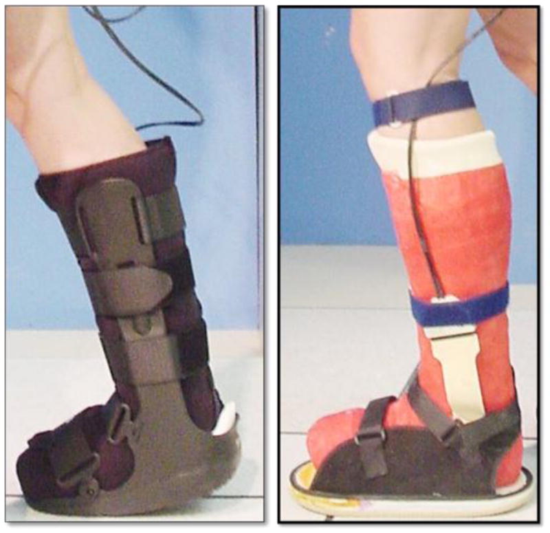
Novel Pedar® pressure insole fitted within removable cast-walker boot (DH Pressure Relief Walker ®) or total contact cast.
2.3 Data processing
All analyses utilized data from the foot with incident ulceration. For the barefoot walking trials on the EMED platform, each trial produced a single step recording, or “map”. Each plantar map was divided into three masks in the anterior-posterior (A-P) direction using the Percent Mask function in Novel Multimask (version 13.3.38) software. These three masks, formed by placing dividing lines at 33% and 63% of the A-P axis of the plantar map, reflect the anatomically distinct regions of the hind foot, midfoot, and forefoot [Sinacore et al. 2008].
For the off-loaded walking trials using the Pedar insoles, Novel Multimask software was used to create similar masks for the hind foot, midfoot, and forefoot based on dividing lines at 33% and 63% of the A-P axis of the plantar map. In the few instances in which the off-loading footwear reduced pressures in the forefoot so much that the plantar map did not reflect the full anterior-posterior foot length, an absolute mask, representing the same percentages as previously described, was applied and fitted to the pressure map to ensure that the mask dividing lines were consistent between barefoot EMED and in-shoe Pedar walking trials. We chose to use three regions for three main reasons. First, the different spatial resolutions of the barefoot (EMED) and off-loaded (Pedar) techniques made it difficult to precisely and accurately match up small regions. Second, given the amount of off-loading observed in the TCC or CWB conditions, particularly in the forefoot region, we did not feel confident that the EMED and Pedar pressure maps could be co-registered with sufficient precision to match up small regions. Third, given that the highest pressures are typically recorded at the ulcer site [Stokes et al., 1976], our choice to sample across a larger spatial area should not change the results compared to an analysis of pressures recorded immediately around the ulcer site.
Due to different subject heights and walking speeds, subjects took a varying number of steps to traverse the 7.6 m walkway. As a result, the total number of steps varied slightly from subject to subject, though there was no difference between groups (TCC = 19 steps [SD 4.5], CWB = 18 steps [SD 6.3]). In all subjects, the total number of steps analyzed exceeded twelve steps, larger than the cut-off (8 steps) which has previously been shown to ensure high reliability of maximum force, force-time integral, peak pressure, and pressure-time integral [Kernozek et al. 1996]. In all cases the first and last steps were omitted so that the average pressure map reflected mid-gait characteristics during steady-state rather than acceleration and deceleration phases of walking [Lavery et al. 1997a].
For both barefoot EMED and in-shoe Pedar data, selected load distribution variables were averaged for each mask over multiple trial steps using GroupMask Evaluation software (Novel Inc., St. Paul, MN, USA). Primary dependent variables of interest for the whole foot and for each mask included maximum force (N), force-time integral (FTI in N·s), peak pressure (PP in kPa), pressure-time integral (PTI in kPa·s), contact area (cm2), and contact time (ms).
2.4 Statistical analyses
The subjects in this analysis were a subset of a larger parent study which was statistically powered to detect a 10% effect size difference in healing proportions and healing times between the TCC and CWB off-loading conditions based on our past research [Mueller et al. 1989, Sinacore et al. 1987, Sinacore 1998]. The power analysis for the larger study assumed a 2-tailed significance level of 0.05 with minimum power of 0.80. No separate a priori power analysis was conducted for the load-related variables assessed in this study, since based on the earlier findings [Lavery et al. 1996b], we did not expect a priori to find a difference in loading variables between the devices.
The distributions between groups for sex, type of diabetes and ulcer location were assessed using Fisher’s Exact Test. Other demographic variables and walking speed were compared between off-loading treatment groups using independent t-tests. In addition, healing times were compared using independent t-tests, and proportions of subjects healed in each device were compared using Chi-Square analysis.
We were interested in assessing the off-loading capabilities of the two treatment conditions in a randomized study design with subjects walking at freely-chosen speed. Thus we considered whether walking speed and the barefoot value of each dependent variable should be included as covariates. For example, we assessed whether peak pressure during barefoot walking should be included as a covariate for peak pressure while wearing the off-loading device. The determination of whether to include barefoot values was made based on a three-step process: (1) assessing the predictive capacity of the barefoot pressure variables by measuring correlation coefficients between the dependent variables and the possible covariates for the entire study sample, with a threshold for inclusion set at α = 0.10; (2) testing for independence of the covariate and the effect of off-loading treatment, evidenced by a non-significant t-test between treatment condition and the possible covariate; and (3) testing for homogeneity of regression slopes between the two treatment conditions, evidenced by a lack of significant interaction effect between the covariate and the treatment condition [Field 2009].
Thus, statistical analysis consisted of a univariate analysis of covariance (ANCOVA) for each of the dependent variables, with footwear condition (TCC or CWB) as the between-groups factor, and both walking speed and the barefoot value of the dependent variable included as covariates. The ANCOVAs were conducted for the foot as a whole and for the hind foot, midfoot, and forefoot regions. In addition, since some subjects had ulcers in the forefoot while others had ulcers in the midfoot, we also assessed load reduction in each subject’s ulcerated region by performing an ANCOVA using only the load variables from whichever region was ulcerated for each subject. For all analyses, the alpha level was set at 0.05. Data are reported in Tables 2 and 3 as adjusted means that account for variability in the covariates. The PASW Statistics version 18 was used for all statistical analyses (SPSS Statistics Inc., Chicago, USA).
Table 2.
Loading and kinematic variables for Total Foot, no masks applied, Mean (95% CI).
| EMED | TCC | CWB | P-value (TCC vs. CWB) | |
|---|---|---|---|---|
| Contact Time (ms) | 1119 (933–1305) | 673 (610–737) | 676 (616–737) | 0.945 |
| Contact Area (cm2) | 147 (132–161) | 141 (129–153) | 143 (132–153) | 0.771 |
| Maximum Force (N) | 995 (895–1095) | 645 (584–705) | 663 (605–721) | 0.659 |
| Force-Time Integral (N·s) | 734 (636–832) | 306 (251–362) | 328 (275–381) | 0.564 |
| Peak Pressure (kPa) | 923 (803–1042) | 245 (195–295) | 209 (161–257) | 0.297 |
| Pressure-Time Integral (kPa·s) | 508 (419–598) | 107 (86–128) | 83 (63–103) | 0.093 |
Table 3.
Loading and kinematic variables for regional masks. Values are Mean (95% CI).
| (a) Hind foot Mask | EMED | TCC | CWB | P-value (TCC vs. CWB) |
|---|---|---|---|---|
| Contact Time (ms) | 725 (533–917) | 624 (561–686) | 676 (617–736) | 0.223 |
| Contact Area (cm2) | 43 (40–46) | 52 (48–55) | 53 (50–57) | 0.511 |
| Maximum Force (N) | 543 (478–608) | 403 (335–471) | 482 (417–547) | 0.098 |
| Force-Time Integral (N·s) | 210 (159–260) | 143 (98–187) | 201 (159–243) | 0.063 |
| Peak Pressure (kPa) | 348 (290–407) | 160 (118–202) | 208 (167–248) | 0.108 |
| Pressure-Time Integral (kPa·s) | 144 (86–201) | 64 (48–80) | 77 (62–92) | 0.234 |
| (b) Mid foot Mask | EMED | TCC | CWB | P-value (TCC vs. CWB) |
| Contact Time (ms) | 881 (705–1058) | 647 (579–715) | 665 (600–730) | 0.701 |
| Contact Area (cm2) | 47 (40–55) | 50 (42–58) | 57 (49–65) | 0.214 |
| Maximum Force (N) | 442 (340–543) | 226 (163–288) | 263 (203–323) | 0.393 |
| Force-Time Integral (N·s) | 201 (153–249) | 89 (62–116) | 108 (83–134) | 0.307 |
| Peak Pressure (kPa) | 476 (337–616) | 180 (134–226) | 111 (67–155) | 0.036 |
| Pressure-Time Integral (kPa·s) | 221 (153–289) | 70 (48–91) | 51 (31–72) | 0.235 |
| (c) Forefoot Mask | EMED | TCC | CWB | P-value (TCC vs. CWB) |
| Contact Time (ms) | 933 (841–1024) | 595 (485–705) | 538 (432–643) | 0.444 |
| Contact Area (cm2) | 54 (47–61) | 40 (31–49) | 32 (24–40) | 0.218 |
| Maximum Force (N) | 723 (594–852) | 183 (141–225) | 99 (59–140) | 0.011 |
| Force-Time Integral (N·s) | 311 (228–395) | 64 (44–84) | 28 (10–47) | 0.017 |
| Peak Pressure (kPa) | 853 (711–996) | 134 (97–170) | 66 (32–101) | 0.011 |
| Pressure-Time Integral (kPa·s) | 359 (263–455) | 53.9 (38.3–69.6) | 19.6 (4.6–34.6) | 0.004 |
| (d) Mask with Ulcer | EMED | TCC | CWB | P-value (TCC vs. CWB) |
| Contact Time (ms) | 969 (177–1760) | 610 (508–712) | 551 (453–648) | 0.392 |
| Contact Area (cm2) | 57 (28–86) | 46 (35–56) | 35 (25–45) | 0.156 |
| Maximum Force (N) | 746 (303–1188) | 229 (159–299) | 123 (57–190) | 0.038 |
| Force-Time Integral (N·s) | 335 (27–643) | 84 (52–116) | 43 (12–74) | 0.073 |
| Peak Pressure (kPa) | 841 (250–1432) | 166 (110–222) | 74 (21–128) | 0.024 |
| Pressure-Time Integral (kPa·s) | 394 (32–820) | 68 (44–92) | 25 (1.6–48) | 0.014 |
3. Results
Twenty-three subjects who met the inclusion criteria and were randomly assigned to either TCC or CWB off-loading footwear had an overall mean age of 54 years (SD 11), a mean body mass index (BMI) of 31.9 (SD 5.2), a mean duration of DM of 17 (SD 14) years, and a mean HbAlc value of 8.7 (SD 2.0). There were no significant differences between the groups in any of the demographic or DM-related variables (Table 1). Walking speed was not significantly different between the groups, and did not meet the criteria for inclusion in the ANCOVA models except for contact time (whole foot and all three regions) and contact area (forefoot only). None of the barefoot EMED variables was significantly different between the groups, but unlike walking speed, the EMED values met the criteria for inclusion in all of the ANCOVA models. Since there were no group differences in EMED values for any dependent variable, for simplicity the barefoot EMED values are collapsed across groups in Tables 2 and 3.
Table 1.
Subject characteristics. Values are Mean (SD).
| TCC group | CWB group | P | |
|---|---|---|---|
| N | 11 | 12 | - |
| Sex (F/M) | 2/9 | 2/10 | 1.00 |
| Type of DM (type 1/type 2) | 1/10 | 2/10 | 1.00 |
| Ulcer Location (forefoot/midfoot) | 8/3 | 11/1 | 0.23 |
| Age (yrs) | 55 (13) 95% CI: 48–63 |
53 (10) 95% CI: 48–59 |
0.69 |
| Height (cm) | 183 (8) 95% CI: 179–188 |
183 (10) 95% CI: 177–188 |
0.83 |
| Mass (kg) | 107 (27) 95% CI: 90–123 |
108 (17) 95% CI: 98–117 |
0.92 |
| BMI | 31.4 (6.2) 95% CI: 27.8–35.1 |
32.3 (4.5) 95% CI: 29.7–34.8 |
0.71 |
| HbA1c | 8.5 (2.3) 95% CI: 7.1–9.8 |
8.9 (1.8) 95% CI: 7.9–10.0 |
0.64 |
| DM duration (years) | 19 (14) 95% CI: 8–26 |
17 (13) 95% CI: 10–24 |
0.79 |
| Walking speed (m/min) | 53 (16) 95% CI: 44–62 |
55 (12) 95% CI: 48–62 |
0.70 |
| Ulcer healing time (days) | 95 (61) | 94 (64) | 0.95 |
| Healing proportion | 9/11 | 5/12 | 0.05 |
Values are mean (SD) and 95% confidence interval
Abbreviations: TCC = total contact cast; CWB = cast walker boot; BMI = body mass index; DM = diabetes mellitus
For the foot as a whole (i.e., no regional masks applied) both off-loading devices produced large reductions in maximum force (33–35%), force-time integral (53–56%), peak pressure (75–77%), and pressure-time integral (81–83%). Contact time and contact area did not differ between the two off-loading devices, as shown in Table 2. In the hind foot mask, no significant differences in adjusted means were observed for any of the six outcome variables, though the CWB condition showed trends (P < 0.10) toward higher maximum force and higher force-time integral compared to the TCC condition. In the midfoot mask, there was a significantly greater reduction in peak pressure in the CWB condition (77%) than in the TCC condition (63%) (P = 0.036). In the forefoot mask, both devices significantly reduced loads compared to the barefoot condition, but there were significantly greater reductions in the CWB compared to the TCC for peak pressure (92% versus 84% reduction), pressure-time integral (94% versus 85%), maximum force (86% versus 75%), and force-time integral (91% versus 79%).
There was a non-significant difference in the distribution of ulcers between the two groups (P = 0.23), with 3/11 midfoot ulcers in the TCC group and 1/12 midfoot ulcers in the CWB group (Table 1). The results of the ANCOVAs for the ulcer region (Table 3) were similar to the reported findings for the forefoot region. The CWB showed greater reductions than TCC with respect to barefoot EMED loads in terms of peak pressure (91% CWB versus 80% TCC), PTI (94% CWB versus 83% TCC), and maximum force (84% CWB versus 69% TCC).
For those subjects whose ulcers healed, there was no difference in healing time between the groups (Table 1), with the TCC group mean of 95 days (SD 61) and the CWB group mean of 94 days (SD 64). However, there was a difference in the proportion of subjects who experienced ulcer healing. Nine out of eleven subjects (82%) who were randomly assigned to the TCC group experienced ulcer healing, compared to five out of twelve (42%) ulcers healed in individuals randomized to the CWB group (χ2 {df=1} = 3.88, P< 0.05).
4. Discussion
Our results suggest that a removable CWB offered equivalent whole-foot and greater forefoot off-loading than TCC when both devices were compared to loads measured during a baseline condition of barefoot walking. Both devices produced large reductions from the barefoot condition for peak pressure, maximum force, pressure-time integral, and force-time integral. Still, despite large load reductions in both devices and greater forefoot off-loading in the CWB, the TCC had a greater healing proportion.
The large pressure reductions seen in the forefoot in both the TCC and removable CWB corroborate previous findings [Lavery et al. 1996b]. However, while Lavery and colleagues found the TCC and removable CWB to be equivalent in reducing peak pressures at the 1st metatarsal head, 2nd–5th metatarsal heads, and hallux [Lavery et al. 1996b], we compared the devices over the entire forefoot and found that the CWB yielded greater reductions in peak pressure, maximum force, pressure-time integral, and force-time integral.
One limitation of this study was the difference in spatial resolution between the barefoot EMED trials (2 sensors/cm2) and the off-loaded Pedar trials (0.45–0.6 sensors/cm2). Spatial averaging in the Pedar trials could lead to lower values for peak pressure and pressure-time integral, thus overestimating the effects of pressure reduction between EMED and Pedar trials. Future studies should aim to have matching spatial resolutions for all pressure-based load assessment. The similarity of results for maximum force and force-time integral, respectively, reinforce our findings of greater reductions in peak pressure and pressure-time integral in the forefoot. It should be noted that while the trends in load reduction were similar between pressure and force variables, the reductions for force were somewhat muted, possibly indicating that the pressure and pressure-time integral reductions were inflated by the difference in spatial resolution between the EMED and Pedar sensors.
In contrast to our findings of superior forefoot load reduction in the removable CWB, we found that the TCC subjects experienced a higher proportion of ulcer healing (9/11, 82%) compared to the CWB subjects (5/12, 42%), though there was no difference in average healing time between the groups for those individuals who healed. Given that both devices produced large, significant reductions in peak pressure, pressure-time integral, maximum force, and force-time integral compared to barefoot walking, and that the removable CWB produced even greater reductions in forefoot loading than the TCC, the fact that TCC off-loading resulted in a higher proportion of ulcer healing than CWB suggests that enhanced load reduction alone may not promote improved healing outcomes.
Along with off-loading, patient compliance to treatment is also important in ulcer healing. Indeed, in previous studies TCC has shown superior healing results compared to the type of removable CWB tested in the current study [Armstrong et al. 2001]. However, in a subsequent study, healing rates were improved by covering the CWB with a layer of fiberglass casting material, thus rendering them irremovable [Armstrong et al. 2005]. In addition, Katz and colleagues showed that irremovable CWB yielded statistically equivalent healing results compared to a standard TCC [Katz et al. 2005]. Thus, our findings of inferior healing rates despite superior pressure reduction in the CWB condition are in agreement with previous findings, further underscoring the need to render a CWB irremovable in order to achieve similar healing rates as a TCC.
In the current study we evaluated the impact of walking speed on various timing, spatial, and load-related dependent variables. We found no significant differences in self-selected walking speed between the TCC and CWB groups, suggesting that the imposed additional weight and joint constraint of the devices did not differentially affect the walking speed in the two treatment groups. Moreover, walking speed only met criteria for inclusion in the ANCOVA models for contact time and contact area, and in these cases accounted for a relatively small portion (6–15%) of the between-subjects variability.
We believe that our methods and findings have important utility for future studies that seek to develop and compare effectiveness of off-loading devices in neuropathic patients. In randomized trials or clinical situations, it may be disadvantageous to test all subjects in all possible off-loading treatments. In these instances, expressing pressure and force variables relative to barefoot loading allows a comparison among devices and allows for a standard expression of similarities and differences of therapeutic devices that can be individualized to the patient’s structural deformities. This method may also have clinical relevance as an educational tool. Clinicians have long recognized that patients with neuropathy regularly walk barefoot with their feet unprotected despite being prescribed an off-loading device [Armstrong et al. 2004]. By showing neuropathic patients the magnitude and location of their barefoot pressures, clinicians can help these at-risk patients to recognize, appreciate, and understand the deleterious consequences of excessive plantar loads while walking barefoot and unprotected. We also propose that future research focuses on patient activity patterns, as excessive cumulative tissue stress can develop even in a device that reduces peak pressure and force if the patient undergoes a rapid increase in weight-bearing activity [Lott et al 2005]. Since we did not record either patient compliance or patient activity, it would be premature to conclude that equivalent or superior healing results would have been found if the CWB had been rendered irremovable. Still, this is a logical hypothesis that warrants further investigation.
5. Conclusion
In subjects with diabetes mellitus, peripheral neuropathy, and plantar ulceration, we found that compared to a baseline condition of barefoot walking, a commercially-available removable CWB provides similar whole-foot load reduction as the “gold standard” total contact casting technique. Moreover, the removable CWB provides greater reduction in the key loading variables of peak pressure, pressure-time integral, maximum force, and force-time integral in the forefoot region of the foot, a frequent site of diabetic foot ulceration. However, our finding of lower healing proportions in the removable CWB despite greater load reductions reinforces the importance of compliance in ulcer healing in the diabetic, neuropathic foot. Thus to improve healing outcomes, clinicians are advised to use irremovable devices such as the TCC, or a CWB that has been rendered irremovable. In the event that irremovable devices are not possible, then patient compliance should be monitored.
Acknowledgments
We acknowledge funding support from the National Institutes of Health:
NIDDK R01 DK 59224 (PI: Sinacore)
NICHD T32 HD 007434 (PI: Mueller)
NIDDK R21 DK 079457 (PI: Prior & Sinacore)
Footnotes
Publisher's Disclaimer: This is a PDF file of an unedited manuscript that has been accepted for publication. As a service to our customers we are providing this early version of the manuscript. The manuscript will undergo copyediting, typesetting, and review of the resulting proof before it is published in its final citable form. Please note that during the production process errors may be discovered which could affect the content, and all legal disclaimers that apply to the journal pertain.
References
- Armstrong DG, Peters EJG, Athanasiou KA, Lavery LA. Is there a critical level of plantar foot pressure to identify patients at risk for neuropathic foot ulceration? J Foot Ankle Surg. 1998a;37:303–307. doi: 10.1016/s1067-2516(98)80066-5. [DOI] [PubMed] [Google Scholar]
- Armstrong DG, Lavery LA, Bushman TR. Peak foot pressures influence the healing time of diabetic foot ulcers treated with total contact casts. J Rehab Res Dev. 1998b;35:1–5. [PubMed] [Google Scholar]
- Armstrong DG, Nguyen HC, Lavery LA, van Schie CH, Boulton AJM, Harkless LB. Offloading the diabetic foot wound: a randomized clinical trial. Diabetes Care. 2001;24:1019–1022. doi: 10.2337/diacare.24.6.1019. [DOI] [PubMed] [Google Scholar]
- Armstrong DG, Lavery LA, Holtz-Neiderer K, Mohler MJ, Wendel CS, Nixon BP, Boulton AJM. Variability in activity may precede diabetic foot ulceration. Diabetes Care. 2004;27:1980–1984. doi: 10.2337/diacare.27.8.1980. [DOI] [PubMed] [Google Scholar]
- Armstrong DG, Lavery LA, Wu S, Boulton AJW. Evaluation of removable and irremovable cast walkers in the healing of diabetic foot wounds: a randomized controlled trial. Diabetes Care. 2005;28:551–554. doi: 10.2337/diacare.28.3.551. [DOI] [PubMed] [Google Scholar]
- Boulton AJ, Hardisty CA, Betts RP, Franks CI, Worth RC, Ward JD, Duckworth T. Dynamic foot pressure and other studies as diagnostic and management aids in diabetic neuropathy. Diabetes Care. 1983;6(1):26–33. doi: 10.2337/diacare.6.1.26. [DOI] [PubMed] [Google Scholar]
- Bus SA, de Lange A. A comparison of the 1-step, 2-step, and 3-step protocols for obtaining barefoot plantar pressure data in the diabetic neuropathic foot. Clinical Biomechanics. 2005;20:892–899. doi: 10.1016/j.clinbiomech.2005.05.004. [DOI] [PubMed] [Google Scholar]
- Field A. Discovering statistics using SPSS. 3. Sage Publications; Thousand Oaks, CA, USA: 2009. [Google Scholar]
- Katz IA, Harlan A, Miranda-Palma B, Prieto-Sanchez L, Armstrong DG, Bowker JH, Mizel MS, Boulton AJM. A randomized trial of two irremovable off-loading devices in the management of plantar neuropathic diabetic foot ulcers. Diabetes Care. 2005;28:555–559. doi: 10.2337/diacare.28.3.555. [DOI] [PubMed] [Google Scholar]
- Kernozek TW, LaMott EE, Dancisak MJ. Reliability of an in-shoe pressure measurement system during treadmill walking. Foot Ankle Int. 1996;17:204–209. doi: 10.1177/107110079601700404. [DOI] [PubMed] [Google Scholar]
- Lavery LA, Armstrong DG, Harkless LB. Classification of diabetic foot wounds. Journal of Foot & Ankle Surgery. 1996a;35(6):528–531. doi: 10.1016/s1067-2516(96)80125-6. [DOI] [PubMed] [Google Scholar]
- Lavery LA, Vela SA, Lavery DC, Quebedeaux TL. Reducing dynamic foot pressures in high-risk diabetic subjects with foot ulcerations: a comparison of treatments. Diabetes Care. 1996b;19:818–821. doi: 10.2337/diacare.19.8.818. [DOI] [PubMed] [Google Scholar]
- Lavery LA, Vela SA, Fleischli JG, Armstrong DG, Lavery DC. Reducing plantar pressure in the neuropathic foot: A comparison of footwear. Diabetes Care. 1997a;20:1706–1710. doi: 10.2337/diacare.20.11.1706. [DOI] [PubMed] [Google Scholar]
- Lavery LA, Vela SA, Lavery DC, Quebedeaux TL. Total contact casts: pressure reduction at ulcer sites and the effect on the contralateral foot. Arch Phys Med Rehabil. 1997b;78:1268–1271. doi: 10.1016/s0003-9993(97)90342-7. [DOI] [PubMed] [Google Scholar]
- Lavery LA, Armstrong DG, Wunderlich RP, Tredwell J, Boulton AJM. Predictive value of foot pressure assessment as part of a population-based diabetes disease management program. Diabetes Care. 2003;26(4):1069–1073. doi: 10.2337/diacare.26.4.1069. [DOI] [PubMed] [Google Scholar]
- Lott DJ, Maluf KS, Sinacore DR, Mueller MJ. Relationship between changes in activity and plantar ulcer recurrence in a patient with diabetes mellitus. Physical Therapy. 2005;85(6):579–588. [PubMed] [Google Scholar]
- Margolis DJ, Allen-Taylor L, Hoffstad O, Berlin JA. Diabetic neuropathic foot ulcers and amputation. Wound Repair Regen. 2005;13:230–236. doi: 10.1111/j.1067-1927.2005.130303.x. [DOI] [PubMed] [Google Scholar]
- McPoil TG, Cornwall MW, Dupuis L, Cornwell M. Variability of plantar pressure data. A comparison of two-step and mid-gait methods. J Am Podiatr Med Assoc. 1999;89:495–501. doi: 10.7547/87507315-89-10-495. [DOI] [PubMed] [Google Scholar]
- Meyers-Rice B, Sugars L, McPoil T, Cornwall MW. Comparison of three methods for obtaining plantar pressures in non-pathologic subjects. J Am Podiatr Med Assoc. 1994;84:499–504. doi: 10.7547/87507315-84-10-499. [DOI] [PubMed] [Google Scholar]
- Mueller MJ, Diamond JE, Sinacore DR, Delitto A, Blair VP, Drury DA, Rose SJ. Total contact casting in treatment of diabetic plantar ulcers. Diabetes Care. 1989;12(6):384–388. doi: 10.2337/diacare.12.6.384. [DOI] [PubMed] [Google Scholar]
- Pecoraro RE, Reiber GE, Burgess EM. Pathways to diabetic limb amputation: basis for prevention. Diabetes Care. 1990;13:513–521. doi: 10.2337/diacare.13.5.513. [DOI] [PubMed] [Google Scholar]
- Rosenbaum D, Hautmann S, Gold M, Claes L. Effects of walking speed on plantar pressure patterns and hindfoot angular motion. Gait & Posture. 1994;2:191–197. [Google Scholar]
- Sinacore DR, Mueller MJ, Diamond JE, Blair VP, Drury DA, Rose SJ. Diabetic plantar ulcers treated by total contact casting. Physical Therapy. 1987;67:1543–1547. doi: 10.1093/ptj/67.10.1543. [DOI] [PubMed] [Google Scholar]
- Sinacore DR. Healing times of diabetic ulcers in the presence of fixed foot deformity using total contact casting. Foot & Ankle International. 1998;19:613–618. doi: 10.1177/107110079801900908. [DOI] [PubMed] [Google Scholar]
- Sinacore DR, Bohnert KL, Hastings MK, Johnson JE. Mid foot kinetics characterize structural polymorphism in diabetic foot disease. Clinical Biomechanics. 2008;23:653–661. doi: 10.1016/j.clinbiomech.2007.05.006. [DOI] [PMC free article] [PubMed] [Google Scholar]
- Stokes IAF, Faris IB, Hutton WC. The neuropathic ulcer and loads on the foot in diabetic patients. Acta Orthop Scand. 1975;46:839–847. doi: 10.3109/17453677508989271. [DOI] [PubMed] [Google Scholar]
- Veves A, Murray HJ, Young MJ, Boulton AJM. The risk of foot ulceration in diabetic patients with high foot pressure: a prospective study. Diabetologia. 1992;35:660–663. doi: 10.1007/BF00400259. [DOI] [PubMed] [Google Scholar]
- Wagner FW. The dysvascular foot: a system for diagnosis and treatment. Foot & Ankle. 1981;2(2):64–122. doi: 10.1177/107110078100200202. [DOI] [PubMed] [Google Scholar]
- Wichmann BA, Hill ID. Generating good pseudo-random numbers. Computational Statistics & Data Analysis. 2006;51:1614–1622. [Google Scholar]