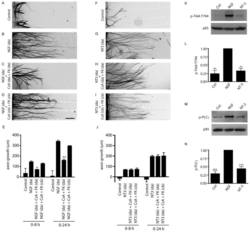
Figure 2. Calcineurin signaling is required in axons for NGF-mediated growth.
(A–D) NGF-mediated axon growth is reduced by addition of calcineurin inhibitors (CsA+FK506) to distal axons (da) (C), but not cell bodies (cb) (D). NGF (100 ng/ml) was added only to distal axons. Axons were stained with β-III-tubulin for visualization. Scale bar, 320μm. (E) Quantification of NGF-mediated axon growth in compartmentalized cultures over 0–8 hr or 0–24 hr. * p<0.05, **p<0.01, n=4 experiments. (F–I) Calcineurin signaling is not required for NT-3-mediated axon growth. (J) Quantification of NT-3-mediated axon growth, n=4. (K) NGF but not NT-3 induces phosphorylation of TrkA on Tyr-794. Neuronal lysates were probed for phospho-TrkA (Y794). Immunoblots were reprobed for p85. (L) Densitometric quantification of phospho-TrkA (Y794). **p<0.01, n=3. (M) NGF treatment selectively promotes tyrosine phosphorylation of PLC-γ. Lysates were immunoprecipitated with anti-phospho-tyrosine and probed for PLC-γ. Supernatants were probed for p85. (N) Densitometric quantification of PLC-γ phosphorylation, ***p<0.001, n=4.

