Abstract
OBJECTIVE
To investigate the effect of 4 weeks of treatment with liraglutide on insulin dose and glycemic control in type 1 diabetic patients with and without residual β-cell function.
RESEARCH DESIGN AND METHODS
Ten type 1 diabetic patients with residual β-cell function (C-peptide positive) and 19 without (C-peptide negative) were studied. All C-peptide–positive patients were treated with liraglutide plus insulin, whereas C-peptide–negative patients were randomly assigned to liraglutide plus insulin or insulin monotherapy. Continuous glucose monitoring with identical food intake and physical activity was performed before (week 0) and during (week 4) treatment. Differences in insulin dose; HbA1c; time spent with blood glucose <3.9, >10, and 3.9–9.9 mmol/L; and body weight were evaluated.
RESULTS
Insulin dose decreased from 0.50 ± 0.06 to 0.31 ± 0.08 units/kg per day (P < 0.001) in C-peptide–positive patients and from 0.72 ± 0.08 to 0.59 ± 0.06 units/kg per day (P < 0.01) in C-peptide–negative patients treated with liraglutide but did not change with insulin monotherapy. HbA1c decreased in both liraglutide-treated groups. The percent reduction in daily insulin dose was positively correlated with β-cell function at baseline, and two patients discontinued insulin treatment. In C-peptide–positive patients, time spent with blood glucose <3.9 mmol/L decreased from 3.0 to 1.0 h (P = 0.03). A total of 18 of 19 patients treated with liraglutide lost weight during treatment (mean [range] −2.3 ± 0.3 kg [−0.5 to −5.1]; P < 0.001). Transient gastrointestinal adverse effects occurred in almost all patients treated with liraglutide.
CONCLUSIONS
Treatment with liraglutide in type 1 diabetic patients reduces insulin dose with improved or unaltered glycemic control.
Glucagon-like peptide-1 (GLP-1) is secreted from the gut after meals (1) and enhances glucose-induced insulin secretion, inhibits glucagon secretion, and delays the gastric-emptying rate (1). GLP-1 receptor agonists improve glycemic control, induce weight loss in overweight subjects with type 2 diabetes (2–4), improve pancreatic β-cell function (5), and have displayed β-cell–protective and β-cell–proliferative effects in some animal studies (6). The glucose-lowering effects resulting from the inhibition of glucagon secretion and the gastric-emptying rate could be of clinical importance in type 1 diabetes (7–14). However, GLP-1 also reduces appetite and spontaneous food intake (15,16). Therefore, potential beneficial effects in terms of reduction of insulin dose, reduced risk of hypoglycemia, and improved glycemic control should be balanced against the occurrence of adverse effects (mainly nausea) and weight loss. We investigated whether 4 weeks of treatment with liraglutide, a once-daily human GLP-1 receptor analog, would reduce insulin dose while preserving or improving glycemic control and decreasing the risk of hypoglycemia in type 1 diabetic patients with and without residual β-cell function.
RESEARCH DESIGN AND METHODS
Ten type 1 diabetic patients with (C-peptide positive) and 20 without (C-peptide negative) residual β-cell function were recruited. All C-peptide–positive patients were treated with liraglutide plus insulin for 4 weeks, whereas C-peptide–negative patients were randomly assigned to either 4 weeks of liraglutide plus insulin or insulin alone (control subjects). We expected treatment effect to be smallest in the C-peptide–negative group, and because eligible C-peptide–positive patients are difficult to recruit, we only included C-peptide–negative patients as control subjects. It was calculated that with nine patients in each group and an SD of 15%, a change in insulin dose of 30% would be detected at a 5% significance level with 80% probability. Patients were recruited through outpatient clinics; they agreed to participate and provided oral and written information, and the study was performed according to the principles of the Helsinki 2 Declaration. The following were the inclusion criteria: age 18–50 years, BMI 18–27 kg/m2, Caucasian descent, diagnosed between the ages of 5 and 40 years, remission period was assumed to be ended, no known late diabetes complications (except microalbuminuria), no use of medication known to affect glucose metabolism, or symptoms of autonomic neuropathy. Participants were excluded if screening revealed not previously recognized late diabetes complications, autonomic neuropathy (see below), anemia, or HbA1c >8.5%. The study (clinical trial reg. no. NCT00993720) was surveyed by the good clinical practice unit, Bispebjerg Hospital, Denmark, and approved by the Danish Medicines Agency and the Danish Data protection Board. Before entering the study, glycemic control was evaluated for 4 days with self-monitored blood glucose measurements six to seven times daily, and, if needed, insulin dose was corrected to ensure the best possible glycemic control before entering the study.
Screening procedures
Screening was performed in the morning after an overnight fast. The night before, usual long-acting insulin was injected, but no insulin was taken in the morning. Blood samples were collected for analysis of HbA1c, liver enzymes, creatinine, total cholesterol, LDL, HDL, VLDL, triglycerides, islet cell antibodies, and GAD-65. Measurements of weight, height, and resting blood pressure were carried out. Autonomic neuropathy (orthostatic systolic blood pressure drop >20 mmHg after 0, 1, or 2 min of standing and/or a beat-to-beat variation during deep breathing of <10 bpm [R-R variation at electrocardiogram] [17]) was assessed. To determine residual β-cell function, blood glucose was raised to 15 mmol/L with intravenous bolus of glucose, and C-peptide was measured before and 6 min after an intravenous bolus of 1 mg glucagon injected 2 min after blood glucose was raised. Patients were classified as C-peptide negative if stimulated plasma C-peptide was below the detection limit (<0.03 nmol/L) and as C-peptide positive if ≥0.06 nmol/L.
Insulin dose, mean blood glucose, 24-h glucose profiles, and blood samples
In week 0 and week 4, subjects carried a blinded continuous glucose-monitoring system (iPro2; Medtronic, København, Denmark) for 3 days. Patients were allowed to live their daily life and followed their own individual routine for food intake and physical activity but had to reproduce meals as well as physical activity between the two study periods. Therefore, for each patient, day 1 in week 0 and day 1 in week 4 were similar with respect to meals and physical activity and so on for days 2 and 3. However, days 1, 2, and 3 were not required to be identical or comparable within or between patients. Patients kept logbook recordings of insulin injections and performed no less than seven daily blood glucose measurements. IPro2 uses a retrospective algorithm to convert sensor signal to glucose values based on self-monitored capillary blood glucose readings (18). Therefore, all patients received a glucose meter (Contour; Bayer Diabetes Care, Lyngby, Denmark) to ensure uniform measurements for conversion of sensor signals. Patients were requested to maintain the same level of glycemic control during the two time periods and to adjust insulin dose accordingly. Changes in mean insulin dose, mean blood glucose, 24-h glucose profiles, fasting, as well as peak postprandial blood glucose during 2 h after breakfast were evaluated from the logbook and continuous glucose-monitoring data. Hypoglycemic time, normoglycemic time, and hyperglycemic time were calculated from the 24-h glucose profiles and defined as time spent with a blood glucose <3.9, between 3.9 and 10, and >10 mmol/L, respectively. The primary end point was change in mean insulin dose, and secondary end points were changes in 24-h glucose profiles, glycemic control, mean blood glucose, and body weight between weeks 0 and 4. Blood samples were collected during weeks 0 and 4.
Randomization procedures
C-peptide–negative patients were randomly assigned to liraglutide plus insulin or insulin alone after completion of the first continuous glucose-monitoring period. Patients opened an envelope containing the randomization code generated according to www.randomization.com and sealed by a person not otherwise involved in the study.
Study medication
The dose for liraglutide was 0.6 mg daily for the first week and 1.2 mg daily for the rest of the study. If severe gastrointestinal adverse effects (vomiting) occurred, dose escalation was postponed or reduced until recovery. The dose of fast-acting insulin was decreased by 50% and long-acting insulin by 0–20% at the start of liraglutide (0% if morning blood glucose was >7 mmol/L and up to 20% if values were lower). Insulin dose was then titrated up or down to meet a target blood glucose of 5–7 mmol/L according to at least 7-point blood glucose profiles (before and after meals and at bedtime) for the next 3 days. All patients received a telephone follow-up once daily for the first 3 days and once in the second treatment week to ensure proper glycemic control and to record adverse effects. Patients also were allowed to adjust their insulin dose during the study, and they all received a chart with target blood glucose and suggested insulin adjustments.
Meal test and bicycle exercise test: C-peptide–negative patients randomly assigned to liraglutide
After an overnight fast, patients consumed a mixed meal consisting of 1,303 KJ (29.2% fat, 14.3% protein, and 52.2% carbohydrates) with 200 mL water plus 300 mL coffee or tea (optional). Before the meal, patients injected their usual dose of fast-acting insulin during week 0, and during week 4 they injected the new dose (if changed) plus liraglutide. After 2 h in a recumbent position, patients performed a 45-min exercise test on a stationary bicycle ergometer. Selected workload was 50% of the maximal watt performance during a max watt test. Frequent blood samples for plasma glucose and glucagon were collected. End points were time to hypoglycemia (plasma glucose ≤2.8 mmol/L) and rate of fall in plasma glucose (mmol/L per min).
Statistical analyses and calculations
Data are means ± SE. Comparisons of differences between normally distributed data were carried out with a two-tailed Student t test (paired within and unpaired between groups) and nonnormally distributed data with a Mann-Whitney U test between groups and Wilcoxon test for paired differences within groups. Spearman correlation analysis was used for calculations of correlation coefficients. Area under the plasma concentration curves were calculated using the trapezoidal rule. Differences resulting in P values <0.05 were considered statistically significant.
RESULTS
One C-peptide–negative patient who was randomly assigned to liraglutide was excluded as a result of protocol violation. The C-peptide–positive (n = 10) and the C-peptide–negative patients treated with liraglutide (n = 9) or insulin (n = 10) were characterized as follows: age 27.0 ± 1.5, 35.7 ± 2.2, and 32.9 ± 1.7 years; male-to-female ratio 9 to 1, 9 to 0, and 9 to 1; BMI 24.6 ± 0.9, 24.6 ± 0.7, and 23.1 ± 0.6 kg/m2; diabetes duration 3.7 ± 0.8, 17.3 ± 2.5, and 23.1 ± 1.6 years; GAD-65 positivity 9/1, 4/5, and 8/2; islet cell antibodies positivity 1/9, 1/8, and 4/6; HbA1c 6.6 ± 0.3, 7.5 ± 0.2, and 7.1 ± 0.3%; and stimulated C-peptide 0.45 ± 0.01 (range 0.08–0.94), <0.03, and <0.03 nmol/L, respectively.
Insulin dose
In liraglutide-treated patients, total daily insulin dose decreased significantly but did not change in patients treated with insulin alone (Table 1). The significance of the absolute change persisted after comparison with patients treated with insulin alone (−0.194 ± 0.03 and −0.13 ± 0.04 vs. +0.017 ± 0.02 units/kg per day; P < 0.001 and P = 0.003 in C-peptide–positive and –negative patients, respectively). In liraglutide-treated C-peptide–positive and –negative patients, fast-acting insulin decreased from 0.26 ± 0.04 to 0.14 ± 0.03 units/kg per day (P < 0.001) and from 0.39 ± 0.05 to 0.28 ± 0.03 units/kg per day (P = 0.01) and long-acting from 0.24 ± 0.03 to 0.17 ± 0.04 units/kg per day (P = 0.01) and from 0.33 ± 0.05 to 0.31 ± 0.05 units/kg per day (P = 0.02) with a mean relative reduction of −47.8 ± 10.7% (range −13 to −100) and −15.8 ± 5.5% (+8.3 to −37.3). Two patients (with stimulated C-peptide of 0.6 and 0.8 nmol/L, respectively) completely discontinued insulin treatment without loss of glycemic control. In C-peptide–positive patients, the relative reduction in insulin dose correlated positively with stimulated C-peptide at week 0 (r = 0.69, P = 0.03). No C-peptide–negative patients discontinued insulin therapy. The change in insulin dose did not correlate with weight loss (r = 0.03, P = 0.8).
Table 1.
Changes in insulin dose, mean BG, HbA1c, and stimulated C-peptide in type 1 diabetic patients with (C-peptide positive) and without (C-peptide negative) residual β-cell function before (week 0) and during (week 4) 4 weeks of treatment with liraglutide or insulin alone
| Treatment | C-peptide positive |
C-peptide negative |
C-peptide negative |
|||
|---|---|---|---|---|---|---|
| Liraglutide + insulin |
Liraglutide + insulin |
Insulin only |
||||
| Week 0 | Week 4 | Week 0 | Week 4 | Week 0 | Week 4 | |
| Insulin dose (units/kg per day) | 0.50 ± 0.06 | 0.31 ± 0.08* | 0.72 ± 0.08 | 0.59 ± 0.06† | 0.62 ± 0.04 | 0.64 ± 0.05 (NS) |
| Mean blood glucose (mmol/L) | 6.0 ± 0.2 | 6.3 ± 0.3 (NS) | 7.5 ± 0.4 | 7.7 ± 0.4 (NS) | 7.5 ± 0.4 | 7.5 ± 0.6 (NS) |
| HbA1c (%) | 6.6 ± 0.3 | 6.4 ± 0.2† | 7.5 ± 0.2 | 7.0 ± 0.1† | 7.1 ± 0.3 | 6.9 ± 0.2 (NS) |
| C-peptide (pmol/L)‡ | 520 ± 106 | 457 ± 79 (NS) | — | — | — | — |
Data are means ± SE. Mean blood glucose levels are derived from continuous glucose monitoring as mean values during 3 days with identical food intake and physical activity in week 0 and week 4. NS, nonsignificant vs. week 0 in the same group.
*P < 0.001 and
†P < 0.05 vs. week 0 in the same group.
‡n = 8.
Glycemic control
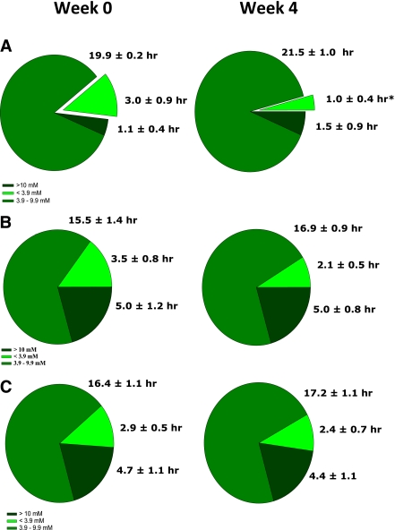
In both groups of liraglutide-treated patients, HbA1c decreased in week 4 (Table 1), but changes were not statistically different from the change in patients treated with insulin alone (−0.26 ± 0.1 and −0.47 ± 0.15 vs. −0.18 ± 0.1%; P = 0.7 and P = 0.12) in C-peptide–positive and –negative patients, respectively, whereas mean blood glucose (during continuous blood glucose monitoring) showed no change in either group (Table 1). In C-peptide–positive patients, time spent in hypoglycemia decreased significantly from 3.0 ± 0.9 to 1.0 ± 0.4 h (P = 0.03) (Fig. 1), but compared with patients treated with insulin alone, changes in hypoglycemia or hyperglycemia were not significant (−2.03 ± 0.70 vs. −0.49 ± 0.72 h, P = 0.17 and +0.38 ± 0.7 vs. −0.28 ± 0.5 h, P = 0.6). C-peptide–negative patients treated with liraglutide experienced no significant changes in time spent with hypo- or hyperglycemia, but normoglycemic time changed from 15.5 to 16.9 h (P = 0.4) as a result of a tendency (P = 0.17) for decreased hypoglycemia (Fig. 1). In patients treated with insulin alone, there were no changes in HbA1c (P = 0.1), mean blood glucose (P = 1.0), or 24-h glucose profiles (Fig. 1). In C-peptide–positive and in C-peptide–negative patients treated with liraglutide and insulin, fasting blood glucose (mmol/L) changed from 5.46 ± 0.46 to 5.95 ± 0.44 (P = 0.30), from 5.44 ± 0.64 to 6.50 ± 0.35 (P = 0.09), and from 5.67 ± 0.61 to 6.54 ± 1.09 (P = 0.35); peak postprandial blood glucose (mmol/L) from 8.09 ± 0.48 to 8.98 ± 0.54 (P = 0.08), from 11.12 ± 0.44 to 10.43 ± 0.86 (P = 0.43), and from 11.32 ± 1.12 to 10.43 (P = 0.54); and differences changed from 2.51 ± 0.62 to 2.76 ± 0.62 (P = 0.43), from 4.64 ± 1.03 to 2.77 ± 0.45 (P = 0.07), and from 4.73 ± 0.79 to 3.58 ± 0.90 (P = 0.31). Thus, the effect of liraglutide was not predominantly exerted via lowering of postprandial glucose excursions.
Figure 1.
Blood glucose evaluated from 24 h continuous glucose monitoring as mean blood glucose during 3 days with self-reported identical meals and physical activity before (week 0) and during (week 4) treatment with liraglutide. A: A total of 10 type 1 diabetic patients with residual β-cell function treated with liraglutide and insulin. B: A total of nine type 1 diabetic patients without residual β-cell function treated with liraglutide and insulin. C: A total of 10 type 1 diabetic patients without residual β-cell function treated with insulin alone. *P < 0.05 between week 0 and week 4 within the same group.
β-Cell function
In eight C-peptide–positive patients, we repeated the glucagon test in week 4 (two patients refused to repeat the test because of nausea during the first test). Blood glucose was 15.8 ± 0.5 and 15.7 ± 0.4 mmol/L before injection of glucagon in week 0 and 4, respectively, but stimulated C-peptide did not change (0.52 ± 0.11 vs. 0.46 ± 0.08 nmol/L; P = 0.4). The C-peptide response after blood glucose was raised to 15 mmol/L but, immediately before injection of glucagon, changed from 0.25 ± 0.05 to 0.33 ± 0.06 nmol/L (P = 0.18)
Meal test and bicycle exercise test
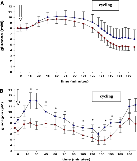
Fasting plasma glucose values were identical between study days (7.97 ± 0.8 mmol/L [week 0] and 8.01 ± 0.7 mmol/L [week 4]; P = 0.9) (Fig. 2A). Patients were injected with 7.44 and 5.44 units of fast-acting insulin in weeks 0 and 4, respectively (P = 0.04). Despite this, there were no differences of incremental or total area under the curve (AUC)0–120 min of plasma glucose between study days. Three patients stopped cycling prematurely because of hypoglycemia in both week 0 and 4 (one patient both times), and one additional patient had to stop prematurely after 25 min of cycling because of vomiting in week 4 (with plasma glucose 5.4 mmol/L). Thus, only six and five patients completed the cycling test in week 0 and 4, respectively. If hypoglycemia occurred (and exercise therefore terminated), the last observed plasma glucose value was carried forward. During cycling, total and incremental AUC120–190 min of plasma glucose changed from 487 ± 109 to 391 ± 55 mmol/L per min (P = 0.9) and from −150 ± 46 to −173 ± 30 mmol/L per min (P = 0.6) in week 0 and 4, respectively. The exercise-induced decline in plasma glucose (120–165 min) did not differ (0.102 ± 0.018 mmol/L per min [week 0] vs. 0.092 ± 0.0107 mmol/L per min [week 4]; P = 0.6). Fasting glucagon were identical (7.9 ± 0.3 vs. 7.1 ± 0.8 pmol/L) in week 0 and 4 (P = 0.4), but total AUC0–120 min of glucagon significantly decreased from 1,106 ± 92 pmol/L per min (week 0) to 845 ± 70 pmol/L per min (week 4) (P = 0.002); however, during exercise-induced decrease in plasma glucose, glucagon increased in both study days (Fig. 2B).
Figure 2.
Time course of plasma glucose (A) and glucagon (B) during a mixed meal followed by 45 min cycling (120–165 min) in nine type 1 diabetic patients before (week 0, blue circles) and during (week 4, red circles) treatment with liraglutide. Arrow: meal served. If two-way repeated-measures ANOVA resulted in significant difference (time × trial), Student paired t tests were performed at each time point. *P < 0.05.
Adverse effects and patient satisfaction
Almost all patients treated with liraglutide complained of initial gastrointestinal adverse effects, among which nausea was most frequent (18 of 19), but vomiting (2 of 19), abdominal distension (5 of 19), diarrhea (3 of 19), and foetor ex ore (3 of 19) also occurred. In most cases, gastrointestinal adverse effects were only present for the first 2–3 days of treatment, except in two patients who only tolerated 0.9 mg because of vomiting and diarrhea at 1.2 mg. Most patients reported loss of appetite (unrelated to nausea) throughout the study. Seven C-peptide–positive and two C-peptide–negative patients wanted to continue liraglutide treatment after the end of the trial. All C-peptide–positive and eight C-peptide–negative patients treated with liraglutide lost weight, amounting to −2.8 ± 0.3 kg and −1.8 ± 0.6 kg, respectively. The mean difference was −2.3 ± 0.3 and +0.2 ± 0.3 kg in liraglutide and in patients treated with insulin alone, respectively (P < 0.001). Weight loss did not correlate with BMI, but no patients became underweight (BMI <20.0 kg/m2). With insulin alone there was no change in weight and no occurrence of gastrointestinal adverse effects. There were no changes in liver enzymes, kidney function, white blood cell count, lipids, or blood pressure between week 0 and week 4 in any group.
CONCLUSIONS
This is the first report that directly compares the effect of 4 weeks of liraglutide treatment on insulin dose and glycemic control in type 1 diabetic patients with and without residual β-cell function. The major finding was a reduction in insulin dose, which was significant in both groups of patients treated with liraglutide and also compared with patients treated with insulin alone. The reduction was accompanied by unchanged time spent with blood glucose >10 mmol/L, whereas HbA1c and time spent with blood glucose <3.9 mmol/L tended to be reduced. Fast-acting insulin accounted for 63 and 85% of the reduction in insulin dose in C-peptide–positive and –negative patients, respectively. This is in accordance with a previous study (13) in which 6 months’ treatment with exenatide in C-peptide–positive patients with longstanding disease decreased total insulin by 13% but prandial insulin by 30% and also with a short-term study in C-peptide–negative adolescents in which exenatide reduced glucose excursions despite a 20% reduction in insulin dose (14). The current study also was inspired by previous short-term trials using intravenous or subcutaneous infusion of native GLP-1 in type 1 diabetic patients. In patients with low or undetectable C-peptide levels, pharmacological concentrations of GLP-1 were shown to reduce fasting plasma glucose from 13.4 to 10 mmol/L, glucagon concentration by 50% (8), and isoglycemic meal–related insulin requirement by 50% (12). Furthermore, subcutaneous exenatide or human GLP-1 administered together with the usual dose of insulin safely improved glucose control in patients without (7,11) as well as with (10) residual β-cell function.
In most cases, the antidiabetes effects were accompanied by a reduction of glucagon levels and/or by delayed gastric emptying. Therefore, inhibition of gastric emptying as well as reduced glucagon levels seem to explain the glucose-regulating effects of GLP-1 during a meal, whereas the importance of residual β-cell function is less well defined. In the current study using liraglutide, inhibition of gastric emptying may be less important because of the development of tachyphylaxis during continuous exposure (19).
In our study, the reduction in insulin dose was larger in C-peptide–positive patients, and two patients completely discontinued insulin treatment without loss of glycemic control. Despite reduced insulin doses, neither fasting nor peak postprandial blood glucose measurements differed significantly between weeks 0 and 4 in any group. This is in accordance with our goal of preserving the same (good) glycemic control during the two periods. However, there were weak trends of higher mean postprandial levels in C-peptide–positive patients and of higher fasting levels in C-peptide–negative patients treated with liraglutide. This could theoretically result from worsening of glycemic control but also, and more likely, from reduced occurrence of hypoglycemia. It may be argued that part of the reduction in insulin dose was attributed to the initial reduction in the liraglutide-treated patients. However, this is highly unlikely because all patients were optimized in insulin dose before entering the study and because insulin dose afterward was titrated up or down to meet the same target blood glucose according to careful examination of at least 7-point blood glucose measurements for 3 consecutive days immediately after start of treatment and in week 2. In C-peptide–positive patients, HbA1c decreased from 6.6 to 6.4% and from 7.5 to 7.0% in C-peptide–negative patients treated with liraglutide, in both instances, with a tendency for decreased hypoglycemia, but this did not change in patients treated with insulin alone. The reduction in HbA1c may partly result from a carry-over effect from the optimization in insulin therapy before entry. However, all patients were dose adjusted if necessary, and random assignment took place after the initial correction, and HbA1c did not change in patients treated with insulin alone.
A limitation of the current study is the lack of blinding because of unavailable placebo pen devices. However, proper blinding would have been difficult because of the necessary reduction in insulin dose (for safety reasons) in liraglutide-treated patients and because of the gastrointestinal adverse effects. The reduction in insulin dose could theoretically also result from reduction in appetite, leading to an unintended (and unreported) decrease in carbohydrate intake in week 4 or from improved insulin sensitivity. However, change in insulin dose was not correlated with weight loss.
The lack of increments in glucose-stimulated as well as glucagon-stimulated C-peptide secretion during liraglutide treatment is puzzling but is in agreement with Rother et al. (13), in which 6 months’ treatment with exenatide did not change the β-cell response to arginine.
The weight loss in the liraglutide-treated patients occurred (in 18 of 19 treated patients) despite encouragements to maintain body weight, but weight loss did not correlate with BMI. Nausea and vomiting occurred only in the first 2–3 treatment days and resolved spontaneously or subsequent to dose reduction, but patients also reported loss of appetite unrelated to nausea. Because almost all liraglutide-treated patients experienced transient gastrointestinal adverse effects, start dose should perhaps have been reduced.
We could not demonstrate a change in risk of hypoglycemia during cycling. However, in week 4, postprandial glucagon was significantly reduced but increased appropriately during exercise, when glucose levels fell; therefore, it appears that liraglutide, like GLP-1 (20), does not inhibit the glucagon response to decreasing glucose levels.
Four weeks’ treatment with liraglutide reduces insulin dose in type 1 diabetic patients along with improved or unaltered glycemic control. Treatment effect is larger in patients with residual β-cell function, and some patients may discontinue insulin treatment. Almost all patients treated with liraglutide lost weight.
Acknowledgments
This study was supported by the Poul and Erna Sehested Hansen Foundation, The Research Cluster for Diabetes and Obesity of the Health Science Faculty of Copenhagen University.
U.K. has received consultancy fees and served as an adviser to Novo Nordisk. T.K. has received consultancy and lecture fees from Lilly, Merck, and Novartis and has received a research grant from Novo Nordisk. J.J.H. has received lecture and/or consultancy fees from GlaxoSmithKline, Merck, Novartis, Novo Nordisk, and Roche. S.M. has served as a consultant or adviser to Novartis, Novo Nordisk, Merck, Sharp and Dome, Pfizer, Abbott Laboratories, sanofi-aventis, AstraZeneca, Johnson & Johnson, Roche, and Mankind; has received a research grant from Novo Nordisk; and has received fees for speaking from Novo Nordisk, Merck, Sharp and Dome, AstraZeneca, Johnson & Johnson, Abbott Laboratories, Pfizer, Roche, Schering-Plough, sanofi-aventis, and Novartis. No other potential conflicts of interest relevant to this article were reported.
U.K. researched data and wrote the manuscript. T.K. contributed to the recruitment of patients and reviewed the manuscript. J.J.H. performed the glucagon analyses, reviewed the manuscript, and contributed to discussion. S.M. reviewed the manuscript and contributed to discussion.
The authors are indebted to the patients for their enthusiastic interest and willingness to participate in this study. The authors thank Jonas Møller Kristensen, Department of Exercise and Sport Sciences, Copenhagen University, for academic and practical support with the max watt test; Leif Breum, Køge University Hospital, Denmark, for allowing the recruitment of patients from the outpatient clinic; and Jette Nymann and Dorthe Baunbjerg, Hvidovre University Hospital, Copenhagen, Denmark, and Sofie Pilgaard, The Panum Institute, Copenhagen, Denmark, for excellent technical assistance. The authors especially thank Henrik Rud, Medtronic, Copenhagen, Denmark, for excellent technical assistance whenever needed. The authors also are grateful to Bayer, Denmark, for kindly providing them with the glucose meters. Finally, the authors thank Aage Voelund, Bagsværd, Denmark, for power calculations and statistical advice.
Footnotes
Clinical trial reg. no. NCT00993720, clinicaltrials.gov.
References
- 1.Holst JJ. The physiology of glucagon-like peptide 1. Physiol Rev 2007;87:1409–1439 [DOI] [PubMed] [Google Scholar]
- 2.Madsbad S, Krarup T, Deacon CF, Holst JJ. Glucagon-like peptide receptor agonists and dipeptidyl peptidase-4 inhibitors in the treatment of diabetes: a review of clinical trials. Curr Opin Clin Nutr Metab Care 2008;11:491–499 [DOI] [PubMed] [Google Scholar]
- 3.Madsbad S. Exenatide and liraglutide: different approaches to develop GLP-1 receptor agonists (incretin mimetics)—preclinical and clinical results. Best Pract Res Clin Endocrinol Metab 2009;23:463–477 [DOI] [PubMed] [Google Scholar]
- 4.Zander M, Madsbad S, Madsen JL, Holst JJ. Effect of 6-week course of glucagon-like peptide 1 on glycaemic control, insulin sensitivity, and beta-cell function in type 2 diabetes: a parallel-group study. Lancet 2002;359:824–830 [DOI] [PubMed] [Google Scholar]
- 5.Vilsbøll T, Toft-Nielsen MB, Krarup T, Madsbad S, Dinesen B, Holst JJ. Evaluation of β-cell secretory capacity using glucagon-like peptide 1. Diabetes Care 2000;23:807–812 [DOI] [PubMed] [Google Scholar]
- 6.Kielgast U, Holst JJ, Madsbad S. Treatment of type 1 diabetic patients with glucagon-like peptide-1 (GLP-1) and GLP-1R agonists. Curr Diabetes Rev 2009;5:266–275 [DOI] [PubMed] [Google Scholar]
- 7.Behme MT, Dupré J, McDonald TJ. Glucagon-like peptide 1 improved glycemic control in type 1 diabetes. BMC Endocr Disord 2003;3:3. [DOI] [PMC free article] [PubMed] [Google Scholar]
- 8.Creutzfeldt WO, Kleine N, Willms B, Orskov C, Holst JJ, Nauck MA. Glucagonostatic actions and reduction of fasting hyperglycemia by exogenous glucagon-like peptide I(7-36) amide in type I diabetic patients. Diabetes Care 1996;19:580–586 [DOI] [PubMed] [Google Scholar]
- 9.Dupre J, Behme MT, Hramiak IM, et al. Glucagon-like peptide I reduces postprandial glycemic excursions in IDDM. Diabetes 1995;44:626–630 [DOI] [PubMed] [Google Scholar]
- 10.Dupré J, Behme MT, Hramiak IM, McDonald TJ. Subcutaneous glucagon-like peptide I combined with insulin normalizes postcibal glycemic excursions in IDDM. Diabetes Care 1997;20:381–384 [DOI] [PubMed] [Google Scholar]
- 11.Dupré J, Behme MT, McDonald TJ. Exendin-4 normalized postcibal glycemic excursions in type 1 diabetes. J Clin Endocrinol Metab 2004;89:3469–3473 [DOI] [PubMed] [Google Scholar]
- 12.Gutniak M, Orskov C, Holst JJ, Ahrén B, Efendic S. Antidiabetogenic effect of glucagon-like peptide-1 (7-36) amide in normal subjects and patients with diabetes mellitus. N Engl J Med 1992;326:1316–1322 [DOI] [PubMed] [Google Scholar]
- 13.Rother KI, Spain LM, Wesley RA, et al. Effects of exenatide alone and in combination with daclizumab on β-cell function in long-standing type 1 diabetes. Diabetes Care 2009;32:2251–2257 [DOI] [PMC free article] [PubMed] [Google Scholar]
- 14.Raman VS, Mason KJ, Rodriguez LM, et al. The role of adjunctive exenatide therapy in pediatric type 1 diabetes. Diabetes Care 2010;33:1294–1296 [DOI] [PMC free article] [PubMed] [Google Scholar]
- 15.Flint A, Raben A, Astrup A, Holst JJ. Glucagon-like peptide 1 promotes satiety and suppresses energy intake in humans. J Clin Invest 1998;101:515–520 [DOI] [PMC free article] [PubMed] [Google Scholar]
- 16.Gutzwiller JP, Göke B, Drewe J, et al. Glucagon-like peptide-1: a potent regulator of food intake in humans. Gut 1999;44:81–86 [DOI] [PMC free article] [PubMed] [Google Scholar]
- 17.Hilsted J, Jensen SB. A simple test for autonomic neuropathy in juvenile diabetics. Acta Med Scand 1979;205:385–387 [DOI] [PubMed] [Google Scholar]
- 18.Medtronic. iPro, Manual Guide København, Denmark, Medtronic, 2011
- 19.Knudsen LB, Jelsing J, Vrang N, Tang-Christensen M, Raun K. Liraglutide: short lived effect on gastric emptying-longlasting effects on body-weight (Abstract). Diabetes 2010;59:A161. [DOI] [PubMed] [Google Scholar]
- 20.Nauck MA, Heimesaat MM, Behle K, et al. Effects of glucagon-like peptide 1 on counterregulatory hormone responses, cognitive functions, and insulin secretion during hyperinsulinemic, stepped hypoglycemic clamp experiments in healthy volunteers. J Clin Endocrinol Metab 2002;87:1239–1246 [DOI] [PubMed] [Google Scholar]


