Dear Editor,
We read with interest the report by Rishi et al.[1] We wish to report our experience in a similar but younger patient with Best's disease complicated by choroidal neovascularization (CNV), being treated by intravitreal bevacizumab (IVBe).
A 10-year-old boy presented to a tertiary eye care hospital in north India with reduced vision in the right eye of 2-week duration. Best-corrected visual acuity (BCVA) was 20/200 in the right eye and 20/25 in the left eye.
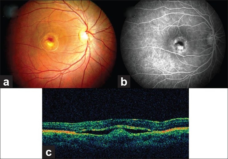
Fundus examination of the right eye revealed a large, hypopigmented, egg yolk-like subfoveal lesion with fresh subretinal hemorrhage [Fig. 1a]. These clinical findings were suggestive of the choroidal neovascular membrane (CNVM) in the right eye. Left eye fundus examination also revealed a hypopigmented, egg yolk-like lesion at the fovea, characteristic of the vitelliform stage of Best's disease. Fundus fluorescein angiography (FFA) revealed early hyperfluorescence with intense late leakage confirmatory of the CNVM in the right eye, along with hypofluorescence corresponding to the subretinal hemorrhage [Fig. 1b]. Optical coherence tomography (OCT) of the right eye confirmed the presence of the subretinal fluid (SRF), cystoid macular edema, disorganization of the retinal pigment epithelium (RPE)–choriocapillaris complex corresponding to the CNVM [Fig. 1c].
Figure 1.

At presentation – (a) fundus examination of the right eye shows a subfoveal vitelliform lesion associated with a yellowish-grey choroidal neovascular membrane with subretinal bleed; (b) right eye fundus fluorescein angiography reveals leakage from the choroidal neovascular membrane. Blocked fluorescence corresponds to the subretinal hemorrhages. (c) Optical coherence tomogram of the right eye shows the presence of subretinal fluid, cystoid macular edema, fresh subretinal hemorrhage, and disorganized retinal pigment epithelium–choriocapillaris complex suggestive of choroidal neovascular membrane
Electro-oculogram showed Arden's ratio to be 1.11 in both the eyes (normal is more than 1.85).
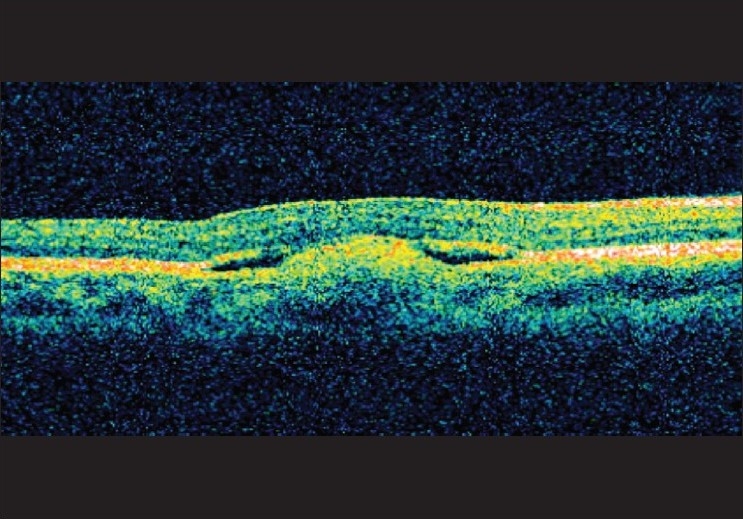
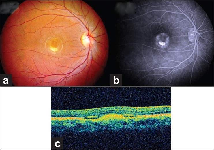
He was treated with 1.25 mg/0.05 ml IVBe after obtaining an informed (written) consent from the parents. At 6-week follow-up, BCVA in the right eye improved to 20/100. Fundus examination of the right eye revealed regression of CNVM with marked resolution of subretinal hemorrhage. OCT revealed markedly reduced SRF and increased fibrosis of CNVM as compared to the previous visit [Fig. 2]. A second IVBe was given at 6 weeks following the first one. His BCVA improved to 20/25 at 6 weeks after the secondinjection and was maintained till 24-month follow-up. Fundus photograph, FFA, and OCT demonstrated regression of the CNV, resolution of subretinal hemorrhage, and SRF [Fig. 3a–c]. BCVA also improved to almost normal without any recurrence during further follow-up (2 years).
Figure 2.

Six weeks after the first injection of bevacizumab – optical coherence tomogram shows involuting choroidal neovascular membrane and significant reduction in subretinalfluid
Figure 3.

Six weeks after the second injection of bevacizumab – (a) fundus examination of the right eye showing a scarred choroidal neovascular membrane; (b) fundus fluorescein angiography shows the absence of leakage and mere staining of the choroidal neovascular membrane; (c) optical coherence tomogram shows fibrosed choroidal neovascular membrane with a complete resolution of subretinal fluid
As discussed by Rishi et al., CNVM is a rare complication of Best's disease in children.[1,2] Various treatment modalities for CNV in Best's disease have been reported in the form of observation,[3] photodynamic therapy, laser photocoagulation, and IVBE with triamcinolone.[4,5] There is an obvious lack of consensus regarding the management of this condition. The most important limitation of laser photocoagulation is being not suitable for subfoveal CNV. On the other hand, PDT with verteporfin with or without intravitreal triamcinolone acetonide though effective, is limited by its high cost.
Leu et al. has reported favorable results of bevacizumab in a 13-year-old boy with Best's disease with CNV.[4] Cakir et al. administered combination therapy of bevacizumab and triamcinolone and reported favorable results without any adverse events.[5]
We fully agree with Rishi et al. that IVBe as a monotherapy is a promising, cost-effective, and most suitable modality of treatment in this disorder with a potential for improvement in visual acuity, although with inherent risks associated with intravitreal injections.
In our case, the vision improved with two injections and was maintained till 2-year follow-up without any adverse events.
References
- 1.Rishi E, Rishi P, Mahajan S. Intravitreal bevacizumab for choroidal neovascular membrane associated with Best's vitelliform dystrophy. Indian J Ophthalmol. 2010;58:160–2. doi: 10.4103/0301-4738.60096. [DOI] [PMC free article] [PubMed] [Google Scholar]
- 2.Miller SA, Bresnick GH, Chandra SR. Choroidal neovascular membrane in Best's vitelliform macular dystrophy. Am J Ophthalmol. 1976;82:252–5. doi: 10.1016/0002-9394(76)90428-1. [DOI] [PubMed] [Google Scholar]
- 3.Viola F, Villani E, Mapelli C, Staurenghi G, Ratiglia R. Bilateral juvenile choroidal neovascularization associated with Best's vitelliform dystrophy: Observation versus photodynamic therapy. J Pediatr Ophthalmol Strabismus. 2010;47:121–2. doi: 10.3928/01913913-20100308-14. [DOI] [PubMed] [Google Scholar]
- 4.Leu J, Schrage NF, Degenring RF. Choroidal neovascularisation secondary to Best's disease in a 13-year-old boy treated by intravitreal bevacizumab. Graefe's Arch Clin Exp Ophthalmol. 2007;245:1723–5. doi: 10.1007/s00417-007-0604-7. [DOI] [PubMed] [Google Scholar]
- 5.Cakir M, Cekiç O, Yilmaz OF. Intravitreal bevacizumab and triamcinolone treatment for choroidal neovascularization in Best disease. J AAPOS. 2009;13:94–6. doi: 10.1016/j.jaapos.2008.06.014. [DOI] [PubMed] [Google Scholar]


