

Abstract
In the title compound, C8H7N3O2·H2O, the 2-methyl-5-nitro-1H-benzimidazole molecule, excluding the methyl H atoms, is approximately planar, with a maximum deviation of 0.137 (1) Å. The crystal structure is stabilized by water molecules via N—H⋯O(water), O(water)—H⋯O and O(water)—H⋯N hydrogen bonds, forming sheets parallel to the (100) plane. A short intermolecular contact between the benzene and imidazole rings, with a centroid–centroid distance of 3.6419 (10) Å, indicates a π–π interaction.
Related literature
For general background to and the potential biological activity of benzimidazole derivatives, see: Puratchikody et al. (2008 ▶); Tonelli et al. (2010 ▶); Shingalapur et al. (2010 ▶); Refaat (2010 ▶); Lazer et al. (1987 ▶). For the preparation of the title compound, see: Umare et al. (2008 ▶); Singh & Pathak (2008 ▶). For the stability of the temperature controller used in the data collection, see: Cosier & Glazer (1986 ▶). For standard bond-length data, see: Allen et al. (1987 ▶). For related structures, see: Eltayeb et al. (2009 ▶); Arumugam et al. (2010 ▶).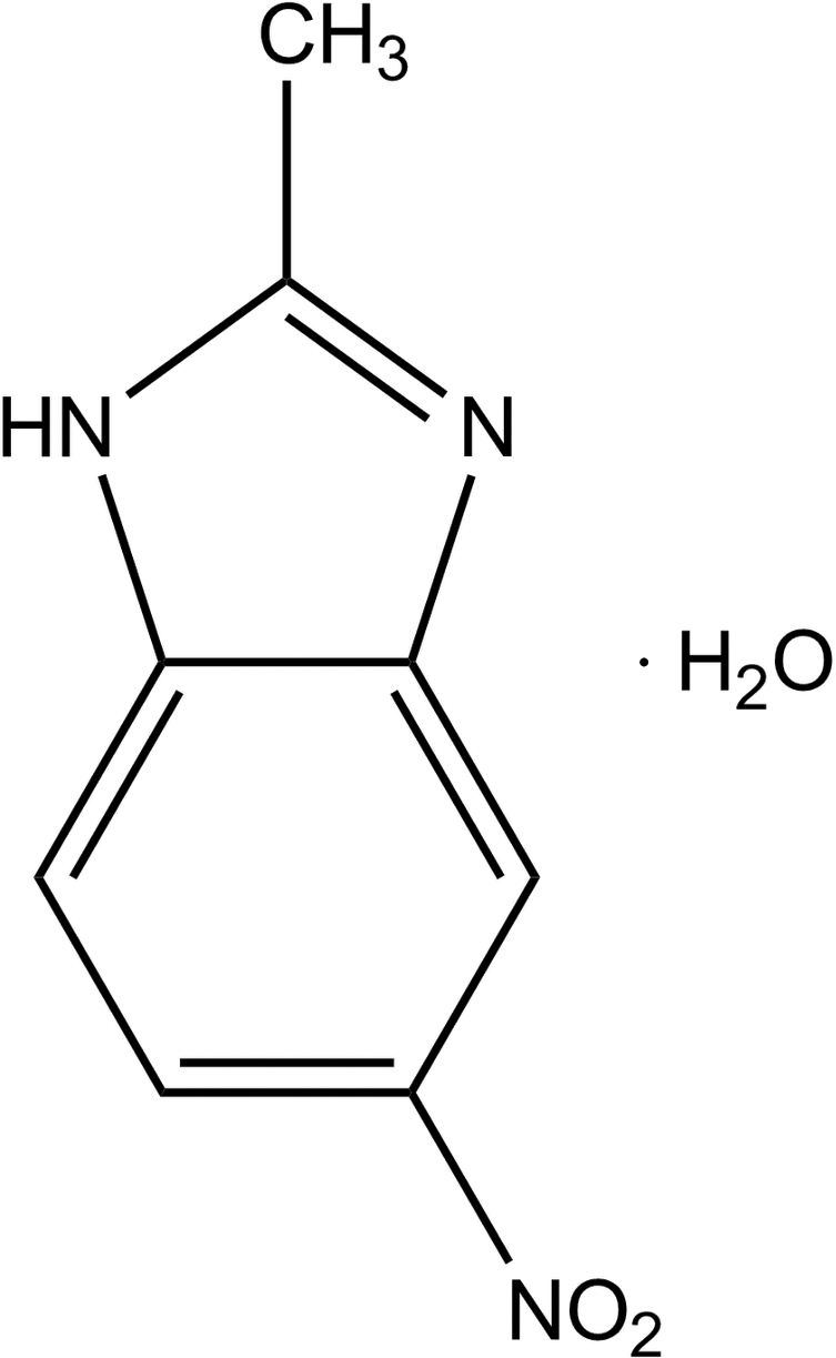
Experimental
Crystal data
C8H7N3O2·H2O
M r = 195.18
Triclinic,

a = 6.9051 (10) Å
b = 7.1309 (11) Å
c = 10.0653 (15) Å
α = 79.421 (3)°
β = 73.062 (3)°
γ = 67.517 (3)°
V = 436.61 (11) Å3
Z = 2
Mo Kα radiation
μ = 0.12 mm−1
T = 100 K
0.52 × 0.19 × 0.14 mm
Data collection
Bruker SMART APEXII DUO CCD area-detector diffractometer
Absorption correction: multi-scan (SADABS; Bruker, 2009 ▶) T min = 0.942, T max = 0.984
6312 measured reflections
1784 independent reflections
1506 reflections with I > 2σ(I)
R int = 0.032
Refinement
R[F 2 > 2σ(F 2)] = 0.040
wR(F 2) = 0.115
S = 1.06
1784 reflections
140 parameters
H atoms treated by a mixture of independent and constrained refinement
Δρmax = 0.30 e Å−3
Δρmin = −0.35 e Å−3
Data collection: APEX2 (Bruker, 2009 ▶); cell refinement: SAINT (Bruker, 2009 ▶); data reduction: SAINT; program(s) used to solve structure: SHELXTL (Sheldrick, 2008 ▶); program(s) used to refine structure: SHELXTL; molecular graphics: SHELXTL; software used to prepare material for publication: SHELXTL and PLATON (Spek, 2009 ▶).
Supplementary Material
Crystal structure: contains datablocks global, I. DOI: 10.1107/S1600536811019027/is2711sup1.cif
Structure factors: contains datablocks I. DOI: 10.1107/S1600536811019027/is2711Isup2.hkl
Supplementary material file. DOI: 10.1107/S1600536811019027/is2711Isup3.cml
Additional supplementary materials: crystallographic information; 3D view; checkCIF report
Table 1. Hydrogen-bond geometry (Å, °).
| D—H⋯A | D—H | H⋯A | D⋯A | D—H⋯A |
|---|---|---|---|---|
| N1—H1N1⋯O1Wi | 0.91 (3) | 1.84 (2) | 2.7347 (18) | 170 (2) |
| O1W—H1W1⋯O2ii | 0.831 (19) | 2.06 (2) | 2.8737 (17) | 168.5 (19) |
| O1W—H2W1⋯N2iii | 0.92 (3) | 1.86 (3) | 2.7808 (18) | 177.2 (17) |
Symmetry codes: (i)  ; (ii)
  ; (iii)
  .
.
Acknowledgments
We would like to acknowledge Universiti Sains Malaysia (USM) for the University Grant 1001/PTEKIND/8140152. HKF and CKQ also thank USM for the Research University Grant (No. 1001/PFIZIK/811160).
supplementary crystallographic information
Comment
benzimidazoles are aromatic heterocylic organic compounds of wide biological importance. They are reported as antimicrobial (Puratchikody et al., 2008), antiviral (Tonelli et al., 2010), anticancer (Refaat, 2010), anti-inflammatory (Lazer et al., 1987) and anti-diabetic (Shingalapur et al., 2010) agents.
The molecular structure of the title compound is shown in Fig. 1. The 2-methyl-5-nitro-1H-benzimidazole molecule (O1/O2/N1-N3/C1-C8), excluding methyl H atoms, is almost planar, with a maximum deviation of 0.137 (1) Å for atom O2. Bond lengths (Allen et al., 1987) and angles are within normal ranges and comparable to related structures (Eltayeb et al., 2009; Arumugam et al., 2010).
The crystal structure (Fig. 2) is stabilized by water molecules via intermolecular N1—H1N1···O1W, O1W—H1W1···O2 and O1W—H2W1···N2 hydrogen bonds to form sheets parallel to the (100) plane. The crystal packing is further consolidated by π–π stacking interactions between the centroids of N1/N2/C2/C3/C8 (Cg1) and C3–C8 (Cg2) rings, with Cg1···Cg2iii distance of 3.6419 (10) Å [symmetry code: (iii) -x, 1 - y, -z].
Experimental
4-Nitro-o-phenylenediamine (1.53 g) was well dissolved in acetic acid and refluxed on a heating mantle for 6 h. The reaction mixture was dried on rotavapor at low pressure to give the solid mass which was then crystallized with alcohol-chloroform (1:1 v/v) mixture to give the brownish crystals of title compound (Umare et al., 2008; Singh & Pathak, 2008), yield 50%, m.p. 496-498 K. Melting point was taken on Thermo Fisher digital melting point apparatus of IA9000 series and is uncorrected.
Refinement
O- and N-bound H atoms were located in a difference Fourier map and refined freely [O—H = 0.83 (2)–0.92 (3) Å, N1—H1N1 = 0.91 (2) Å]. The remaining H atoms were positioned geometrically and refined using a riding model with C—H = 0.93 or 0.96 Å, and with Uiso(H) = 1.2 or 1.5Ueq(C). A rotating-group model was applied for the methyl group. The highest residual electron density peak is located at 0.66 Å from C3 and the deepest hole is located at 0.67 Å from N3.
Figures
Fig. 1.

The molecular structure of the title compound showing 50% probability displacement ellipsoids for non-H atoms.
Fig. 2.

The crystal structure of the title compound, viewed along the a axis. H atoms not involved in hydrogen bonds (dashed lines) have been omitted for clarity.
Crystal data
| C8H7N3O2·H2O | Z = 2 |
| Mr = 195.18 | F(000) = 204 |
| Triclinic, P1 | Dx = 1.485 Mg m−3 |
| Hall symbol: -P 1 | Mo Kα radiation, λ = 0.71073 Å |
| a = 6.9051 (10) Å | Cell parameters from 2800 reflections |
| b = 7.1309 (11) Å | θ = 3.1–29.8° |
| c = 10.0653 (15) Å | µ = 0.12 mm−1 |
| α = 79.421 (3)° | T = 100 K |
| β = 73.062 (3)° | Needle, brown |
| γ = 67.517 (3)° | 0.52 × 0.19 × 0.14 mm |
| V = 436.61 (11) Å3 |
Data collection
| Bruker SMART APEXII DUO CCD area-detector diffractometer | 1784 independent reflections |
| Radiation source: fine-focus sealed tube | 1506 reflections with I > 2σ(I) |
| graphite | Rint = 0.032 |
| φ and ω scans | θmax = 26.5°, θmin = 2.1° |
| Absorption correction: multi-scan (SADABS; Bruker, 2009) | h = −8→8 |
| Tmin = 0.942, Tmax = 0.984 | k = −8→8 |
| 6312 measured reflections | l = −12→12 |
Refinement
| Refinement on F2 | Primary atom site location: structure-invariant direct methods |
| Least-squares matrix: full | Secondary atom site location: difference Fourier map |
| R[F2 > 2σ(F2)] = 0.040 | Hydrogen site location: inferred from neighbouring sites |
| wR(F2) = 0.115 | H atoms treated by a mixture of independent and constrained refinement |
| S = 1.06 | w = 1/[σ2(Fo2) + (0.0635P)2 + 0.1355P] where P = (Fo2 + 2Fc2)/3 |
| 1784 reflections | (Δ/σ)max = 0.001 |
| 140 parameters | Δρmax = 0.30 e Å−3 |
| 0 restraints | Δρmin = −0.35 e Å−3 |
Special details
| Experimental. The crystal was placed in the cold stream of an Oxford Cryosystems Cobra open-flow nitrogen cryostat (Cosier & Glazer, 1986) operating at 100.0 (1) K. |
| Geometry. All esds (except the esd in the dihedral angle between two l.s. planes) are estimated using the full covariance matrix. The cell esds are taken into account individually in the estimation of esds in distances, angles and torsion angles; correlations between esds in cell parameters are only used when they are defined by crystal symmetry. An approximate (isotropic) treatment of cell esds is used for estimating esds involving l.s. planes. |
| Refinement. Refinement of F2 against ALL reflections. The weighted R-factor wR and goodness of fit S are based on F2, conventional R-factors R are based on F, with F set to zero for negative F2. The threshold expression of F2 > 2sigma(F2) is used only for calculating R-factors(gt) etc. and is not relevant to the choice of reflections for refinement. R-factors based on F2 are statistically about twice as large as those based on F, and R- factors based on ALL data will be even larger. |
Fractional atomic coordinates and isotropic or equivalent isotropic displacement parameters (Å2)
| x | y | z | Uiso*/Ueq | ||
| O1 | 0.23733 (19) | −0.19984 (17) | 0.30176 (12) | 0.0281 (3) | |
| O2 | 0.2283 (2) | 0.0307 (2) | 0.41865 (11) | 0.0350 (3) | |
| N1 | 0.26105 (19) | 0.47231 (19) | −0.17804 (12) | 0.0176 (3) | |
| N2 | 0.2160 (2) | 0.17912 (19) | −0.18388 (12) | 0.0190 (3) | |
| N3 | 0.2355 (2) | −0.0294 (2) | 0.30934 (13) | 0.0220 (3) | |
| C1 | 0.2320 (3) | 0.4036 (2) | −0.40419 (15) | 0.0248 (4) | |
| H1A | 0.1500 | 0.3383 | −0.4285 | 0.037* | |
| H1B | 0.3773 | 0.3594 | −0.4608 | 0.037* | |
| H1C | 0.1669 | 0.5486 | −0.4198 | 0.037* | |
| C2 | 0.2349 (2) | 0.3487 (2) | −0.25514 (15) | 0.0186 (3) | |
| C3 | 0.2308 (2) | 0.1917 (2) | −0.05118 (14) | 0.0167 (3) | |
| C4 | 0.2203 (2) | 0.0559 (2) | 0.06588 (15) | 0.0183 (3) | |
| H4A | 0.1994 | −0.0652 | 0.0652 | 0.022* | |
| C5 | 0.2426 (2) | 0.1109 (2) | 0.18391 (15) | 0.0185 (3) | |
| C6 | 0.2731 (2) | 0.2924 (2) | 0.19023 (15) | 0.0189 (3) | |
| H6A | 0.2881 | 0.3206 | 0.2726 | 0.023* | |
| C7 | 0.2808 (2) | 0.4286 (2) | 0.07407 (15) | 0.0187 (3) | |
| H7A | 0.2995 | 0.5504 | 0.0760 | 0.022* | |
| C8 | 0.2594 (2) | 0.3764 (2) | −0.04673 (15) | 0.0168 (3) | |
| O1W | 0.28031 (19) | 0.83785 (17) | 0.68930 (12) | 0.0244 (3) | |
| H1N1 | 0.265 (3) | 0.598 (4) | −0.212 (2) | 0.036 (5)* | |
| H1W1 | 0.269 (3) | 0.877 (3) | 0.608 (2) | 0.036 (5)* | |
| H2W1 | 0.257 (4) | 0.949 (4) | 0.734 (2) | 0.052 (6)* |
Atomic displacement parameters (Å2)
| U11 | U22 | U33 | U12 | U13 | U23 | |
| O1 | 0.0388 (7) | 0.0180 (6) | 0.0275 (6) | −0.0098 (5) | −0.0114 (5) | 0.0032 (5) |
| O2 | 0.0605 (8) | 0.0352 (7) | 0.0160 (6) | −0.0209 (6) | −0.0151 (5) | 0.0003 (5) |
| N1 | 0.0242 (6) | 0.0138 (7) | 0.0168 (6) | −0.0068 (5) | −0.0075 (5) | −0.0017 (5) |
| N2 | 0.0259 (6) | 0.0153 (6) | 0.0177 (6) | −0.0062 (5) | −0.0089 (5) | −0.0028 (5) |
| N3 | 0.0248 (7) | 0.0214 (7) | 0.0194 (7) | −0.0065 (5) | −0.0076 (5) | −0.0004 (5) |
| C1 | 0.0349 (8) | 0.0219 (8) | 0.0186 (8) | −0.0083 (7) | −0.0104 (6) | −0.0017 (6) |
| C2 | 0.0212 (7) | 0.0157 (7) | 0.0191 (7) | −0.0040 (6) | −0.0069 (6) | −0.0042 (6) |
| C3 | 0.0185 (7) | 0.0161 (7) | 0.0162 (7) | −0.0041 (6) | −0.0064 (5) | −0.0036 (5) |
| C4 | 0.0211 (7) | 0.0137 (7) | 0.0211 (8) | −0.0056 (6) | −0.0062 (5) | −0.0033 (6) |
| C5 | 0.0197 (7) | 0.0179 (8) | 0.0163 (7) | −0.0043 (6) | −0.0061 (5) | 0.0000 (6) |
| C6 | 0.0203 (7) | 0.0206 (8) | 0.0165 (7) | −0.0049 (6) | −0.0066 (5) | −0.0051 (6) |
| C7 | 0.0203 (7) | 0.0166 (7) | 0.0216 (7) | −0.0061 (6) | −0.0066 (6) | −0.0057 (6) |
| C8 | 0.0179 (7) | 0.0146 (7) | 0.0177 (7) | −0.0041 (5) | −0.0053 (5) | −0.0030 (5) |
| O1W | 0.0428 (7) | 0.0166 (6) | 0.0191 (6) | −0.0125 (5) | −0.0127 (5) | −0.0008 (5) |
Geometric parameters (Å, °)
| O1—N3 | 1.2271 (18) | C3—C4 | 1.384 (2) |
| O2—N3 | 1.2334 (17) | C3—C8 | 1.414 (2) |
| N1—C2 | 1.3660 (18) | C4—C5 | 1.383 (2) |
| N1—C8 | 1.3710 (19) | C4—H4A | 0.9300 |
| N1—H1N1 | 0.91 (2) | C5—C6 | 1.404 (2) |
| N2—C2 | 1.320 (2) | C6—C7 | 1.378 (2) |
| N2—C3 | 1.3905 (18) | C6—H6A | 0.9300 |
| N3—C5 | 1.4609 (19) | C7—C8 | 1.3973 (19) |
| C1—C2 | 1.483 (2) | C7—H7A | 0.9300 |
| C1—H1A | 0.9600 | O1W—H1W1 | 0.83 (2) |
| C1—H1B | 0.9600 | O1W—H2W1 | 0.92 (3) |
| C1—H1C | 0.9600 | ||
| C2—N1—C8 | 107.18 (12) | N2—C3—C8 | 109.57 (13) |
| C2—N1—H1N1 | 122.1 (13) | C5—C4—C3 | 116.11 (13) |
| C8—N1—H1N1 | 130.4 (13) | C5—C4—H4A | 121.9 |
| C2—N2—C3 | 104.83 (12) | C3—C4—H4A | 121.9 |
| O1—N3—O2 | 123.11 (13) | C4—C5—C6 | 124.00 (14) |
| O1—N3—C5 | 119.20 (13) | C4—C5—N3 | 117.95 (13) |
| O2—N3—C5 | 117.69 (13) | C6—C5—N3 | 118.05 (13) |
| C2—C1—H1A | 109.5 | C7—C6—C5 | 119.80 (13) |
| C2—C1—H1B | 109.5 | C7—C6—H6A | 120.1 |
| H1A—C1—H1B | 109.5 | C5—C6—H6A | 120.1 |
| C2—C1—H1C | 109.5 | C6—C7—C8 | 117.33 (14) |
| H1A—C1—H1C | 109.5 | C6—C7—H7A | 121.3 |
| H1B—C1—H1C | 109.5 | C8—C7—H7A | 121.3 |
| N2—C2—N1 | 113.13 (13) | N1—C8—C7 | 132.77 (14) |
| N2—C2—C1 | 124.88 (13) | N1—C8—C3 | 105.30 (12) |
| N1—C2—C1 | 121.98 (13) | C7—C8—C3 | 121.93 (14) |
| C4—C3—N2 | 129.61 (13) | H1W1—O1W—H2W1 | 109 (2) |
| C4—C3—C8 | 120.82 (13) | ||
| C3—N2—C2—N1 | −0.04 (16) | O2—N3—C5—C6 | 9.3 (2) |
| C3—N2—C2—C1 | 179.09 (13) | C4—C5—C6—C7 | 0.4 (2) |
| C8—N1—C2—N2 | 0.05 (16) | N3—C5—C6—C7 | 179.98 (12) |
| C8—N1—C2—C1 | −179.10 (13) | C5—C6—C7—C8 | −0.6 (2) |
| C2—N2—C3—C4 | 179.60 (14) | C2—N1—C8—C7 | 179.43 (15) |
| C2—N2—C3—C8 | 0.01 (15) | C2—N1—C8—C3 | −0.04 (15) |
| N2—C3—C4—C5 | 179.45 (13) | C6—C7—C8—N1 | −179.40 (14) |
| C8—C3—C4—C5 | −1.0 (2) | C6—C7—C8—C3 | 0.0 (2) |
| C3—C4—C5—C6 | 0.4 (2) | C4—C3—C8—N1 | −179.62 (12) |
| C3—C4—C5—N3 | −179.17 (12) | N2—C3—C8—N1 | 0.02 (15) |
| O1—N3—C5—C4 | 9.0 (2) | C4—C3—C8—C7 | 0.8 (2) |
| O2—N3—C5—C4 | −171.14 (13) | N2—C3—C8—C7 | −179.52 (12) |
| O1—N3—C5—C6 | −170.60 (13) |
Hydrogen-bond geometry (Å, °)
| D—H···A | D—H | H···A | D···A | D—H···A |
| N1—H1N1···O1Wi | 0.91 (3) | 1.84 (2) | 2.7347 (18) | 170 (2) |
| O1W—H1W1···O2ii | 0.831 (19) | 2.06 (2) | 2.8737 (17) | 168.5 (19) |
| O1W—H2W1···N2iii | 0.92 (3) | 1.86 (3) | 2.7808 (18) | 177.2 (17) |
Symmetry codes: (i) x, y, z−1; (ii) x, y+1, z; (iii) x, y+1, z+1.
Footnotes
Supplementary data and figures for this paper are available from the IUCr electronic archives (Reference: IS2711).
References
- Allen, F. H., Kennard, O., Watson, D. G., Brammer, L., Orpen, A. G. & Taylor, R. (1987). J. Chem. Soc. Perkin Trans. 2, pp. S1–19.
- Arumugam, N., Abdul Rahim, A. S., Osman, H., Quah, C. K. & Fun, H.-K. (2010). Acta Cryst. E66, o2412–o2413. [DOI] [PMC free article] [PubMed]
- Bruker (2009). APEX2, SAINT and SADABS Bruker AXS Inc., Madison, Wisconsin, USA.
- Cosier, J. & Glazer, A. M. (1986). J. Appl. Cryst. 19, 105–107.
- Eltayeb, N. E., Teoh, S. G., Quah, C. K., Fun, H.-K. & Adnan, R. (2009). Acta Cryst. E65, o1613–o1614. [DOI] [PMC free article] [PubMed]
- Lazer, E. S., Matteo, M. R. & Possanza, G. J. (1987). J. Med. Chem. 30, 726–729. [DOI] [PubMed]
- Puratchikody, A., Nagalakshmi, G. & Doble, M. (2008). Chem. Pharm. Bull. 56, 273–281. [DOI] [PubMed]
- Refaat, H. M. (2010). Eur. J. Med. Chem. 45, 2949–2956. [DOI] [PubMed]
- Sheldrick, G. M. (2008). Acta Cryst. A64, 112–122. [DOI] [PubMed]
- Shingalapur, R. V., Hosamani, K. M., Keri, R. S. & Hugar, M. H. (2010). Eur. J. Med. Chem. 45, 1753–1759. [DOI] [PubMed]
- Singh, J. & Pathak, D. P. (2008). Orient. J. Chem. 24, 175–180.
- Spek, A. L. (2009). Acta Cryst. D65, 148–155. [DOI] [PMC free article] [PubMed]
- Tonelli, M., Simone, M., Tasso, B., Novelli, F., Boido, V., Sparatore, F., Paglietti, G., Pricl, S., Giliberti, G., Blois, S., Ibba, C., Sanna, G., Loddo, R. & La Colla, P. (2010). Bioorg. Med. Chem. 18, 2937–2953. [DOI] [PubMed]
- Umare, V. D., Ingle, V. N. & Wanare, R. K. (2008). Indian J. Heterocycl. Chem. 17, 253–256.
Associated Data
This section collects any data citations, data availability statements, or supplementary materials included in this article.
Supplementary Materials
Crystal structure: contains datablocks global, I. DOI: 10.1107/S1600536811019027/is2711sup1.cif
Structure factors: contains datablocks I. DOI: 10.1107/S1600536811019027/is2711Isup2.hkl
Supplementary material file. DOI: 10.1107/S1600536811019027/is2711Isup3.cml
Additional supplementary materials: crystallographic information; 3D view; checkCIF report