Abstract
Reconstruction of an exenterated orbit remains a challenge. Orbital prostheses are nowadays are made of silicone elastomers. A major limitation with silicone orbital prostheses is their relatively short life span. This case report describes the treatment of a patient with an exenterated orbit using a combined surgical and prosthetic approach. The upper and lower eyelids were reconstructed surgically using a deltopectoral flap. A sectional eye prosthesis was made and placed in the modified bottle-neck shaped defect to restore the patient’s appearance and confidence.
Keywords: Exenteration, Ocular prosthesis, Maxillofacial prosthesis, Psychology, Silicone elastomers, Case report, India
Orbital exenteration is a radical procedure consisting of removal of the orbital contents, including orbital fat, conjunctival sac, globe, and a part or whole of the eyelids1. Orbital defects are injurious to a person’s self-concept and sense of body image. Treatment should be provided as soon as possible to raise morale and ease the mind of the afflicted person.
Orbital prostheses are made of silicone elastomers, acrylic resin, or a combination of these.2 Most maxillofacial elastomers perform well initially, but deterioration, associated with either degradation of mechanical properties or changes in appearance, commonly occurs subsequently.3–8 This deterioration limits the service life of extraoral prostheses, and refabrication of these prostheses is time consuming, labour intensive, and costly.
Moreover, extraoral prostheses are exposed to mucosa and skin secretions; subsequently, multilayer biofilm formation can occur on the silicone surfaces. Problems such as black stains on the surface of prostheses, offensive odours, and tissue infection can arise from microbial colonisation.9
Karakoca et al. reported a mean life span of 14.5 to 14.7 months for a patient’s first and second implant-retained extraoral prostheses, respectively. The primary reasons for making a new prosthesis were discoloration, tear of the prosthesis, and mechanical failures of the acrylic resin substructure or retentive elements.10 Jebreil reported that adhesive-retained orbital prostheses last for 6–9 months and need to be refabricated subsequently.11
Problems with silicone orbital prosthesis can be avoided by reconstructing the upper and lower eyelids surgically and then rehabilitating with custom-made ocular prostheses. This clinical report describes the management of a patient with an orbital defect by a combined surgical and prosthetic approach.
Case Report
A 24 year-old woman, who had undergone a right orbital exenteration for orbital meningioma, presented to the Department of Prosthodontics and Implantology, at the Government Dental College and Research Institute (GDCRI), Bangalore, India, for rehabilitation of an orbital defect [Figure 1]. It was decided to reconstruct the eyelids surgically in collaboration with the department of Plastic Surgery, Victoria Hospital, Bangalore.
Figure 1:
Orbital defect following exenteration
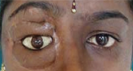
A deltopectoral flap from the second and third intercostal region was raised and then rotated into the exenterated orbit and sutured to the free skin margins. After 3 weeks, the pedicle was separated from the intercostal region. Four weeks later the flap was divided to construct the upper and lower eyelids. After 6 months, the patient presented to the Department with reconstructed upper and lower eyelids [Figure 2]. Although plastic surgery resulted in marked improvement, yet the resultant eyelids were thick, tense, and scarred. Moreover, the modified orbital defect was bottle-neck shaped. The palpebral aperture was 15 mm whereas the cavity inside was approximately 32 mm; hence, it was virtually impossible to fabricate a single piece eye prosthesis. This necessitated a unique treatment modality for the rehabilitation of the modified exenterated orbit.
Figure 2:
Reconstructed upper and lower eyelids
Separate impressions were made of the superior and inferior surfaces of the defect using irreversible hydrocolloid material (Hydrogum, Zhermack SpA, Italy). After that, irreversible hydrocolloid was injected directly into the remaining socket using an ocular impression tray. Multiple parts of the impression were assembled outside to pour a two-piece dental stone cast. Three pieces of wax template were fabricated to be assembled in a specific spatial configuration in the reconstructed orbit and processed using heat cure acrylic (Trevalon, India). A notch was made in the middle piece for orientation of the fourth piece. After placing the multiple pieces of eye prosthesis in the modified orbit, an impression for the fourth piece was made according to the Allen and Webster technique.12 The irreversible hydrocolloid material was injected into the remaining socket through the attached hollow stem of the impression tray.
Using this impression and a small plastic tumbler, an irreversible hydrocolloid mould was prepared, which was cut using a sharp blade to remove the impression. The mould space was subsequently filled with molten inlay wax. Once the wax was hard, it was removed from the mould, and the external surface was smoothed for a try-in on the patient’s face. The wax form and its corneal prominence were modified wherever necessary to duplicate the shape of the natural eye. After a try-in the wax template was processed using white acrylic. The acrylic shell was removed and trimmed for about 1–2mm on the external surface.
During iris orientation patient was asked to gaze straight ahead. The distance from the pupil of the normal eye to the midline was used in establishing the horizontal position of the prosthetic pupil’s centre. Its vertical position was determined by the canthus relationships. Marked coordinates of the pupil were used to circumscribe the diameter of the iris. Iris and scleral painting were carried out using acrylic colors and mono-poly. As the palpebral aperture of the reconstructed eyelids was larger than that of the natural eye, it was camouflaged during scleral painting. Subsequently, using the same plaster mould, the eye shell was packed with transparent acrylic to give a natural appearance. Later, it was removed from the mould, trimmed, finished and polished. The prosthesis was placed into the ocular defect and critically evaluated [Figure 3]. Spectacles were used to camouflage the scarred tissue [Figure 4]. At the time of writing, the prosthesis had been in service for 9 months without complications.
Figure 3:
Multiple unit eye prosthesis placed in reconstructed exenterated orbit
Figure 4:
Spectacles used to camouflage the scarred tissue
Discussion
This case report describes the treatment of a patient with exenterated orbit using a combined surgical and prosthetic approach. Upper and lower eyelids were reconstructed surgically using a deltopectoral flap from the second and third intercostal region to avoid the fabrication of a silicone prosthesis.
Silicones have been used for over 50 years in the field of maxillofacial prosthetics, with desirable material properties including flexibility, biocompatibility, ability to accept intrinsic and extrinsic colorants, chemical and physical inertness and mouldability.9,13 A major limitation with silicone orbital prostheses is their relatively short life span (on average 1.5 to 2 years). The main reasons for the refabrication of orbital prostheses are discoloration, problems with the attachment of the acrylic resin clip carrier to the silicone, rupture or deterioration of the silicone material and a poor fit. Moreover, meticulous hygiene is mandatory to prevent peri-implant problems, including inflammation of the skin in the implant retained orbital prosthesis.14
The resultant eyelids were thick, tense, and scarred and the modified orbital defect was bottle-neck shaped. A sectional eye prosthesis was fabricated to overcome this limitation. The impression was made with irreversible hydrocolloid which helped in retrievability of the impression from undercut area. Heat cure polymethyl methacrylate was used for fabrication of the prosthesis which has better biocompatibility.4 Multiple pieces of eye prosthesis were fabricated to be assembled in a specific spatial configuration in the reconstructed exenterated orbit. The prosthesis, although static, helped restore the patient’s appearance and confidence. In the absence of recurrent orbital meningioma, this prosthesis can be a definitive treatment for the patient.
Conclusion
Reconstruction of the exenterated orbit remains a challenge. Patients in this situation can be treated by reconstructing the upper and lower eyelids surgically and then rehabilitation with custom-made ocular prostheses.
Acknowledgments
The authors would like to thank the patient for granting consent for her case and photographs to be published.
References
- 1.Perman KI, Baylis HI. Evisceration, enucleation and exenteration. Otolaryngol Clin North Am. 1988;21:171–82. [PubMed] [Google Scholar]
- 2.Kurunmaki H, Kantola R, Hatamleh MM, Watts DC, Vallittu PK. A fiber-reinforced composite prosthesis restoring a lateral midfacial defect:A clinical report. J Prosthet Dent. 2008;100:348–52. doi: 10.1016/S0022-3913(08)60235-8. [DOI] [PubMed] [Google Scholar]
- 3.Han Y, Kiat-amnuay S, Powers JM, Zhao Y. Effect of nano-oxide concentration on the mechanical properties of a maxillofacial silicone elastomer. J Prosthet Dent. 2008;100:465–73. doi: 10.1016/S0022-3913(08)60266-8. [DOI] [PubMed] [Google Scholar]
- 4.Dootz ER, Koran A, 3rd, Craig RG. Physical properties of three maxillofacial materials as a function of accelerated aging. J Prosthet Dent. 1994;71:379–83. doi: 10.1016/0022-3913(94)90098-1. [DOI] [PubMed] [Google Scholar]
- 5.Haug SP, Moore BK, Andres CJ. Color stability and colorant effect on maxillofacial elastomers. Part II: Weathering effect on physical properties. J Prosthet Dent. 1999;81:423–30. doi: 10.1016/s0022-3913(99)80009-2. [DOI] [PubMed] [Google Scholar]
- 6.Frangou MJ, Polyzois GL, Tarantili PA, Andreopoulos AG. Bonding of silicone extraoral elastomers to acrylic resin: The effect of primer composition. Eur J Prosthodont Restor Dent. 2003;11:115–8. [PubMed] [Google Scholar]
- 7.Lemon JC, Chambers MS, Jacobsen ML, Powers JM. Color stability of facial prostheses. J Prosthet Dent. 1995;74:613–8. doi: 10.1016/s0022-3913(05)80314-2. [DOI] [PubMed] [Google Scholar]
- 8.Polyzois GL, Tarantili PA, Frangou MJ, Andreopoulos AG. Physical properties of a silicone prosthetic elastomer stored in simulated skin secretions. J Prosthet Dent. 2000;83:572–7. doi: 10.1016/s0022-3913(00)70017-5. [DOI] [PubMed] [Google Scholar]
- 9.Kiat-amnuay S, Johnston DA, Powers JM, Jacob RF. Color stability of dry earth pigmented maxillofacial silicone A-2186 subjected to microwave energy exposure. J Prosthodont. 2005;14:91–6. doi: 10.1111/j.1532-849X.2005.00017.x. [DOI] [PubMed] [Google Scholar]
- 10.Karakoca S, Aydin C, Yilmaz H, Bal BT. Retrospective study of treatment outcomes with implant-retained extraoral prostheses: Survival rates and prosthetic complications. J Prosthet Dent. 2010;103:118–126. doi: 10.1016/S0022-3913(10)60015-7. [DOI] [PubMed] [Google Scholar]
- 11.Jebreil K. Acceptability of orbital prostheses. J Prosthet Dent. 1980;43:82–5. doi: 10.1016/0022-3913(80)90358-3. [DOI] [PubMed] [Google Scholar]
- 12.Allen L, Webster HE. Modified impression method of artificial eye fitting. Am J Ophthalmol. 1969;67:189. doi: 10.1016/0002-9394(69)93148-1. [DOI] [PubMed] [Google Scholar]
- 13.Jani RM, Schaaf NG. An evaluation of facial prostheses. J Prosthet Dent. 1978;39:546–50. doi: 10.1016/s0022-3913(78)80191-7. [DOI] [PubMed] [Google Scholar]
- 14.Anita V, Raghoebar GM, Oort RP, Vissink A. Fate of implant-retained craniofacial prostheses: Life span and aftercare. Int J Oral Maxillofac Implants. 2008;23:89–98. [PubMed] [Google Scholar]


