Abstract
Ophthalmomyiasis rarely occurs worldwide, and has not been reported in Korea. We present here a case of ophthalmomyiasis caused by Phormia sp. fly larva in an enucleated eye of a patient. In June 2010, a 50-year-old man was admitted to Dankook University Hospital for surgical excision of a malignant melanoma located in the right auricular area. He had a clinical history of enucleation of his right eye due to squamous cell carcinoma 5 years ago. During hospitalization, foreign body sensation developed in his right eye, and close examination revealed a fly larva inside the eye, which was evacuated. The larva was proved to be Phormia sp. based on the morphology of the posterior spiracle. Subsequently, no larva was found, and the postoperative course was uneventful without any complaints of further myiasis. This is the first case of ophthalmomyiasis among the literature in Korea, and also the first myiasis case caused by Phormia sp. in Korea.
Keywords: Phormia sp., ophthalmomyiasis, melanoma, enucleation, fly larva
INTRODUCTION
The most common site of fly larva infestation is skin wounds. However, cases of ophthalmomyiasis have rarely been reported worldwide, accounting for <5% of all cases [1]. Most of the patients with ophthalmomyiasis were reported from tropical countries with low socioeconomic status; however, sporadic cases have occurred in developed countries [2]. Ophthalmomyiasis is classified into the external and the internal type, with the former being more common [3]. External ophthalmomyiasis is a limited infestation of superficial periocular tissues, including conjunctival myiasis. Internal ophthalmomyiasis is characterized by the presence of the larva within the eye, which occurs when dipterous larvae penetrate the conjunctiva and sclera, and migrate into the subretinal space [2]. Symptoms, such as severe eye irritation, redness, foreign body sensation, pain, lacrimation, and swelling of the lids, have been reported in patients with ophthalmomyiasis, but complications, including corneal ulcers and decreased vision, are not uncommon [4].
In Korea, 4 cases of myiasis have been reported [5]; however, myiasis of the eye has never been reported. We present here a case of ophthalmomyiasis caused by Phormia sp. larva in a Korean man.
CASE RECORD
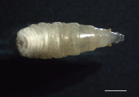
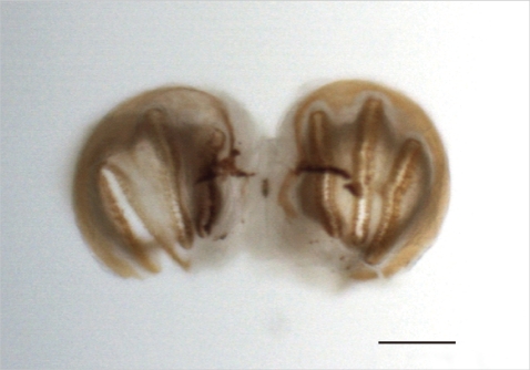
A 50-year old male residing in Cheonan-si, Chungcheongnam-do, the Republic of Korea visited the Department of Ear, Nose, and Throat of Dankook University Hospital on 1 June 2010 for a mass in the right auricular area. The mass was diagnosed as a malignant melanoma with metastatic lymphadenopathy, and wide excision was scheduled. He was a day laborer, and had an enucleation of his right eye due to squamous cell carcinoma 5 years ago. During hospitalization, foreign body sensation developed in his right eye on 9 June, and close examination revealed a moving fly larva inside the eye cavity (external type). The larva was removed and sent to the Department of Parasitology for analysis. The larva was white, wriggling, and 9.0 mm×2.0 mm in size (Fig. 1). Examination of posterior spiracles on a glass slide under light microscopy showed that the larva belongs to a species of Phormia (Fig. 2), which is endemic in Korea [6]. Identification of the larva was done by In-Yong Lee, the Department of Environmental Medical Biology, Yonsei University College of Medicine, Seoul, Korea. A wide excision was performed on 7 June 2010. The postoperative course was uneventful without any complaints of further myiasis.
Fig. 1.
The larva of Phormia sp. extracted from the patient's eye. Bar=2 mm.
Fig. 2.
Posterior spiracles of the larval stage of Phormia sp. sampled in the right eye of the patient. Bar=100 µm.
DISCUSSION
This is the first report of ophthalmomyiasis, the external type, in Korea. In humans, ophthalmomyiasis is commonly caused by the ovine nasal botfly Oestrus ovis [1]. The Russian botfly Rhinoestrus purpureus which is found in sheep-farming communities [7] and Hypoderma spp., including H. bovis, have also been reported to be the cause of ophthalmomyiasis [8]. In addition, ophthalmomyiases caused by Cochliomyia hominivorax larvae has been described in Brazil [9].
In our case, it is interesting that the causative fly was Phormia sp., a species of the blowfly. Blowflies have considerable medical and veterinary importance because of their capacity of causing traumatic myiasis and they are utilized to determine the postmortem interval in human deaths [10]. However, reported myiasis cases due to Phormia sp. are rare in the world, and human myiasis due to Phormia regina was reported mainly in the damaged tissues, such as the necrotic hip wound, scalp with necrosis, malignant wound of lower extremities, and ulcers of the ankle [11-15]. In our case, the predisposing factor for the orbital infestation was probably the necrotic tissue offered by malignant melanoma in the auricular area. The existence of P. regina flies in Korea was previously confirmed by DNA-based identification [6]. We could not perform species identification by DNA study because only 1 larva was discovered which was used for a morphological study. However, the present case is worthy of report as the first case caused by a Phormia sp. fly, and all myiasis cases reported in Korea were caused by Lucilia spp. flies (Diptera: Calliphoridae), possibly L. sericata [5].
In recent years, myiasis has been uncommon in Korea. However, a nasal myiasis was reported last year at Dankook University Hospital [5], and the present case occurred again. Judging from the developmental period of the larval stage of Phormia sp. and the warm climate, it could have been possible that he was infested with fly larvae during hospitalization [11,16]. Control of fly populations is needed at Dankook University Hospital, and prevention from myiasis is recommended in patients with open, draining wounds through continuous use of dressings. Since it has been suggested that hospital-acquired myiasis has been under-reported [17], careful observation should be performed on the presence of flies in other hospitals.
References
- 1.Baliga MJ, Davis P, Rai P, Rajasekhar V. Orbital myiasis: a case report. Int J Oral Maxillofac Surg. 2001;30:83–84. doi: 10.1054/ijom.2000.0007. [DOI] [PubMed] [Google Scholar]
- 2.Caça I, Unlü K, Cakmak SS, Bilek K, Sakalar YB, Unlü G. Orbital myiasis: case report. Jpn J Ophthalmol. 2003;47:412–414. doi: 10.1016/s0021-5155(03)00077-7. [DOI] [PubMed] [Google Scholar]
- 3.Jun BK, Shin JC, Woog JJ. Palpebral myiasis. Korean J Ophthalmol. 1999;13:138–140. doi: 10.3341/kjo.1999.13.2.138. [DOI] [PubMed] [Google Scholar]
- 4.Masoodi M, Hosseini K. External ophthalmomyiasis caused by sheep botfly (Oestrus ovis) larva: a report of 8 cases. Arch Iran Med. 2004;7:136–139. [Google Scholar]
- 5.Kim JS, Seo PW, Kim JW, Go JH, Jang SC, Lee HJ, Seo M. A nasal myiasis in a 76-year-old female in Korea. Korean J Parasitol. 2009;47:405–407. doi: 10.3347/kjp.2009.47.4.405. [DOI] [PMC free article] [PubMed] [Google Scholar]
- 6.Park SH, Zang Y, Yu DH, Jung JH, Yoo GY, Jo TH, Hwang JJ. DNA-based identification of forensically important blow fly species in Korea; Aldrichina graham, Calliphora lata, Calliphora vicina and Phormia regina. Korean J Leg Med. 2006;30:140–146. [Google Scholar]
- 7.Dar MS, Benamer M, Dar FK, Parazotos V. Ophthalmomyiasis caused by the sheep nasal botfly Oestrus ovis (Oestridae) larvae, in Bengazi area of eastern Libya. Trans R Soc Trop Med Hyg. 1980;74:303. doi: 10.1016/0035-9203(80)90087-5. [DOI] [PubMed] [Google Scholar]
- 8.Lagace-Wiens PR, Dookeran R, Skinner S, Leicht R, Colwell DD, Galloway TD. Human ophthalmomyiasis interna caused by Hypoderma tarandi, Northern Canada. Emerg Infect Dis. 2008;14:64–66. doi: 10.3201/eid1401.070163. [DOI] [PMC free article] [PubMed] [Google Scholar]
- 9.Saraiva Vda S, Amaro MH, Blfort R, Jr, Burnier MN., Jr A case of anterior internal ophthalmomyiasis: case report. Arq Bras Oftalmol. 2006;69:741–743. doi: 10.1590/s0004-27492006000500023. [DOI] [PubMed] [Google Scholar]
- 10.Szpila K, Pape T, Rusknek A. Morphology of the first instar of Calliphora vicina, Phormia regina and Lucilia illustris (Diptera, Calliphoridae) Med Vet Entomol. 2008;22:16–25. doi: 10.1111/j.1365-2915.2008.00715.x. [DOI] [PubMed] [Google Scholar]
- 11.Ali-Khan FEA, Ali-Khan Z. A case of traumatic dermal myiasis in Quebec caused by Phormia regina (Meigen) (Diptera: Calliphoridae) Can J Zool. 1975;53:1472–1476. doi: 10.1139/z75-178. [DOI] [PubMed] [Google Scholar]
- 12.Alexis JB, Mittleman RE. An unusual case of Phormia regina myiasis of the scalp. Am J Clin Pathol. 1988;90:734–737. doi: 10.1093/ajcp/90.6.734. [DOI] [PubMed] [Google Scholar]
- 13.Seaquist ER, Henry TR, Cheong E, Theologieds A. Phormia regina myiasis in a malignant wound. Minn Med. 1983;66:409–410. [PubMed] [Google Scholar]
- 14.Hall RD, Anderson PC, Clark DP. A case of human myiasis caused by Phormia regina (Diptera: Calliphoridae) in Missouri, USA. J Med Entomol. 1986;23:578–579. doi: 10.1093/jmedent/23.5.578. [DOI] [PubMed] [Google Scholar]
- 15.Miller KB, Hribar LJ, Sanders LJ. Human myiasis caused by Phormia regina in Pennsylvania. J Am Podiatr Med Assoc. 1990;80:600–602. doi: 10.7547/87507315-80-11-600. [DOI] [PubMed] [Google Scholar]
- 16.Byrd JH, Allen JC. The development of the black blow fly, Phormia regina (Meigen) Forensic Sci Int. 2001;120:79–88. doi: 10.1016/s0379-0738(01)00431-5. [DOI] [PubMed] [Google Scholar]
- 17.Joo CY, Kim JB. Nosocomial submandibular infections with dipterous fly larvae. Korean J Parasitol. 2001;39:255–260. doi: 10.3347/kjp.2001.39.3.255. [DOI] [PMC free article] [PubMed] [Google Scholar]


