Commotio retinae is a self-limited opacification of the retina secondary to direct blunt ocular trauma. Histologic studies of monkeys and humans relate this clinical observation to damaged photoreceptor outer segments and receptor cell bodies.1–3 Reports using time-domain optical coherence tomography (OCT) and spectral-domain OCT support the involvement of the photoreceptor layer, but these techniques lack the resolution necessary to confirm results of histologic analysis.4–6 Prototype high-speed ultra–high-resolution OCT (hs-UHR-OCT) images demonstrate these anatomical changes in a patient with acute commotio retinae.
Report of a Case
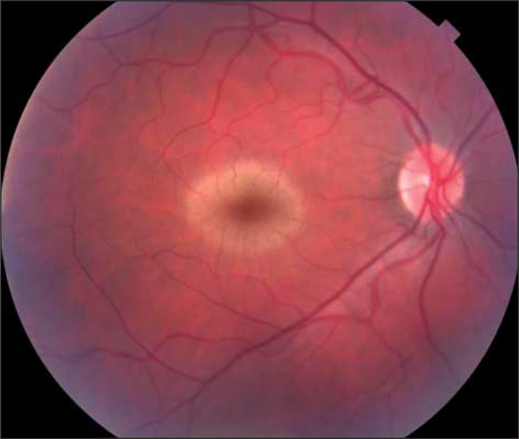
A 46-year-old man visited the emergency department with pain and blurry vision in the right eye after blunt ocular trauma. Uncorrected visual acuities were 20/30 OD and 20/25 OS. External examination showed periorbital ecchymosis and laceration. Pupil examination results were normal with relative afferent pupillary defect. Intraocular pressures were 14 mm Hg OD and 13 mm Hg OS. Slitlamp examination revealed a subconjunctival hemorrhage in the right eye. Orbital computed tomography demonstrated fracture of the right inferior and medial orbital walls. Dilated examination of the right eye showed a central, annular area of opacification of the retina surrounding the fovea consistent with commotio retinae (Figure 1). Retinal imaging was performed using spectral-domain OCT (Cirrus HD-OCT, software version 3.0; Carl Zeiss Meditec, Dublin, California) and prototype hs-UHR-OCT.
Figure 1.
Color fundus photograph of the right eye showing annular opacification surrounding the macula of commotio retinae after blunt trauma. Corresponding spectral-domain and ultra–high-resolution optical coherence tomographic images are shown in Figure 2A and C.
Comment
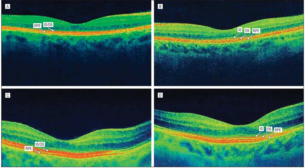
The spectral-domain OCT image suggests hyperreflectivity at the level of the photoreceptors (Figure 2A). However, the hs-UHR-OCT image better demonstrates increased backscattering at the level of the outer photoreceptor layer, with an obscuration of the inner segment–outer segment junction of the photoreceptor layers and hyperreflectivity of the retinal pigment epithelial layer (Figure 2C).
Figure 2.
Optical coherence tomographic (OCT) images. IS indicates inner segment; OS, outer segment; and RPE, retinal pigment epithelium. A, A spectral-domain OCT image of the right eye showing acute commotio retinae. B, A spectral-domain OCT image of a normal internal control (the left eye). C, A high-speed ultra–high-resolution OCT image showing higher-resolution disruption between the IS and OS photoreceptor layers and the RPE. D, A high-speed ultra–high-resolution OCT image of a normal internal control (the left eye).
Improved visualization of these changes is likely owing to the greater axial resolution of the hs-UHR-OCT at 3.5 µm compared with the 5-µm axial resolution of the spectral-domain OCT as well as denser A-scan acquisition by the hs-UHR-OCT. The hs-UHROCT uses a broader bandwidth light source to achieve higher axial resolution. Normal internal controls ofthe left eye (Figure 2B and D) are included for comparison.
Retinal disruption in commotion retinae is demonstrated at the level of the outer and inner photoreceptor layers and retinal pigment epithelial layer using prototype hs-UHR-OCT. These in vivo findings are consistent with results of previous histologic studies of commotio retinae.1–3
Acknowledgments
Funding/Support: This work was supported in part by a Challenge grant to the New England Eye Center and Department of Ophthalmology, Tufts University School of Medicine from Research to Prevent Blindness, by contracts RO1-EY11289-23 and R01-EY13178-07 from the National Institutes of Health, by grants FA9550-07-1-0101 and FA9550-07-1-0014 from the Air Force Office of Scientific Research, and by the Lions Club of Massachusetts.
Footnotes
Financial Disclosure: Dr Duker receives research support from Carl Zeiss Meditech, Optovue, and Topcon Medical Systems. Dr Fujimoto receives royalties from Carl Zeiss Meditech and has stock options in Optovue.
References
- 1.Sipperley JO, Quigley HA, Gass DM. Traumatic retinopathy in primates: the explanation of commotio retinae. Arch Ophthalmol. 1978;96(12):2267–2273. doi: 10.1001/archopht.1978.03910060563021. [DOI] [PubMed] [Google Scholar]
- 2.Mansour AM, Green WR, Hogge C. Histopathology of commotio retinae. Retina. 1992;12(1):24–28. doi: 10.1097/00006982-199212010-00006. [DOI] [PubMed] [Google Scholar]
- 3.Liem AT, Keunen JE, van Norren D. Reversible cone photoreceptor injury in commotio retinae of the macula. Retina. 1995;15(1):58–61. doi: 10.1097/00006982-199515010-00011. [DOI] [PubMed] [Google Scholar]
- 4.Sony P, Venkatesh P, Gadaginamath S, Garg SP. Optical coherence tomography findings in commotio retina. Clin Experiment Ophthalmol. 2006;34(6):621–623. doi: 10.1111/j.1442-9071.2006.01290.x. [DOI] [PubMed] [Google Scholar]
- 5.Meyer CH, Rodrigues EB, Mennel S. Acute commotio retinae determined by cross-sectional optical coherence tomography. Eur J Ophthalmol. 2003;13(9–10):816–818. doi: 10.1177/1120672103013009-1017. [DOI] [PubMed] [Google Scholar]
- 6.Ismail R, Tanner V, Williamson TH. Optical coherence tomography imaging of severe commotio retinae and associated macular hole. Br J Ophthalmol. 2002;86(4):473–474. doi: 10.1136/bjo.86.4.473-a. [DOI] [PMC free article] [PubMed] [Google Scholar]


