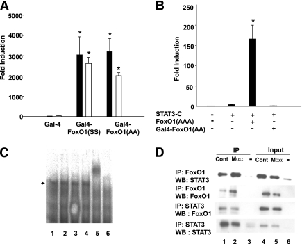
FIG. 5.
FoxO1 domains involved in suppression of FoxO1 by Mαα. A: Cos7 cells were transfected as described in research design and methods with (UAS)5-GAL4 reporter plasmid and with GAL4, GAL4-FoxO1(SS) (Δaa211–655, S256, S319), or GAL4-FoxO1(AA) (Δaa211–655, S256A, S319A) expression plasmids as indicated, in the presence of vehicle (black bars) or 120 μmol/L Mαα (empty bars). Luciferase activity of GAL4-transfected cells normalized to β-galactosidase was defined as 1.0. Mean ± SE for three independent experiments. *Significant as compared with GAL4-transfected cells (P < 0.05). Nonsignificant as compared with respective vehicle-treated cells. B: Cos7 cells were transfected with STAT3(M67-TATA-TK-Luciferase) reporter plasmid and with empty (–), STAT3-C, FoxO1(AAA), or GAL4-FoxO1(AA) (Δaa211–655, S256A, S319A) as indicated. Luciferase activity of empty-transfected cells normalized to β-galactosidase was defined as 1.0. Mean ± SE of three independent experiments. *Significant as compared with STAT3-C–transfected cells (P < 0.05). C: Cos7 cells lacking (lane 6) or overexpressing FoxO1(AAA) (lanes 1–5) were cultured in the presence of vehicle (lanes 1–2) or 150 μM Mαα (lanes 3–5) and in the presence of added anti-FoxO1 antibody (lane 5) as indicated. Labeled double-stranded oligonucleotide containing the FoxO1 binding site of the IGFBP-1 promoter (sense: 5′- cactaGCAAAACAAACTTATTTTGAACAC-3′, antisense: 5′- GTGTGCAAAATAAGTTTGTTTTGctagtg-3′) was prepared by the Klenow fragment of DNA polymerase I and [32P]dCTP. Nuclear extracts prepared from COS7 cells overexpressing FoxO1(AAA) were incubated with the labeled oligonucleotide and subjected to 5% PAGE (29). FoxO1-DNA complex is indicated by an arrow. D: Cos7 cells transfected with empty (–) (lanes 3, 6) or with FoxO1(AAA), and STAT3-C expression plasmids (lanes 1, 4) were cultured in the presence of added 150 μmol/L Mαα (lanes 2, 5) as indicated. Nuclear extracts were immunoprecipitated as described in research design and methods with anti-STAT3 or -FoxO1 antibodies as indicated. Immunoprecipitates (lanes 1–3) and nuclear lysates (input) (lanes 4–6) were subjected to SDS-PAGE followed by Western blotting with anti-STAT3 or -FoxO1 antibodies as indicated. Representative blots of three independent experiments.

