Abstract
OBJECTIVE
There is interest in using cortical auditory evoked potentials (CAEPs) to evaluate hearing aid fittings and experience-related plasticity associated with amplification; however, little is known about hearing aid signal processing effects on these responses. The purpose of this study was to determine the effect of clinically relevant hearing aid gain settings, and the resulting in-the-canal signal-to-noise ratios (SNRs), on the latency and amplitude of P1, N1, and P2 waves.
DESIGN & SAMPLE
Evoked potentials and in-the-canal acoustic measures were recorded in nine normal-hearing adults in unaided and aided conditions. In the aided condition, a 40-dB signal was delivered to a hearing aid programmed to provide four levels of gain (0, 10, 20, and 30 dB). As a control, unaided stimulus levels were matched to aided condition outputs (i.e., 40, 50, 60, and 70 dB) for comparison purposes.
RESULTS
When signal levels are defined in terms of output level, aided CAEPs were surprisingly smaller and delayed relative to unaided CAEPs, likely resulting from increases to noise levels caused by the hearing aid.
DISCUSSION
These results reinforce the notion that hearing aids modify stimulus characteristics such as SNR, which in turn affects the CAEP in a way that does not reliably reflect hearing aid gain.
Keywords: Cortical auditory evoked potentials (CAEPs), Event-related potentials (ERPs), signals in noise, signal-to-noise ratio (SNR), N1, Auditory cortex, Hearing aids, Amplification
1. Introduction
There is growing interest in cortical auditory evoked potentials (CAEPs) as a measure of cortical function in hearing aid users. Aided CAEPs1, or evoked potentials recorded from individuals while wearing their hearing aids, may be of use to evaluate hearing aid fittings as well as experience-related plasticity associated with amplification. Aided CAEPs are not new, in fact reports of recording CAEPs from aided individuals date back to 1967 (Rapin & Graziani, 1967), and the idea has been revisited many times since (Billings et al., 2007; Gatehouse & Robinson, 1996; Golding et al., 2007; Gravel et al., 1989; Korczak et al., 2005; Kraus & McGee, 1994; Kurtzberg, 1989; Marynewich, 2010; Purdy et al., 2005; Rapin & Graziani, 1967; Sharma et al., 2004; Stapells & Kurtzberg, 1991; Tremblay, Billings et al., 2006; Tremblay, Kalstein et al., 2006). However, more than 40 years later, the utility of aided evoked potentials has yet to be established. One reason for this is conflicting evidence showing that CAEPs are affected by amplification in some individuals/studies but not others. Given these conflicting results, it is important to consider how the hearing aid signal processing alters the acoustic content of the stimulus and, in turn, affects the evoked responses. Hearing aid signal processing causes many acoustic modifications to a stimulus (e.g., rise-fall time, signal level, etc) that are likely to affect CAEPs; however, it remains unclear if and how these acoustic modifications affect the aided CAEP. Before aided CAEPs are to be of use clinically, it must be determined what hearing aid factors contribute to the aided evoked potential.
One can assume that signal level is an important factor, because decades of literature indicate that when signals are presented in quiet, the latency of P1-N1-P2 components increases (i.e., neural conduction time increases) and amplitude decreases (i.e., magnitude or synchrony of the response decreases) as signal level is reduced (e.g., Adler & Adler, 1989; Picton et al., 1977). When signals are presented in background noise, however, signal-to-noise ratio (SNR) is a key contributor to the morphology of CAEPs, including the P1, N1, and P2 waves (Billings et al, 2009; Kaplan-Neeman et al., 2006; Whiting et al, 1998).
Noise is always present in an amplified signal and contributors may range from amplified ambient noise to circuit noise generated by the hearing aid. Therefore, it is important to consider the effects of noise on aided CAEPs. In a previous study (Billings et al., 2009) we examined the effect of SNR on CAEPs using computer generated signal changes that varied in SNR. Results demonstrated that CAEPs were primarily sensitive to SNR, rather than absolute signal level. A hearing aid was not used so it is unclear how the results generalize to wearable hearing aids when programmed clinically and gain settings are altered. Clinically, it is important to understand the effect of SNR in cases where aided CAEPs are recorded, because hearing aids amplify ambient noise in addition to the signal of interest. Clinicians might encounter problems when fitting a hearing aid if they assume that increasing the signal level (adjusting the gain) will improve the morphology of the evoked response. If increases in gain increase signal and noise levels together, CAEP patterns might not change as expected (Billings et al., 2009) and over amplification could result. The contributions of SNR to the aided CAEP may also help to explain a significant portion of the variability in the aided CAEP literature. For example, if a study tested individuals near threshold where amplified ambient noise was inaudible, then amplification effects would likely be present; while in contrast, if individuals were tested at suprathreshold levels, amplification effects may be absent because SNRs remained the same.
In this experiment, we set out to determine: (1) the effect of output level on aided CAEPs, by manipulating hearing aid gain, and (2) the contribution of in-the-canal SNR levels to resulting CAEP measures. To do this, signal input level was held constant while hearing aid gain settings were manipulated. We hypothesized that amplification and increments in gain would affect latencies and amplitudes of the aided CAEP, but only to the extent that SNRs changed across unaided and aided conditions and different gain settings.
2. Methods
Using a repeated measures design, participants were tested under nine conditions. Five were unaided conditions (30, 40, 50, 60, and 70 dB SPL) and four were aided conditions (40 dB SPL with 0, 10, 20, and 30 dB gain provided by the hearing aid). Amplitude and latency values for evoked responses P1, N1, and P2 were determined and analyzed. In addition, in-the-canal acoustic measures were completed for each participant and all conditions.
2.1. Participants
Nine young normal-hearing individuals participated in this study (mean age = 24.1 years, SD = 2.8; 3 male and 6 females; all right-handed). Participants had normal hearing from 250 to 8000 Hz (<20 dB HL) and normal immittance measures (single admittance peak between ± 50 daPa to a 226 Hz tone and present ipsilateral acoustic reflexes). Tympanometry and air conduction testing were completed prior to each session to ensure stability of middle ear function and hearing sensitivity. As in our previous studies (Billings et al., 2007; Tremblay, Billings et al., 2006) we elected to use normal-hearing individuals to control for the effects of hearing impairment that would also affect the evoked response. All participants were in good general health with no report of significant history of otologic or neurologic disorders. All participants provided informed consent and research was completed with approval from the pertinent institutional review board.
2.2. Stimuli
The stimulus was a 1000 Hz tone with rise/fall times of 7.5 ms and duration of 756 ms. Although CAEP stimuli need not be longer than 50 ms, a longer stimulus was used for two reasons: (1) there is a movement toward using ecologically relevant speech sounds for CAEP research (Ostroff et al., 1998; Martin et al. 2007) and a longer stimulus more closely approximates syllables and words; and (2) this duration was used in our previous research (Billings et al., 2007, 2009), which enables us to compare our results to previously published findings.
In the unaided conditions, the signal was presented at five stimulus levels: 30, 40, 50, 60, and 70 dB SPL. In the aided condition, the stimulus was presented at one level: 40 dB SPL, and the overall hearing aid gain was adjusted to provide 0, 10, 20, or 30 dB gain at 1000 Hz. By design, the four aided in-the-canal output levels matched four of the unaided in-the-canal levels (i.e., 40, 50, 60, 70 dB SPL). Table 1 illustrates the design of the study. The unaided 30 dB SPL condition was included because it resulted in an in-the-canal SNR that was slightly less than that measured in the aided 0 dB gain condition thus ensuring that all aided SNRs fell within the range of unaided SNRs. An output-level design was used for this study (i.e., the level after amplification) because it was useful for comparisons with our previous studies where input level (i.e., the level prior to amplification) was the independent variable (Billings et al, 2007). For all conditions, stimuli were presented in the sound field (6.5′ × 6′ double-walled, sound-treated booth) through a speaker (JBL Professional LSR25P). The participant was seated in the center of the room, 1 meter from the speaker at 0° azimuth. Distance measurements were repeated during and between each condition to ensure minimal movement. Sound field root mean square stimulus levels were calibrated using a sound level meter placed at ear level with linear weighting and using a fast time constant (125 ms). For all conditions, the left ear was plugged with a foam ear plug. It should be noted that all participants reported a subjective change in signal level and were able to recognize changes in gain.
Table 1.
Experimental design (left) and mean in-the-canal output (right) for the 9 participants displayed as 1/3 octave band values of the band centered at 1000 Hz. All signal and noise values are dB SPL stimulus levels.
| Experimental Design | In-the-Canal Output | |||||
|---|---|---|---|---|---|---|
| Signal | Gain | Output | Signal mean (SD) | Noise mean (SD) | SNR | |
| Unaided | ||||||
| 30 | -- | 30 | 32.5 (1.6) | 26.5 (0.7) | 6.0 | |
| 40 | -- | 40 | 44.0 (1.6) | 26.4 (0.8) | 17.6 | |
| 50 | -- | 50 | 54.4 (1.4) | 26.6 (0.8) | 27.8 | |
| 60 | -- | 60 | 64.4 (1.5) | 26.6 (0.6) | 37.8 | |
| 70 | -- | 70 | 74.3 (2.2) | 26.4 (0.7) | 47.9 | |
| Aided | ||||||
| 40 | 0 | 40 | 43.5 (2.0) | 31.9 (1.0) | 11.6 | |
| 40 | 10 | 50 | 55.2 (2.4) | 37.1 (1.5) | 18.1 | |
| 40 | 20 | 60 | 66.7 (2.1) | 47.0 (1.4) | 19.7 | |
| 40 | 30 | 70 | 77.3 (2.5) | 56.8 (2.6) | 20.5 | |
2.3. In-the-Canal Measurement Procedures
Similar to our previous studies, acoustic recordings for each individual were made using the Etymotic ER7c probe microphone to measure stimulus level in the ear canal (Billings et al., 2007; Tremblay, Billings et al., 2006). Output of the ER7c probe microphone was digitized by a Tucker-Davis Technologies real-time processor (RP2) and then recorded and analyzed in Matlab. Tone and noise levels were calculated as 1/3 octave band measures of the band centered at 1000 Hz. Noise measurements were taken from 400 ms analysis windows immediately preceding or following the 1000-Hz tone.
In-the-canal acoustic measurements were used for two purposes. First, prior to testing, in-the-canal measures were used to adjust overall gain of the hearing aid with the purpose of matching output in the ear canal for unaided and aided conditions for each subject. Second, in the canal measures were made during electrophysiological testing for an online measure of levels at the eardrum. This was done as there was the chance of head movement over time by the subjects despite instructions to remain still during testing. Online measurements were completed at the beginning and end of each of the two blocks (i.e., four times for each condition tested). For all conditions, the four values did not vary by more than 2 dB SPL. These four values were averaged for each individual, and then the mean of the individual averages was taken resulting in a grand mean measure of all nine participants for each condition (Table 1).
2.4. Hearing Aid
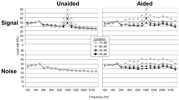
A digitally programmable analog behind-the-ear hearing aid coupled to a foam stock earmold was used. It was the same hearing aid programmed to the same frequency response used in our previous study (Billings et al., 2007). According to the manufacturer’s published specifications, the frequency range of this hearing aid extends from 210 to 6500 Hz. The hearing aid was set to amplify omnidirectionally with a deactivated volume control. No other signal processing algorithms were active during testing (e.g., noise reduction, feedback suppression, etc.). Electroacoustic verification using a 1000-Hz tone demonstrated a compression knee point of 65 dB SPL, a compression ratio of approximately 2: 1, and attack and release times of 5 and 30 ms, respectively. With an input level of 40 dB SPL, hearing aid processing was likely linear for all conditions tested. Processing delay measurements using the Fonix 7000 (Frye Electronics; Tigard, OR) revealed a signal processing delay of 0.5 ms across the four gain settings. To differentiate sources of background noise measured in the ear canal, we completed aided coupler measurements comparing an unplugged microphone condition with a plugged condition to try and eliminate as much ambient noise as possible (Zakis & Wise 2006). Measurements for the two conditions were within 4 dB SPL at all gain settings, indicating that the majority of background noise added by the hearing aid was due to internal circuit noise rather than amplified ambient noise. Figure 1 displays the in-the-canal 1/3 octave band levels of the tone and underlying noise as measured using a Brüel & Kjær Ear Simulator (Type 4157) and Ear Canal Extension (DB 2012) for four of the stimulus levels tested. Increases in background noise can be seen as hearing aid gain is increased.
Figure 1.
Signal and noise spectra (in 1/3 octave bands) in unaided and aided conditions as measured in a B&K Ear Simulator positioned in the sound field. Overall signal levels at 1000 Hz are approximately equivalent in unaided and aided conditions. In contrast, background noise in the 1000 Hz octave band is very different in the unaided and aided conditions as a result of circuit noise produced at the four gain settings.
2.5. Electrophysiology
Each stimulus was presented in a homogeneous train for a total of 500 stimulus presentations for each stimulus condition; this was done across two blocks of 250 presentations. This block design was used because it is representative of the type of stimulus presentation method used when estimating hearing thresholds in clinical settings. Five-minute listening breaks were given between blocks and between recording conditions. An inter-stimulus interval (offset to onset) of 1910 ms was used. Stimulus presentation order within unaided or aided condition was randomized. Unaided conditions were presented one day and aided conditions were presented on the other day with order randomized across subjects. Subjects were instructed to ignore the stimuli and watch a silent close-captioned movie of their choice.
Evoked potential activity was recorded using an Electro-Cap International, Inc. cap which housed 64 tin electrodes. The ground electrode was located on the forehead and Cz was the reference electrode. Data were re-referenced offline to the nose electrode. Horizontal and vertical eye movement was monitored with electrodes located inferiorly and at the outer canthi of both eyes. The recording window consisted of a 100 ms pre-stimulus period and a 700 ms post-stimulus time. Evoked responses were analog band-pass filtered on-line from 0.15 to 100 Hz (12 dB/octave roll off). Using a Neuroscan™ recording system, all channels were amplified with a gain × 500, and converted using an analog-to-digital sampling rate of 1000 Hz. Trials containing ocular artifacts exceeding +/− 70 microvolts were rejected from averaging. Following ocular artifact rejection, the remaining sweeps were averaged and filtered off-line from 1 Hz (high-pass filter, 24 dB/octave) to 30 Hz (low-pass filter, 12 dB/octave).
2.6. Data Analysis and Interpretation
To compare our results with the published literature, responses were analyzed from electrode Cz. Electrode site Cz was analyzed because frontal-central sites such as this are typically used to estimate hearing thresholds, and other experience-related changes, both in research and in clinic. Our results would therefore apply to clinical procedures using similar methods. In addition, global field power measures were used to quantify simultaneous activity from all electrode sites (Skrandies, 1989). Global field power (GFP) is the standard deviation across channels as a function of time. Wave P1, N1, and P2 were analyzed at electrode site Cz, and for GFP, waves N1 and P2 were analyzed from the GFP waveform. The P1 component was not included because it is not robust enough to be present in GFP measures. Peak amplitudes were calculated relative to baseline, and peak latencies were calculated relative to stimulus onset. Latency and amplitude values of each wave were determined by agreement of two judges. Each judge used temporal electrode inversion, global field power traces, and grand averages to determine peaks for a given condition.
Repeated-measures analyses of variance (ANOVA) were completed on amplitude and latency measures of each component of the evoked response (P1, N1, and P2). The 2 × 4 analysis included the factors of amplification (unaided and aided) and output level (40, 50, 60, 70 dB SPL). Greenhouse-Geisser corrections (Greenhouse & Geisser, 1959) were used where an assumption of sphericity was not appropriate. In addition, linear regression analysis was completed on latency and SNR.
3. Results
3.1. Effect of Output Level & Hearing Aid Gain on CAEP Latencies & Amplitudes
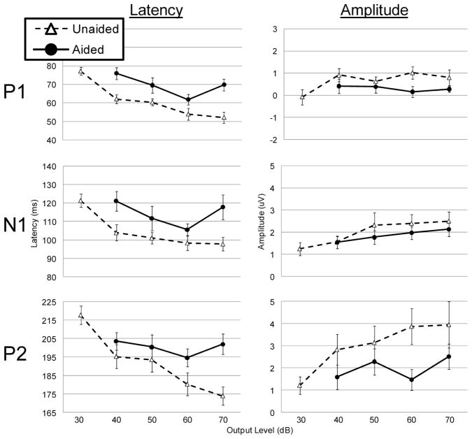
Figure 2 illustrates the latency and amplitude growth functions for Cz waveforms. Repeated measures ANOVA results, summarized in Table 2, demonstrate significant effects of output level for many of the peaks. In general, as would be expected, latency decreased and amplitude increased as output level increased. There was one exception to the general pattern of decreased latency with increasing output; the aided 70 dB output condition tended to have longer latencies than would be expected, especially at electrode Cz. Only P2 amplitude resulted in a significant output by amplification interaction, indicating that for all measures except P2 amplitude, the significant effect of output on amplitudes and latencies was similar across unaided and aided conditions.
Figure 2.
Output level growth functions at electrode Cz for P1, N1, and P2 waves. Latency (left) and amplitude (right) measures are displayed for the aided (solid line, filled circles) and unaided (dotted line, open triangles) conditions. Error bars represent 1 standard error of the mean. In the unaided conditions, latencies decrease as the stimulus level increases. Aided response show a similar function except at the highest stimulus level. Most important is the difference between unaided and aided responses despite similar signal levels being present in the ear canal.
Table 2.
ANOVA Results.
Repeated-measures analyses of variance (ANOVA) results for data collected at electrode Cz and for global field power (GFP). Results for latency and amplitude across components P1, N1, and P2 are included.
| Effect of Output Level | Effect of Amplification (unaided vs. aided) | Amplification × Output | |||||
|---|---|---|---|---|---|---|---|
| F-statistic (df) | P-value | F-statistic (df) | P-value | F-statistic (df) | P-value | ||
| Cz | |||||||
| Latency | |||||||
| P1 | 5.028 (3,24) | 0.008 | 20.682 (1,8) | 0.002 | 1.160 (3,24) | 0.331 | |
| N1 | 2.799 (3,24) | 0.062 | 18.854 (1,8) | 0.002 | 1.263 (3,24) | 0.309 | |
| P2 | 3.394 (3,24) | 0.034 | 19.192 (1,8) | 0.002 | 1.914 (3,24) | 0.154 | |
| Amplitude | |||||||
| P1 | 0.087 (3,24) | 0.967 | 13.371 (1,8) | 0.006 | 0.985 (3,24) | 0.417 | |
| N1 | 1.880 (3,24) | 0.160 | 1.435 (1,8) | 0.265 | 0.419 (3,24) | 0.741 | |
| P2 | 2.637 (3,24) | 0.073 | 7.772 (1,8) | 0.024 | 4.184 (3,24) | 0.016 | |
| GFP | |||||||
| Latency | |||||||
| N1 | 6.124 (3,24) | 0.003 | 75.569 (1,8) | < 0.001 | 3.543 (3,24) | 0.059 | |
| P2 | 2.452 (3,24) | 0.088 | 71.03 (1,8) | < 0.001 | 0.341 (3,24) | 0.796 | |
| Amplitude | |||||||
| N1 | 5.462 (3,24) | 0.005 | 66.280 (1,8) | 0.266 | 1.103 (3,24) | 0.367 | |
| P2 | 6.665 (3,24) | 0.002 | 39.146 (1,8) | < 0.001 | 4.761 (3,24) | 0.010 | |
3.2. Effect of Amplification (Unaided versus Aided Comparisons)
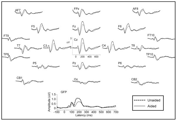
There were significant differences in evoked brain activity when sounds were presented through a hearing aid compared to the unaided conditions in which output levels were the same. That is, even though in-the-canal levels were equal, raising the expectation that CAEP morphology obtained under the two conditions to be equal, aided CAEP latencies were generally prolonged and smaller in amplitude compared to unaided conditions for all measures except N1 amplitude (see Table 2). Figure 3 illustrates this effect in a way analogous to the main effect of amplification in the repeated measures ANOVA; the four aided output conditions and the four unaided conditions were collapsed to make one aided and one unaided waveform respectively.
Figure 3.
Grand mean waveforms (n=9) for aided (solid line) and unaided (dotted line) conditions obtained using signals that were equal in signal level according to in-the-canal recordings. The four aided and four unaided signal level conditions were collapsed into one aided and one unaided waveform respectively. Scalp topography for a small subset of electrodes is shown and GFP (below) is also illustrated. Despite signals being similar in stimulus level in the ear canal, unaided peak latencies are earlier and amplitudes are larger when compared to the aided condition. These results suggest that the hearing aid is affecting more than just signal level, otherwise there would be no difference between unaided and aided conditions.
3.3. Acoustic Recordings and Effect of SNR
Table 1 (right) illustrates the average online in-the-canal levels recorded for all participants and Figure 4 shows representative unaided and aided ER7c acoustic recordings from one individual, demonstrating obvious modifications to the noise floor relative to the signal. In order to understand the effect of SNR on CAEP morphology, in-the-canal SNRs were computed for each individual and condition. To determine the effect of SNR and condition variables, linear regression of logarithmic latency on SNR and condition (unaided vs. aided) was completed for P1, N1, and P2 using the following model: log(latency) = alpha + beta1(SNR) + beta2(condition) + beta3(interaction). Interaction terms were initially included, but were subsequently removed because they were not significant. The SNR coefficients were significant for all waves [P1 (beta1 = −.009, standard error = .001, d.f. = 80, t value = −6.06, p value < .001); N1 (beta1 = −.005, standard error = .001, d.f. = 80, t value = −3.57, p value = .001), P2 (beta1 = −.005, standard error = .001, d.f. = 80, t value = −5.92, p value < .001)]. Coefficients for the amplification condition (unaided vs. aided) were not significant for any of the waves. These results are in agreement with Figure 5 which displays a scatter plot of P1, N1, and P2 latencies taken from Cz and plotted as a function of in-the-canal SNRs for the unaided (triangles) and aided (circles) data. The effect of hearing aid signal processing on SNR is also apparent: the SNR range is much larger for the unaided condition (4.5 to 49.9 dB) than it is for the aided condition (8.8 to 22.2 dB), suggesting a clearer separation between signal and noise floor in the unaided condition. It is also apparent that the distribution of aided latencies falls within the same range as the unaided latencies indicating no difference between conditions when SNR is taken into account.
Figure 4.
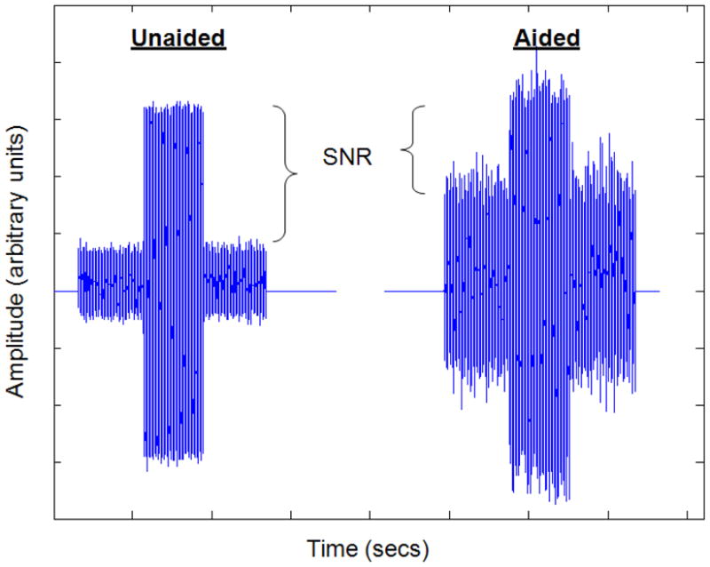
Time waveforms of in-the-canal acoustic recordings for one individual. The unaided (left) and aided (right) conditions are shown together. Signal output as measured at the 1000 Hz centered 1/3 octave band was approximately equivalent at 73 and 74 dB SPL for the unaided and aided conditions. However, noise levels in the same 1/3 octave band were approximately 26 and 54 dB SPL, demonstrating the significant change in SNR.
Figure 5.
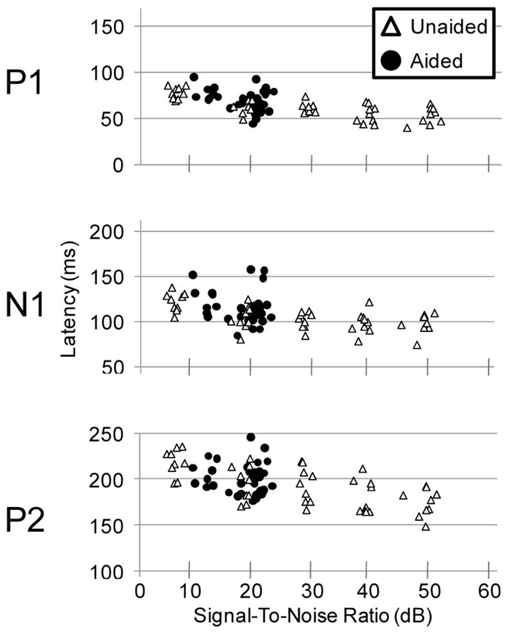
Scatter plots of P1, N1, and P2 latency taken from Cz and plotted as a function of SNR. Data for the nine participants demonstrate that as SNR increases, latency decreases. Also note the wide range of unaided SNRs (open triangles) in contrast to the limited range of aided SNRs (filled circles). Therefore, these aided CAEPs do not differ across hearing aid gain settings, because SNR is similar across the different gain conditions.
4. Discussion & Conclusions
The purposes of this experiment were to determine the effects of: (1) variations in output level on aided CAEPs, resulting from manipulations to hearing aid gain, and (2) in-the-canal SNR levels on CAEPs. The design of this study complemented our previous work, in which input stimulus level (i.e., levels delivered to the hearing aid) was varied systematically while hearing aid gain was held constant (Billings et al., 2007). In the current experiment, we kept input level constant in the aided condition and varied hearing aid gain. This experiment simulates a recording condition that might take place in a clinical setting where hearing aid gain might be altered to estimate hearing thresholds, or evoke a desired response.
4.1 Effect of Output Level & Hearing Aid Gain on CAEP Latencies & Amplitudes
The results reported here clearly demonstrate the expected outcome of CAEP latency and amplitudes being affected by overall signal level (i.e., as signal level increases, latencies decrease and amplitudes increase). These results are consistent with our previous published findings (Billings et al., 2007). When level is changed by the hearing aid (i.e., sound field speaker level is fixed and gain is varied) as in the current study, this pattern of decreasing latency and increasing amplitude with increasing output also applies. However, there were exceptions. For example, the 70 dB SPL aided condition (i.e., 30 dB gain condition) did not fit the general pattern as latencies were longer than expected (see Figure 2). The general change in function shape is likely due to a combination of SNR and other signal processing effects.
4.2 Effect of Amplification (unaided vs. aided) & Contributions of SNR
Given that CAEPs are sensitive to signal level, it follows that equal signal levels in the ear canal during unaided and aided conditions should result in similar CAEPs. However, our results demonstrate that this assumption is not necessarily true. Even though the sound levels (in the ear canal) were similar in the unaided and aided conditions, CAEP morphology differed. Aided condition latencies were delayed and amplitudes were smaller than the unaided condition at equivalent in-the-canal levels. This finding is important because if CAEP morphology was determined primarily by signal level, one would hypothesize that there would be no difference between aided and unaided growth functions because in-the-canal output levels were the same.
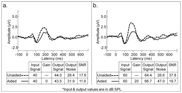
When SNRs were compared in this study, large differences between aided and unaided stimuli were found (Table 1). For example SNRs in the aided condition varied between 11.6 and 20.5 dB, while unaided SNRs varied between 6 and 47.9 dB. The resulting aided CAEP morphology (see Figure 3) was generally weaker (longer in latency and smaller in amplitude) than unaided CAEPs. The relationship between SNR and CAEP morphology is further illustrated in Figure 6. Despite a similar overall signal level in the ear canal, evoked CAEP patterns were dramatically different, as were the SNRs. The contribution of SNR to CAEP morphology is further emphasized by the significant SNR regression coefficients. When SNR was taken into account in the regression, no significant effect of amplification (unaided vs. aided) was found.
Figure 6.
Two examples showing grand mean CAEPs recorded with similar mean output signal levels. Panels a. (40 dB input signals) and b. (60 dB input signals) show unaided and aided grand mean waveforms evoked with corresponding in-the-canal acoustic measures. Despite similar input and output signal levels, unaided and aided brain responses are quite different. Aided responses are smaller than unaided responses, perhaps because the SNRs are poorer in the aided condition.
We speculate that the reduction of SNR in the aided condition is likely due to the hearing aid’s introduction of circuit noise and some amplification of ambient noise. The effect of gain setting on noise levels is clearly demonstrated in Figure 1 where background noise levels, measured in the ear canal with the hearing aid on, increased between 5–30 dB SPL depending on the gain condition and the frequency in question. It is noteworthy that at least for amplitude, effects of amplification were greatest on the P2 wave rather than N1 wave. The significance of these P2 findings is not clear at this time given our limited understanding of the functional significance of P2; however, recent studies demonstrate that the P2 amplitude changes may represent higher level neural processes beyond the encoding of stimulus acoustics such as those related to stimulus exposure or auditory training (Tremblay et al., 2001; Tremblay et al., 2009).
Only normal-hearing individuals were tested in this study. This was done so that the relationship between signal level and evoked brain activity could be determined in the absence of confounding variables that are present in people with hearing loss. They include variables such as age, duration of hearing loss, and varied audibility across frequencies. In addition, the impact of stimulus SNR on CAEPs will be very important to study in people with hearing loss because noise levels may or may not be audible to the individual. It would also be important to consider how SNR varies across frequency and time. For example, the frequency response of the hearing aid may lead to audible background noise in certain frequency ranges but not in others as a result of the programmed frequency response. Furthermore, the effects of compression can result in differing SNRs over time depending on characteristics of the incoming signal and noise and the compression settings of the hearing aid (Naylor & Johannesson, 2009). Collectively, these issues reinforce the point that CAEPs recorded through hearing prostheses should not be regarded in the same way as those obtained without prostheses.
4.3 Other contributing variables introduced by the hearing aid
While it is clear that SNR is an important contributor to the results of the current study, there may be other important signal processing contributors to aided CAEP morphology, especially when non-linear amplification is used. In particular, hearing aid signal processing characteristics that affect the first 30–50 ms of the stimuli would be especially important factors in determining the morphology of the evoked response (Onishi & Davis, 1968). Compression characteristics, for example, would likely affect stimulus rise slope, rise time, and introduce overshoot of the onset. Such modifications to the first 30–50 ms of a stimulus have been demonstrated recently by Marynewich and colleagues (2010) even when linear hearing aids are used. Because of the potential effects of the hearing device settings on the acoustic content of the signal, we cannot be certain that results described here will generalize to all wearable hearing aids. This point further illustrates the current limitations of our understanding of aided CAEPs and cautions against the clinical use of aided CAEPs at this time. At a minimum, future aided CAEPs studies should include in-the-canal acoustic recordings to allow for analysis of SNR and onset modifications made by the hearing aid. In addition, the interaction between device settings and hearing loss must be determined before clinical application could take place.
4.4 Conclusions
Results from this experiment demonstrate that hearing aid gain settings alter the latency and amplitude of aided CAEP responses. Because of noise in the output of the hearing aid, aided SNRs are considerably smaller than unaided SNRs when absolute signal level is equivalent. Therefore, signal-to-noise ratio is an important factor to be aware of when recording aided CAEP measures. While the use of CAEPs can provide valuable information to determine if an audible signal reached the level of the cortex, an important realization from this experiment is that signal processing of a hearing aid contributes to the latency and amplitude of evoked responses. For this reason, it is important to question how amplification alters other aspects of the stimulus (e.g., rise slope, rise time) and how these effects vary across instruments, and interact with a person’s sensitivity threshold at different frequencies. More importantly, it is necessary to acknowledge that CAEPs recorded through hearing prostheses should not be regarded in the same way as those obtained without prostheses. And finally, it is time to critically question if it will be possible to control all of the potential variables that might arise in a clinical setting when using aided CAEPs.
Acknowledgments
The authors wish to thank Pamela Souza for helpful discussions about these data and Wendy Tolin for assistance with data processing. This work was supported by the National Institutes of Health through the National Institute on Deafness and Other Communication Disorders and the Department of Veterans Affairs through the Rehabilitation Research and Development Service [F31-DC007296 and C6971M (CJB); R01-DC007705 (KLT); P30-DC004661].
Abreviations
- CAS
central auditory system
- SNR
signal-to-noise ratio
- SD
standard deviation
- GFP
global field power
- CAEPs
cortical auditory evoked potentials
- ANOVA
analysis of variance
Footnotes
We use the term “aided CAEPs” to refer to CAEPs recorded from an individual who is wearing a hearing aid at the time of testing, such that sound field stimuli are processed by the hearing aid and delivered to the individual’s ear canal.
References
- Adler G, Adler J. Influence of stimulus intensity on AEP components in the 80- to 200- millisecond latency range. Audiology. 1989;28(6):316–324. doi: 10.3109/00206098909081638. [DOI] [PubMed] [Google Scholar]
- Billings CJ, Tremblay KL, Souza PE, Binns MA. Effects of hearing aid amplification and stimulus intensity on cortical auditory evoked potentials. Neurootol. 2007;12(4):234–246. doi: 10.1159/000101331. [DOI] [PubMed] [Google Scholar]
- Billings CJ, Tremblay KL, Stecker GC, Tolin WM. Human evoked cortical activity to signal-to-noise ratio and absolute signal level. Hear Res. 2009;254(1–2):15–24. doi: 10.1016/j.heares.2009.04.002. [DOI] [PMC free article] [PubMed] [Google Scholar]
- Gatehouse S, Robinson K. Acclimatisation to monaural hearing aid fitting - effects on loudness functions and preliminary evidence for parallel electrophysiological and behavioural effects. In: Kollmeier B, editor. Psychoacoustics, speech and hearing aids. 1996. pp. 319–330. [Google Scholar]
- Golding M, Pearce W, Seymore J, Cooper A, Ching T, Dillon H. The relationship between obligatory cortical auditory evoked potentials (CAEPs) and functional measures in young infants. J Am Acad Audiol. 2007;18:117–125. doi: 10.3766/jaaa.18.2.4. [DOI] [PubMed] [Google Scholar]
- Gravel J, Kurtzberg D, Stapells DR, Vaughan HG, Wallace IF. Case Studies. Semin Hear. 1989;10(3):272–287. [Google Scholar]
- Greenhouse WW, Geisser S. On methods in the analysis of profile data. Psychometrika. 1959;24:95–112. [Google Scholar]
- Kaplan-Neeman R, Kishon-Rabin L, Henkin Y, Muchnik C. Identification of syllables in noise: electrophysiological and behavioral correlates. J Acoust Soc Am. 2006;120(2):926–933. doi: 10.1121/1.2217567. [DOI] [PubMed] [Google Scholar]
- Korczak PA, Kurtzberg D, Stapells DR. Effects of sensorineural hearing loss and personal hearing aids on cortical event-related potential and behavioral measures of speech-sound processing. Ear Hear. 2005;26(2):165–185. doi: 10.1097/00003446-200504000-00005. [DOI] [PubMed] [Google Scholar]
- Kraus N, McGee TJ. Mismatch negativity in the assessment of central auditory function. Am J Audio. 1994;3:39–51. doi: 10.1044/1059-0889.0302.39. [DOI] [PubMed] [Google Scholar]
- Kurtzberg D. Cortical event-related potentials assessment of auditory system function. Semin Hear. 1989;10:252–261. [Google Scholar]
- Martin BA, Tremblay KL, Korczak P. Speech evoked potentials: from the laboratory to the clinic. Ear Hear. 2008;29 (3):285–313. doi: 10.1097/AUD.0b013e3181662c0e. [DOI] [PubMed] [Google Scholar]
- Marynewich SL. Master of Science Thesis. University of British Columbia; Vancouver, Canada: 2010. Slow cortical potential measures of amplification. [Google Scholar]
- Naylor G, Johannesson RB. Long-term signal-to-noise ratio at the input and output of amplitude-compression systems. J Am Acad Audiol. 2009;20:161–171. doi: 10.3766/jaaa.20.3.2. [DOI] [PubMed] [Google Scholar]
- Onishi S, Davis H. Effects of duration and rise time of tone bursts on evoked V potentials. J Acoust Soc Am. 1968;44(2):582–591. doi: 10.1121/1.1911124. [DOI] [PubMed] [Google Scholar]
- Ostroff JM, Martin BA, Boothroyd A. Cortical evoked responses to spectral change within a syllable. Ear Hear. 1998;19:290–297. doi: 10.1097/00003446-199808000-00004. [DOI] [PubMed] [Google Scholar]
- Picton TW, Woods DL, Baribeau-Braun J, Healey TM. Evoked potential audiometry. J Otolaryngol. 1977;6:90–119. [PubMed] [Google Scholar]
- Purdy SC, Katsch R, Dillon H, Storey L, Sharma M, Agung K. Aided cortical auditory evoked potentials for hearing instrument evaluation in infants. In: Phonak A, editor. A Sound Foundation Through Early Amplification. Chicago, Illinois: 2005. pp. 115–127. [Google Scholar]
- Rapin I, Graziani LJ. Auditory-evoked responses in normal, brain-damaged, and deaf infants. Neurology. 1967;17(9):881–894. doi: 10.1212/wnl.17.9.881. [DOI] [PubMed] [Google Scholar]
- Sharma A, Tobey E, Dorman M, Bharadwaj S, Martin K, Gilley P, Kunkel F. Central auditory maturation and babbling development in infants with cochlear implants. Arch Otolaryngol Head Neck Surg. 2004;130(5):511–516. doi: 10.1001/archotol.130.5.511. [DOI] [PubMed] [Google Scholar]
- Skrandies W. Data reduction of multichannel fields: global field power and principal component analysis. Brain Topogr. 1989;2(1–2):73–80. doi: 10.1007/BF01128845. [DOI] [PubMed] [Google Scholar]
- Stapells DR, Kurtzberg D. Evoked potential assessment of auditory system integrity in infants. Clin Perinatol. 1991;18(3):497–518. [PubMed] [Google Scholar]
- Tremblay KL, Billings CJ, Friesen LM, Souza PE. Neural representation of amplified speech sounds. Ear Hear. 2006;27(2):93–103. doi: 10.1097/01.aud.0000202288.21315.bd. [DOI] [PubMed] [Google Scholar]
- Tremblay KL, Kalstein L, Billings CJ, Souza PE. The neural representation of consonant-vowel transitions in adults who wear hearing aids. Trends Amplif. 2006;10(3):155–162. doi: 10.1177/1084713806292655. [DOI] [PMC free article] [PubMed] [Google Scholar]
- Tremblay K, Kraus N, McGee T, Ponton C, Otis B. Central auditory plasticity: changes in the N1–P2 complex after speech-sound training. Ear Hear. 2001;22:79–90. doi: 10.1097/00003446-200104000-00001. [DOI] [PubMed] [Google Scholar]
- Tremblay KL, Shahin AJ, Picton T, Ross B. Auditory training alters the physiological detection of stimulus-specific cues in humans. Clin Neurophysiol. 2009;120:128–135. doi: 10.1016/j.clinph.2008.10.005. [DOI] [PMC free article] [PubMed] [Google Scholar]
- Whiting KA, Martin BA, Stapells DR. The effects of broadband noise masking on cortical event-related potentials to speech sounds /ba/ and /da. Ear Hear. 1998;19(3):218–231. doi: 10.1097/00003446-199806000-00005. [DOI] [PubMed] [Google Scholar]
- Zakis JA, Wise C. The acoustic and perceptual effects of two noise-suppression algorithms. J Acoust Soc Am. 2006;121(1):433–441. doi: 10.1121/1.2401656. [DOI] [PubMed] [Google Scholar]


