



Abstract
Recurrence of Giant cell tumor of the tendon sheath (GCTTS) is an unresolved issue, though it is a non malignant condition. The authors operated on fourteen cases of GCTTS, after fine needle aspiration cytology confirmation and using a magnifying loupe for complete excision of the lesion including the satellite nodules. In only one case recurrence was noted which was successfully managed by a second wide excision. Preoperative diagnosis and meticulous surgical technique were found the only predictive factor of recurrence. During the 5 year period from 2002, 12 patients [11 females, 1 male, mean age 29.5, ranging from 10–53 years] underwent excision of giant cell tumor of tendon sheath of the hand. The lesions were found over the thumb [n = 7], ring finger [n = 1], index finger [n = 1], and over the hand [n = 2]. The lesions were classified using the Al-Qattan classification. The most common presentation was with a mass over the hand, with a predilection to the thumb [n = 7]. Radiological changes in the form of bony indentation was seen in only 2 cases. FNAC was inconclusive in 2 out of the 12 cases. Due to the high incidence of recurrence, pre-operative planning aided by a tissue diagnosis with fine needle aspiration cytology, wide surgical exposure, and meticulous dissection with help of magnification are imperative for a successful outcome in GCTTS.
Keywords: Hand, Tendon sheath, Giant cell tumor, Recurrence
Introduction
Giant cell tumor of tendon sheath (GCTTS) is clinically a slow growing soft tissue mass that develops over a period of months to years. It is the second commonest tumor of the hand [1]. Trauma, inflammation, metabolic disease and a neoplastic etiology are considered as etiological factors [2, 3]. Many factors are considered as causing recurrence, including proximity to the distal interphalangeal joints, presence of degenerative joint disease, pressure erosions in the radiographs, increased mitotic activity, and type 2 lesions described by Al-Qattan [1, 3–5]. But the only consistent observation by various authors in preventing recurrence is complete surgical excision with removal of all satellite nodules if present [6–8]. The use of an operating microscope [7] or magnifying loupe [6] ensures radical excision. Few authors suggest that the surgeons are able to take precautions if during surgery frozen section [2, 6] or a Fine Needle Aspiration Cytology (FNAC) is available [9].
Materials and Methods
This study was conducted in the Orthopaedic department of a Regional Referral Hospital over a period of 5 years (2002–2006). A total of 14 cases of GCTTS were operated by the senior surgeon, after confirmation with FNAC and using magnifying loupe for meticulous dissection. All patients who were operated following the above protocol were contacted over telephone and were asked to come for follow-up. Follow up ranged from 3–9 years [mean 4.3 years].
Two cases were not available for follow up and are excluded from the study.
Age of patients ranged from 10 to 53 years (mean age 29.5 years). Of the 12 cases available for follow up seven involved the thumb, one each in the ring, index and little fingers, and two were over the hand. History of antecedent trauma was present in only two cases (Figs. 1 and 2).
Fig. 1.
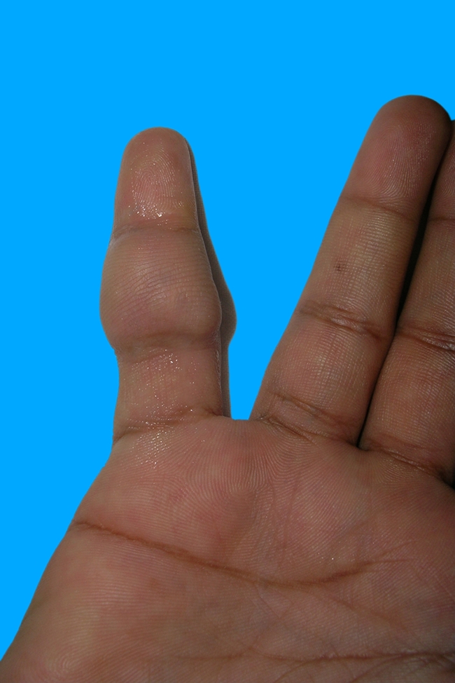
Mass over the little finger in a patient with antecedent trauma
Fig. 2.

Type I c lesion: Multi-lobulated lesion surrounded by a common pseudocapsule, excised specimen from volar aspect of the ring finger, in a patient with antecedent trauma
Patients were investigated with routine radiographs of the involved part and an ultra sound scan. All patients were subjected to FNAC. The procedure was performed by the attending surgeon. The slides were analyzed by the histopathologist for giant cells to make sure preoperatively the probability of GCTTS so that extra caution could be taken during the dissection (Figs. 3 and 4).
Fig. 3.

Mass over the fifth metacarpal
Fig. 4.

Postero anterior radiograph of the patient in Fig. 3, showing the soft tissue shadow [white arrow heads] and scalloping of the shaft of the 5th metacarpal [black arrow]
All cases were operated under Bier’s block, and tourniquet control, using a magnifying loupe. Special care was taken to excise the tumor in total, retaining the capsule, with margin of normal tissue. The operating field is searched for presence of satellite lesions or daughter cysts. The entire specimen was then subjected to histopathological examination, and the margins were observed for clearance.
All tumors were classified as per the new classification by Al-Qattan (Table 1).
Table 1.
Demographic details of patients
| No | Age | Sex | Site of swelling | FNAC | Al Qattan type | X-ray changes | Recurrence |
|---|---|---|---|---|---|---|---|
| 1 | 32 | F | Left thumb[dorsum] | Positive | Ib | Nil | 0 |
| 2 | 39 | M | Right thumb[dorsum] | Positive | Ia | Nil | 0 |
| 3 | 53 | F | Thumb near interphalangeal joint | Inconclusive | Ia | Nil | 0 |
| 4 | 29 | F | Thumb[dorsum] | Positive | Ib | Nil | 0 |
| 5 | 27 | F | Dorsum of thumb[near interphalangeal joint] | Positive | Ib | Bony indentation | 1 |
| 6 | 31 | F | Thumb[dorsum] | Positive | Ia | Nil | 0 |
| 7 | 10 | F | Thumb[dorsum] | Positive | Ib | Nil | 0 |
| 8 | 15 | F | Dorsum of hand[Fig. 3] | Positive | Ia | Bony indentation[Fig. 4] | 0 |
| 9 | 35 | F | Wrist in relation to FCU | Inconclusive | II b | Nil | 0 |
| 10 | 30 | F | Volar aspect of Ring finger[Fig. 2] | Positive | Ic | Soft tissue shadows | 0 |
| 11 | 28 | F | Volar aspect of Little finger[Fig. 1] | Positive | Ia | Nil | 0 |
| 12 | 25 | F | Volar aspect of Index finger | Positive | Ia | Nil | 0 |
Six cases were single nodule surrounded by one thick pseudo capsule (Al Qattan Ia), four were single nodule within a thin capsule (Ib). One lesion in the ring finger (Fig. 2) was found to be multi-lobulated surrounded by a common pseudo-capsule(Ic). Another lesion found in relation to the flexor carpi ulnaris was found diffuse with multiple granular like lesions without a pseudo-capsule (IIb) (Fig. 5).
Fig. 5.

Fine needle aspiration cytology slide showing scattered groups of mononuclear cells with occasional osteoclast like giant cells
There was only one case of recurrence, in a thumb lesion, which recurred after a period of 2 months. The mass was over the dorsum of the thumb near the interphalangeal joint, and there was indentation of the terminal phalanx over the radial aspect in the frontal radiograph. The patient underwent re-exploration and there was no recurrence on his review after 5 years.
Results
Out of the 12 cases FNAC was positive for giant cells in 10 cases, whereas in 2 cases it was inconclusive. Two cases showed skeletal changes in the form of bony indentation, due to pressure effect of the mass. Recurrence was noted in one case [8.3%], with mass over the dorsal aspect of the thumb. This was successfully managed with a second excision. The cause of recurrence was thought to be due to inadequate resection of the margins. There was superficial wound infection in 2 cases which responded to antibiotic cover. Three cases in the thumb and one in the little finger developed post operative stiffness, and regained full range of movements with mobilization in the hand therapy clinic.
Discussion
Reported rate of recurrence of GCTTS is 9–44% [3, 5, 6] and some authors have reported recurrence rate as high as 45% [2, 4, 8].
Various factors have been described predictive of recurrence. These include pressure erosions on radiographs, location at the interphalangeal joint and presence of degenerative joint disease. Lowyck and De Smet [2] found no significant correlation of recurrence and presence of pressure erosions, or degenerative joint disease. They also found no association of recurrence and lesion in the distal interphalangeal joint [2]. Al-Qattan commented that bony indentation due to pressure from the overlying tumor should not be considered as intra osseous invasion and is not associated with a higher recurrence rate [1]. The higher incidence of recurrence in the distal joint may be due to difficulties in excising the lesion completely due to limited space [3].
Tumor classification has helped in analyzing the recurrence pattern. Byers classified GCTTS into localized nodular type (common in hand) and diffuse type (common in joints) [1, 7]. Al Qattan has analyzed the recurrence after surgical excision. He has proposed a new classification for GCTTS, where he classified Type I- as single tumor which is round or multi lobulated, and Type II, where there are two or more distinct tumors which are not joined together [1]. The new classification by Al-Qattan is useful in predicting the recurrence as satellite lesions are often missed when a magnifying loupe is not used especially in Type IIa and Type II b lesions [1, 10].
In a nodular tumor the tumor could be easily excised since there is clear margin, where as in diffuse tumor excision is difficult due to infiltrating nature. Even when the tumor may appear nodular, there may be a small part which is diffuse, and hence Ikeda et al. recommends microscopic excision [7]. Ikeda infers that there are more diffuse tumors in the hand than described [7]. They associate diffuse tumors with adjacent degenerative joint disease, location at the distal interphalangeal joint, and presence of pressure erosions of bone.
Microscopic excision has been described to prevent recurrence [7]. Ikeda reported recurrence in only one case of 18 reported when they used the operating microscope. In the only case of recurrence, the case recurred as microscope was not used. Ozalp [8] recommends using a magnifying loupe to excise the satellite lesions after excision of the nodules. Complete excision with help of operating microscope or a magnifying loupe has been advocated by many authors who got promising results [7, 8], as complete surgical excision is the only factor which has been proved to prevent recurrence.
Sonography can detect whether the tumor is solid or cystic, and to note if there are satellite lesions. Sonography also describes the relationship of the lesion to the surrounding structures [11]. Information regarding the extent of contact with underlying tendon and the percentage of circumferential involvement is possible with sonography [11].
Histologically these tumors are composed of multinucleated giant cells, polyhedral histiocytes, fibrosis and hemosiderin deposits [8, 12, 13]. Histological features of cellularity and mitosis, though previously found significant, were not found to influence recurrence [1, 10]. But Rao and Vigorita [5] has found high incidence of recurrence in tumors with increased mitotic activity. There was no relation between the number of mitosis found initially and recurrence, but all recurrent lesions showed increased mitotic activity [5]. However studies by Monaghan et al. has shown that mitotic figures do not indicate recurrence and advocate complete local excision as the treatment of choice [14].
Grover et al. has found that absence of a gene nm23 is associated with high rate of recurrence [15]. These authors described nm23-H1 as an independent prognostic factor.
However a report by Lorea et al. in 2004 found no correlation between nm23-H1 expression and recurrence [16].
Complete surgical excision remains the mainstay of treatment, assisted either with an operating microscope or a magnifying loupe. Radiotherapy has been suggested after inadequate excision and in patients with high mitotic activity to prevent recurrence [4]. They reported a recurrence rate of only 4% with this method of management. Excision is suggested even after multiple recurrences, by Ozalp et al., and recurrent tumors were not managed by radiotherapy [8].
There is a recent trend to use FNAC as a primary diagnostic aid and helps is proceeding with care in soft tissue masses [17]. As per the authors in most of the cases management decisions in soft tissue masses can be made from the cytological analysis [17]. Diagnosis of GCTTS is possible preoperatively with a FNAC and helps in preoperative planning to prevent recurrence [9]. Patient should be counseled in the preoperative meeting with regard to the probability of recurrence. Incomplete excision and leaving behind satellite nodules is considered as the most important factor deciding recurrence pattern. Adequate surgical exposure, meticulous dissection and use of magnification are necessary to reduce recurrence and should remain the mainstay of surgical management. Following the above surgical protocol the authors were able to achieve complete clearance of the tumor with recurrence in only one case [8.3%].
Acknowledgments
Conflict of interest Nil
Financial support Nil
References
- 1.Al-Qattan M. Giant cell tumors of tendon sheath: classification and recurrence rate. J Hand Surg. 2001;26B:72–75. doi: 10.1054/jhsb.2000.0522. [DOI] [PubMed] [Google Scholar]
- 2.Lowyck H, Smet L. Recurrence rate of giant cell tumors of the tendon sheath. Eur J Plast Surg. 2006;28:385–388. doi: 10.1007/s00238-005-0791-6. [DOI] [Google Scholar]
- 3.Reilly KE, Stern PJ, Dale JA. Recurrent giant cell tumors of the tendon sheath. J Hand Surg Am. 1999;24:1298–1302. doi: 10.1053/jhsu.1999.1298. [DOI] [PubMed] [Google Scholar]
- 4.Kotwal PP, Gupta V, Malhotra R. Giant cell tumour of the tendon sheath- is radiotherapy indicated to prevent recurrence after surgery? J Bone Joint Surg. 2000;82B:571–573. doi: 10.1302/0301-620X.82B4.10328. [DOI] [PubMed] [Google Scholar]
- 5.Rao AS, Vigorita VJ. Pigmented villonodular synovitis (Giant-Cell tumor of the tendon sheath and synovial membrane): a review of 81 cases. J Bone Joint Surg. 1984;66A:76–94. [PubMed] [Google Scholar]
- 6.Gholve PA, Hosalkar HS, Kreiger PA, et al. Giant cell tumor of tendon sheath: largest single series in children. J Pediatr Orthop. 2007;27:67–74. doi: 10.1097/01.bpo.0000242380.95348.8b. [DOI] [PubMed] [Google Scholar]
- 7.Ikeda K, Osamura N, Tomita K. Giant cell tumour in the tendon sheath of the hand: importance of the type of lesion. Scand J Plast Reconstr Surg Hand Surg. 2007;41:138–142. doi: 10.1080/02844310601159766. [DOI] [PubMed] [Google Scholar]
- 8.Ozalp T, Yercan H, Kurt C, et al. Giant cell tumor of tendon sheath of the hand. Acta Orthop Traumatol Turc. 2004;38:120–124. [PubMed] [Google Scholar]
- 9.Iyer KV, Kusum K, Kusum V. Fine-needle aspiration cytology of giant cell tumor of the tendon sheath. Diagn Cytopathol. 2003;29:105–110. doi: 10.1002/dc.10319. [DOI] [PubMed] [Google Scholar]
- 10.Rodriques C, Desai S, Chinoy R. Giant cell tumor of the tendon sheath: a retrospective study of 28 cases. J Surg Oncol. 1998;68:100–103. doi: 10.1002/(SICI)1096-9098(199806)68:2<100::AID-JSO5>3.0.CO;2-A. [DOI] [PubMed] [Google Scholar]
- 11.Middleton WD, Patel V, Teefey SA, et al. Giant cell tumors of the tendon sheath: an analysis of sonographic findings. Am J Roentgenol. 2004;183:337–339. doi: 10.2214/ajr.183.2.1830337. [DOI] [PubMed] [Google Scholar]
- 12.Messoudi A, Fnini S, Labsaili N, et al. Giant cell tumors of the tendon sheath of the hand: 32 cases. Chir Main. 2007;26:165–169. doi: 10.1016/j.main.2007.03.008. [DOI] [PubMed] [Google Scholar]
- 13.Liu PT. Radiological reasoning: acutely painful swollen finger. Am J Roentgenol. 2007;188:A13–S17. doi: 10.2214/ajr.188.3_supplement.0s13. [DOI] [PubMed] [Google Scholar]
- 14.Monaghan H, Salter DM, Al-Nafussi A. Giant cell tumour of tendon sheath (localized nodular tenosynovitis): clinicopathological features of 71 cases. J Clin Pathol. 2001;54:404–407. doi: 10.1136/jcp.54.5.404. [DOI] [PMC free article] [PubMed] [Google Scholar]
- 15.Grover R, Grobbelaar AO, Richman PI, et al. Measurement of invasive potential provides an accurate prognostic marker for giant cell tumor of tendon sheath. J Hand Surg. 1998;23B:728–731. doi: 10.1016/s0266-7681(98)80084-3. [DOI] [PubMed] [Google Scholar]
- 16.Lorea P, Walle H, Kinnen L. Giant cell tumours of tendon sheath: lack of correlation between NM 23-H1 expression and recurrence. J Hand Surg. 2004;29B:67–70. doi: 10.1016/s0266-7681(03)00222-5. [DOI] [PubMed] [Google Scholar]
- 17.Ng VY, Thomas K, Crist M, et al. Fine needle aspiration for clinical triage of extremity soft tissue masses. Clin Orthop Rel Res. 2010;468:1120–1128. doi: 10.1007/s11999-009-1100-7. [DOI] [PMC free article] [PubMed] [Google Scholar]