Abstract
Modulation of human NK cell function by killer cell immunoglobulin-like receptors (KIR) and MHC class I is dominated by the bipartite interactions of inhibitory lineage III KIR with the C1 and C2 epitopes of HLA-C. In comparison, the ligand specificities and functional contributions of the activating lineage III KIR remain poorly understood. Using a robust, sensitive assay of KIR binding and a representative panel of 95 HLA class I targets, we show that KIR2DS1 binds C2 with ∼50% the avidity of KIR2DL1, whereas KIR2DS2, 2DS3 and 2DS5 have no detectable avidity for C1, C2 or any other HLA class I epitope. In contrast, the chimpanzee has activating C1 and C2-specific lineage III KIR with strong avidity, comparable to those of their paired inhibitory receptors. One variant of chimpanzee Pt-KIR3DS2, the activating C2-specific receptor, has the same avidity for C2 as inhibitory Pt-KIR3DL4, and a second variant has ∼73% the avidity. Chimpanzee Pt-KIR3DS6, the activating C1-specific receptor, has avidity for C1 that is ∼70% that of inhibitory Pt-KIR2DL6. In both humans and chimpanzees we observe an evolutionary trend toward reducing the avidity of the activating C1- and C2-specific receptors through selective acquisition of attenuating substitutions. However, the extent of attenuation has been extreme in humans as exemplified by KIR2DS2, an activating C1-specific receptor that has lost all detectable avidity for HLA class I. Supporting such elimination of activating C1-specific receptors as a uniquely human phenomenon is the presence of a high avidity activating C1-specific receptor (Gogo-KIR2DSa) in gorilla.
Keywords: Natural Killer Cells, Cell Surface Molecules, Comparative Immunology/ Evolution, MHC
Introduction
The killer cell immunoglobulin-like receptors (KIR) are a family of diverse immunoreceptors that modulate natural killer (NK) cell function through recognition of MHC class I (1). In higher primates the role of KIR is analogous to that of rodent Ly49 receptors (2, 3). Of four human KIR lineages, lineages II and III include the KIR specific for polymorphic HLA-A, B and C. Dominant are the C1 and C2 epitopes borne by HLA-C and recognized by lineage III KIR; the most numerous lineage, comprising two genes encoding inhibitory C1 (KIR2DL2/3) and C2 (KIR2DL1) receptors, and five genes encoding activating receptors (KIR2DS1, 2, 3, 4, and 5). Although the importance of inhibitory KIR for NK cell biology is well established, the contributions of the activating KIR remain poorly understood. Pointing to their relevance are correlations with a variety of clinical conditions (4).
The five activating lineage III KIR comprise three groups. One consists of KIR2DS1 and 2DS2 that have similar extracellular domains to inhibitory KIR2DL1 and 2DL2/3, respectively. Consistent with this relationship, KIR2DS1 recognizes C2 but has lower avidity than KIR2DL1 due to replacement of proline 70 with lysine for (5-7). Despite this attenuation, KIR2DS1 influences NK-cell education and response in a C2-dependent manner (8, 9). In contrast, KIR2DS2 has no demonstrable interaction with C1+ HLA-C, an abrogation due to replacement of phenylalanine 45 with tyrosine (10, 11). Because activating KIR were derived from their paired inhibitory receptors by recombination, the subsequent acquisition of substitutions that reduce ligand binding was likely a consequence of natural selection (12).
The C1 and C2 specificities of KIR2DL1 and 2DL2/3 are determined by lysine and methionine at position 44 (13). The second group of activating lineage III KIR, formed by KIR2DS3 and KIR2DS5, has threonine at position 44. These divergent KIR are not paired with inhibitory receptors, but are similar to each other. Distinguishing KIR2DS3 is its absence from the NK cell surface and retention inside the cell (14). KIR2DS5 reaches the NK cell surface and transduces activating signals when crosslinked with anti-KIR antibody, but a physiological ligand has yet to be identified (15). KIR2DS4, the most divergent lineage III KIR, forms the third group. This activating receptor binds HLA-A11 and subsets of C1+ and C2+ HLA-C with moderate avidity, and can activate NKL cells on engagement of HLA-A11 (16).
MHC-C mediated regulation of NK cells evolved in hominids (humans and great apes), with C1-mediated regulation arising several million years before C2-mediated regulation. In modern human populations both activating and inhibitory C2 receptors are functional, whereas natural selection has reduced C1-mediated regulation to the inhibitory receptor. To see if this asymmetry in NK-cell regulation by C1 and C2 is human-specific, or a more general phenomenon, we studied activating lineage III KIR in the most closely related hominid species: chimpanzee and gorilla (17, 18).
Materials and Methods
Single-Ag bead analysis of KIR-Fc specificity
KIR-Fc fusion proteins were made, assessed for conformational integrity, and assayed for binding to LABScreen single-Ag bead sets (One Lambda) as described (16, 19, 20). Binding of W6/32 antibody was used as a positive control and to normalize for bead-to-bead variation in the amount of HLA class I displayed. Each KIR-Fc protein was assayed a minimum of three times in independent experiments for binding to 95 HLA class I allotypes.
The HLA class I proteins used to coat the beads were isolated from a panel of transfected cell lines, each made from the class I deficient B cell line 221 and expressing a different HLA class I allele (21). Consequently the target HLA class I molecules carry a repertoire of bound peptides that is representative of those presented by HLA on the surface of the human B cell lines commonly used as NK cell targets. The bead sets were initially developed for high-resolution analysis of the HLA class I specificity of monoclonal antibodies and human alloantisera (21). The 95 HLA class I allotypes present in the bead sets were chosen to represent the variation in structure and serology as well as the common variants of HLA-A, B and C. It is well established that there is a good correlation between the results obtained with direct binding to single-antigen beads and those obtained with conventional serology using the complement-dependent cytotoxicity of human lymphocytes and with direct antibody binding to cells (22). Such correlations strongly indicate that the representation of complexes of class I HLA and peptide by the beads is comparable to that of normal human cells.
Cytotoxicity assays
Killing of 221 target cells (expressing a single HLA class I variant) by NKL effector cells (expressing a single KIR) was assayed as described (19, 20). Inhibitory versions of short-tailed receptors were constructed by fusing exons encoding their extracellular domains with exons encoding the transmembrane and cytoplasmic portions of their related inhibitory receptors. Redirected cytotoxicity assays using mouse P815 target cells were performed as described (16) with antibody pre-incubation for 30 min. Mab specific for KIR (NKVFS1), NKG2D and 2B4 (CD244) (Becton Dickinson) were included, singly and in combination, at 10 μg/ml final concentration for each antibody. Untransfected 221 target cells and untransduced NKL cells were controls. Assays were replicated three or more times and included triplicates for each condition.
Results
Exploration of KIR specificity and avidity for HLA class I has been previously carried out using small numbers of target HLA class I variants (7, 10, 13). Recently, we developed a new, sensitive and quantitative assay that measures the binding of KIR-Fc fusion proteins to beads coated with a representative set of 95 HLA class I variants (20). The target HLA class I molecules are purified from transfectants of the 221 HLA class I-deficient B cell line and have a heterogeneity of bound peptides that is comparable to that of normal human cells. The advantage of this assay is it allows KIR specificity and avidity to be defined with much greater precision and confidence than was previously possible (16). Consequently, it was essential to reevaluate the HLA class I specificities of the human activating lineage III KIR, before comparing them to their chimpanzee counterparts.
KIR2DS1 is C2-specific but 2DS2, 2DS3 and 2DS5 have little or no avidity for HLA class I
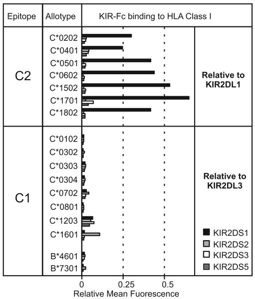
KIR2DS1, 2DS2, 2DS3 and 2DS5 Fc-fusion proteins were tested for binding to 29 HLA-A, 50 HLA-B and 16 HLA-C allotypes, and compared to the high avidity inhibitory receptors: C1-specific KIR2DL3 and C2-specific KIR2DL1 (Fig. 1). Of the four activating receptors, only 2DS1 gave strong reactions. It bound the seven C2+ HLA-C allotypes at 20-65% the KIR2DL1 level. In contrast, 2DS2, 2DS3 and 2DS5 gave very weak reactions with no general preference for C1+ or C2+ allotypes. Weak binding of the four KIR2DS to HLA-C*1203 is not significant because the normalizing KIR2DL3 control also binds this allotype poorly (20). KIR2DS2-Fc selectively bound to HLA-C*1601, albeit weakly, suggesting this reaction is specific. No reactions with HLA-A and –B were observed. We see that KIR2DS1 has significant avidity for the range of C2+ HLA-C, whereas 2DS2, 2DS3, and 2DS5 have little or no detectable avidity for any HLA class I. These results affirm and extend the observations from previous analyses of limited numbers of HLA class I variants (7, 10, 13).
Fig. 1. Binding of human activating lineage III KIR to C2+ HLA-C and to C1+ HLA-B and -C.
C2 binding is normalized to that of inhibitory KIR2DL1; C1 binding is normalized to that of inhibitory KIR2DL3.
Activating chimpanzee lineage III KIR exhibit high avidity for C1 or C2
Chimpanzee Pt-KIR3DS6 and Pt-KIR3DS2 Fc-fusion proteins were analyzed for binding to the 95 HLA class I variants and compared to the closely related inhibitory receptors: Pt-KIR2DL6v and Pt-KIR3DL4, respectively. Activating Pt-KIR3DS6 and inhibitory Pt-KIR2DL6v both showed a specificity like human KIR2DL3 for the C1 epitope carried by eight HLA-C and two HLA-B allotypes, consistent with all three KIR having lysine 44. Except C*1601, for which the binding was comparable, activating Pt-KIR3DS6 bound less strongly to the C1 epitopes (∼70%) than inhibitory Pt-KIR2DL6v (Fig. 2A). This reduced avidity must arise from the single aminoacid difference in their ligand-binding domains, replacement of leucine 83 for valine in Pt-KIR3DS6; of note, a second allotype of the inhibitory receptor, Pt-KIR2DL6, has leucine 83 like Pt-KIR3DS6, but differs at position 68 (Supplemental Fig. S1). In comparison to its human counterpart, KIR2DS2, chimpanzee Pt-KIR3DS6 is seen to be a strong C1 receptor. Pt-KIR3DS6 and Pt-KIR2DL6 are adjacent genes in the chimpanzee KIR locus, and in strong linkage disequilibrium (Fig.3). This arrangement is similar to that of human KIR2DS2 with KIR2DL2, the latter being allelic to KIR2DL3 and also encoding an inhibitory C1-specific receptor.
Fig. 2. Binding of chimpanzee and gorilla activating lineage III KIR to C1 and C2.
Panels A and B show the binding of chimpanzee KIR with C1 and C2 specificity, respectively. Data is normalized as described in the legend to Fig. 1. Panel C compares the binding of gorilla Gg-KIR2DSa and human KIR2DL3 to HLA-A, B and C allotypes.
Fig. 3. The genomic organization of the chimpanzee and human KIR loci.
Shown are composite haplotypes containing all known KIR genes. Inhibitory and activating lineage III genes are colored red and blue, respectively. Conserved framework genes are colored green, other genes and the 2DP1 pseudogene are white. Double-headed arrows connect the paired receptors. †, indicates loss of reactivity by 2DS2.
Similar comparison of activating Pt-KIR3DS2 and inhibitory Pt-KIR3DL4 showed they both bound strongly to C2+ HLA-C allotypes and to no other HLA class I (Fig. 2B), their C2 specificity being consistent with presence of methionine 44 in both KIR. In addition to identical specificity, Pt-KIR3DS2 and Pt-KIR3DL4 bound individual C2+ allotypes with equivalent avidity. Thus the single difference in their ligand-binding domains, at position 51 (Supplemental Fig. S1), was not selected to reduce the activating receptor's avidity for ligand. The Pt-KIR3DS2v allotype differs from Pt-KIR3DS2 by two substitutions in D1 and four in D2 (Supplemental Fig. S1). The Pt-KIR3DS2v fusion protein has the same C2 specificity as Pt-KIR3DS2, but its avidity for each of the C2+ HLA-C allotypes was reduced to ∼73% that of Pt-KIR3DS2. Thus KIR3DS2v is likely to be a product of natural selection for reduced receptor avidity. Although Pt-KIR3DS2 and Pt-KIR3DL4 are paired high avidity C2 receptors, their genes are separated by up to six other KIR genes and lack linkage disequilibrium (Fig. 3). This arrangement resembles that of the human KIR2DL1 and KIR2DS1 genes, present in different halves of the human KIR locus and separated by a comparable number of other KIR genes. Pt-KIR3DS2 is now adjacent and in linkage disequilibrium with Pt-KIR2DL9 (Fig. 3), a divergent form of inhibitory C2 receptor with glutamate 44 and differing from Pt-KIR3DS2 by 32 substitutions in the Ig-like domains (Fig 2B).
In conclusion, the chimpanzee has pairs of activating and inhibitory receptors that recognize C1 and C2. One variant of the activating C2 receptor has the same strong avidity as the inhibitory receptor, while a second variant is about ∼27% less avid. To similar extent the activating C1 receptor is less avid than its inhibitory counterpart. This trend, in which natural selection acts to weaken the activating receptor relative to the inhibitory receptors, has been observed for other paired immunoreceptors (23).
Gorilla Gg-KIR2DSa is an activating receptor with high avidity for C1
Of six gorilla lineage III KIR, Gg-KIR2DSa with lysine 44 is the only activating KIR and a candidate C1 receptor (18). As seen in Figure 2C, the Gg-KIR2DSa fusion protein binds only to C1+ HLA-B and HLA-C allotypes, and it thus has C1 specificity like KIR2DL3, Pt-KIR2DL6 and Pt-KIR3DS6. However, Gg-KIR2DSa and KIR2DL3 exhibit significant avidity differences for individual C1+ allotypes, the extreme cases being HLA-C*1203 that binds weakly to KIR2DL3 but strongly to Gg-KIR2DSa, and HLA-C*0102 that binds weakly to Gg-KIR2DSa but strongly to KIR2DL3. In our hands, Gg-KIR2DSa is the only KIR that binds well to HLA-C*1203. This unique reactivity correlates with alanine 73 in HLA-C (present only in C*12 and the structurally divergent C*07) and arginine 71 in Gg-KIR2DSa, a unique residue at a position that influences KIR specificity (16). On average, the binding of Gg-KIR2DSa to C1+ allotypes was about half that of KIR2DL3. But in comparison to human KIR2DS2 (Fig.1), Gg-KIR2DSa is an activating receptor with high avidity for C1.
Pt-KIR3DS2 and Pt-KIR3DS6 Recognize MHC-C to Control NK Cell Function
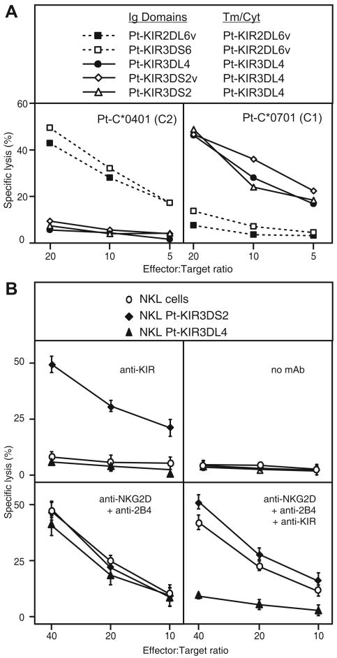
To assess the functional influence of interactions between MHC class I and activating chimpanzee KIR, the ligand-binding domains of Pt-KIR3DS2 and Pt-KIR3DS6 were grafted onto the signaling domains of their paired inhibitory receptors: Pt-KIR3DL4 and Pt-KIR2DL6v, respectively. NKL cells expressing these chimeric KIR were tested for their capacity to kill class I deficient 221 cells and 221 transfectants expressing C1+ and C2+ chimpanzee MHC class I allotypes. Comparison was made with NKL cells expressing the natural inhibitory receptors Pt-KIR3DL4 and Pt-2DL6v (Fig. 4).
Fig.4. Functional analysis of chimpanzee activating KIR.
Panel A shows the results of cytotoxic assays in which the target cells are 221 cells expressing either C1+ or C2+ MHC class I and the effector cell are NKL cells expressing inhibitory chimpanzee KIR or chimeric proteins carrying the D0, D1 and D2 domains of activating KIR with the transmembrane and cytoplasmic domains of the paired inhibitory receptor. Shown is the capacity of the C2 (left) and C1 (right) epitopes to engage the chimeric receptors and prevent NKL cell mediated lysis of 221 target cells. Panel B shows the results of cytotoxic assays in which the target cells are P815 cells and the effector cells are NKL cells and NKL cell transduced with activating Pt-KIR3DS2 or inhibitory Pt-KIR3DL4 that react with the anti-KIR antibody, NKVSF1. Assays were carried out in the presence or absence of monoclonal antibodies directed against KIR, NKG2D and 2B4.
The killing of 221 target cells by NKL transfected with chimeric Pt-KIR3DS2 was inhibited by the presence of C2+ MHC-C at the target cell surface (Fig.4A, left panel) but not by presence of C1+ MHC-C (Fig. 4A, right panel). This pattern of C2-mediated inhibition mimics that of NKL cells expressing Pt-KIR3DL4. Conversely, killing of 221 target cells by NKL cells expressing chimeric Pt-KIR3DS6 was inhibited by presence of C1 on the target cell surface but not C2 (Fig 4A, right panel). These results show that the extracellular domains of Pt-KIR3DS6 and 3DS2 are binding sites for cell-surface associated MHC-C ligands that can signal an inhibitory NK cell response; they also imply that natural Pt-KIR3DS2 and 3DS6 can engage their cognate ligands on target cells and generate an activating signal in chimpanzee NK cells.
Pt-KIR3DS2 activates NKL cells in a redirected cytotoxicity assay
Pt-KIR3DS2 was expressed in NKL cells and tested for its potential to induce killing in a redirected cytotoxicity assay using P815 target cells and monoclonal anti-KIR antibody (NKVFS1). NKL cells expressing Pt-KIR3DS2 killed P815 target cells in the presence of anti-KIR antibody (Fig.4B, upper left panel) but not in its absence (Fig. 4B, upper right). The extent of activation was comparable to that induced by the combination of anti-NKG2D and anti-2B4 antibodies (Fig. 4B, lower left panel). In contrast, parental NKL or NKL expressing inhibitory Pt-KIR3DL4 were not induced to kill P815 cells in the presence of anti-KIR antibody. Neither did the combination of all three antibodies significantly increase cytotoxicity over that seen with anti-KIR alone, and was only slightly higher than that observed for parental NKL cells, suggesting that anti-KIR is sufficient to induce maximal killing in this assay (Fig. 4, lower right panel). Conversely, for NKL cells expressing Pt-KIR3DL4 the inhibitory signals induced by anti-KIR antagonized the activating effects of antibody-mediated cross-linking of NKG2D and 2B4.
Discussion
Our study definitively shows that no human activating KIR is a C1 receptor, while KIR2DS1 recognizes C2 with reduced avidity (∼50%) compared to inhibitory KIR2DL1. The corresponding chimpanzee receptor (Pt-KIR3DS2) has two variants, one as strong as its paired inhibitory C2-specific receptor (Pt-KIR3DL4), the other with reduced affinity (73%), like KIR2DS1. Whereas human KIR2DS2 has no detectable avidity for C1, its chimpanzee counterpart (Pt-KIR3DS6) binds C1 with ∼70% of the avidity of its paired inhibitory receptor (Pt-KIR2DL6). In both human and chimpanzee the trend is for activating receptors to have reduced avidity relative to the inhibitory receptors. But only for the human activating C1 receptor has this reached the extreme where the function appears lost. Like chimpanzee, gorilla has a strong activating C1 receptor, while orangutan has paired activating and inhibitory receptors with identical strong avidities for C1 (A.M.O.A manuscript submitted). Thus human appears unique among hominid species in having no functional C1-specific activating receptor. A weak reactivity with HLA-C*1601 shows that KIR2DS2 has a narrowed specificity for HLA-C, raising the possibility that it is also highly dependent on the peptide bound by MHC-C. Thus we cannot rule out the possibility that KIR2DS2 functions in the presence of peptides presented by HLA-C in infected or malignant cells but not B cell lines.
The C1 epitope and its cognate KIR arose several million years before C2 and C2-specific KIR, an ancestral state retained in the modern orangutan (24). That chimpanzee retains strong activating C1- and C2-specific receptors shows that loss of activating C1 receptor is a product of human-specific evolution. A key feature of KIR2DS2 is the almost total linkage disequilibrium with KIR2DL2, a divergent allele of the KIR2DL2/3 gene having higher avidity for C1 than KIR2DL3 (20). Thus the inactivation of the activating C1 receptor is associated with increased avidity of the inhibitory C1 receptor. Studies showing that KIR2DS1 counterbalances KIR2DL1 in NK cell development and function (8), suggest that elimination of the equivalent KIR2DL2/3 counterbalance could have an effect on NK cell education in C1-expressing individuals that increases the frequency and potency of NK cells expressing KIR2DL2.
KIR2DS2 and KIR2DL2 are characteristic centromeric genes of the group B KIR haplotypes that are substituted by KIR2DL3 on group A haplotypes. Differences between the group A and B KIR haplotypes correlate with a wide range of clinical conditions. For example, the beneficial effect of donor B haplotypes in unrelated HLA-matched bone marrow transplantation for acute myelogenous leukemia (25) is principally due to presence of centromeric motifs containing KIR2DS2 and KIR2DL2 (26). A further feature of these centromeric B haplotype motifs are variants of KIR2DL, such as KIR2DL1*004, that are reduced in their capacity to educate KIR2DL1-expressing NK cells in C2-bearing individuals (27). Such linkage disequilibrium points to a process of co-evolution that has modified the functions of both the C1 and C2 receptors encoded on the B haplotypes. This process also appears human-specific because in no other primate species examined do the KIR haplotypes divide into two functionally distinctive groups analogous to human A and B.
Supplementary Material
Footnotes
This work was supported by NIH grant AI022039 (to P.P.), a Ruth L. Kirschstein National Research Service Award from NIH (to A.M.O.A.), and a National Science Foundation Graduate Fellowship (to A.K.M.).
References
- 1.Moretta A, Bottino C, Vitale M, Pende D, Biassoni R, Mingari MC, Moretta L. Receptors for HLA class-I molecules in human natural killer cells. Annu Rev Immunol. 1996;14:619–648. doi: 10.1146/annurev.immunol.14.1.619. [DOI] [PubMed] [Google Scholar]
- 2.Carlyle JR, Mesci A, Fine JH, Chen P, Belanger S, Tai LH, Makrigiannis AP. Evolution of the Ly49 and Nkrp1 recognition systems. Semin Immunol. 2008;20:321–330. doi: 10.1016/j.smim.2008.05.004. [DOI] [PubMed] [Google Scholar]
- 3.Natarajan K, Dimasi N, Wang J, Mariuzza RA, Margulies DH. Structure and function of natural killer cell receptors: multiple molecular solutions to self, nonself discrimination. Annu Rev Immunol. 2002;20:853–885. doi: 10.1146/annurev.immunol.20.100301.064812. [DOI] [PubMed] [Google Scholar]
- 4.Kulkarni S, Martin MP, Carrington M. The Yin and Yang of HLA and KIR in human disease. Semin Immunol. 2008;20:343–352. doi: 10.1016/j.smim.2008.06.003. [DOI] [PMC free article] [PubMed] [Google Scholar]
- 5.Biassoni R, Pessino A, Malaspina A, Cantoni C, Bottino C, Sivori S, Moretta L, Moretta A. Role of amino acid position 70 in the binding affinity of p50.1 and p58.1 receptors for HLA-Cw4 molecules. Eur J Immunol. 1997;27:3095–3099. doi: 10.1002/eji.1830271203. [DOI] [PubMed] [Google Scholar]
- 6.Stewart CA, Laugier-Anfossi F, Vely F, Saulquin X, Riedmuller J, Tisserant A, Gauthier L, Romagne F, Ferracci G, Arosa FA, Moretta A, Sun PD, Ugolini S, Vivier E. Recognition of peptide-MHC class I complexes by activating killer immunoglobulin-like receptors. Proc Natl Acad Sci U S A. 2005;102:13224–13229. doi: 10.1073/pnas.0503594102. [DOI] [PMC free article] [PubMed] [Google Scholar]
- 7.Vales-Gomez M, Reyburn HT, Erskine RA, Strominger J. Differential binding to HLA-C of p50-activating and p58-inhibitory natural killer cell receptors. Proc Natl Acad Sci U S A. 1998;95:14326–14331. doi: 10.1073/pnas.95.24.14326. [DOI] [PMC free article] [PubMed] [Google Scholar]
- 8.Fauriat C, Ivarsson MA, Ljunggren HG, Malmberg KJ, Michaelsson J. Education of human natural killer cells by activating killer cell immunoglobulin-like receptors. Blood. 2010;115:1166–1174. doi: 10.1182/blood-2009-09-245746. [DOI] [PubMed] [Google Scholar]
- 9.Morvan M, David G, Sebille V, Perrin A, Gagne K, Willem C, Kerdudou N, Denis L, Clemenceau B, Follea G, Bignon JD, Retiere C. Autologous and allogeneic HLA KIR ligand environments and activating KIR control KIR NK-cell functions. Eur J Immunol. 2008;38:3474–3486. doi: 10.1002/eji.200838407. [DOI] [PubMed] [Google Scholar]
- 10.Winter CC, Gumperz JE, Parham P, Long EO, Wagtmann N. Direct binding and functional transfer of NK cell inhibitory receptors reveal novel patterns of HLA-C allotype recognition. J Immunol. 1998;161:571–577. [PubMed] [Google Scholar]
- 11.Saulquin X, Gastinel LN, Vivier E. Crystal structure of the human natural killer cell activating receptor KIR2DS2 (CD158j) J Exp Med. 2003;197:933–938. doi: 10.1084/jem.20021624. [DOI] [PMC free article] [PubMed] [Google Scholar]
- 12.Abi-Rached L, Parham P. Natural selection drives recurrent formation of activating killer cell immunoglobulin-like receptor and Ly49 from inhibitory homologues. J Exp Med. 2005;201:1319–1332. doi: 10.1084/jem.20042558. [DOI] [PMC free article] [PubMed] [Google Scholar]
- 13.Winter CC, Long EO. A single amino acid in the p58 killer cell inhibitory receptor controls the ability of natural killer cells to discriminate between the two groups of HLA-C allotypes. J Immunol. 1997;158:4026–4028. [PubMed] [Google Scholar]
- 14.VandenBussche CJ, Mulrooney TJ, Frazier WR, Dakshanamurthy S, Hurley CK. Dramatically reduced surface expression of NK cell receptor KIR2DS3 is attributed to multiple residues throughout the molecule. Genes Immun. 2009;10:162–173. doi: 10.1038/gene.2008.91. [DOI] [PMC free article] [PubMed] [Google Scholar]
- 15.Della Chiesa M, Romeo E, Falco M, Balsamo M, Augugliaro R, Moretta L, Bottino C, Moretta A, Vitale M. Evidence that the KIR2DS5 gene codes for a surface receptor triggering natural killer cell function. Eur J Immunol. 2008;38:2284–2289. doi: 10.1002/eji.200838434. [DOI] [PubMed] [Google Scholar]
- 16.Graef T, Moesta AK, Norman PJ, Abi-Rached L, Vago L, Older Aguilar AM, Gleimer M, Hammond JA, Guethlein LA, Bushnell DA, Robinson PJ, Parham P. KIR2DS4 is a product of gene conversion with KIR3DL2 that introduced specificity for HLA-A*11 while diminishing avidity for HLA-C. J Exp Med. 2009;206:2557–2572. doi: 10.1084/jem.20091010. [DOI] [PMC free article] [PubMed] [Google Scholar]
- 17.Khakoo SI, Rajalingam R, Shum BP, Weidenbach K, Flodin L, Muir DG, Canavez F, Cooper SL, Valiante NM, Lanier LL, Parham P. Rapid evolution of NK cell receptor systems demonstrated by comparison of chimpanzees and humans. Immunity. 2000;12:687–698. doi: 10.1016/s1074-7613(00)80219-8. [DOI] [PubMed] [Google Scholar]
- 18.Rajalingam R, Parham P, Abi-Rached L. Domain shuffling has been the main mechanism forming new hominoid killer cell Ig-like receptors. J Immunol. 2004;172:356–369. doi: 10.4049/jimmunol.172.1.356. [DOI] [PubMed] [Google Scholar]
- 19.Moesta AK, Abi-Rached L, Norman PJ, Parham P. Chimpanzees use more varied receptors and ligands than humans for inhibitory killer cell Ig-like receptor recognition of the MHC-C1 and MHC-C2 epitopes. J Immunol. 2009;182:3628–3637. doi: 10.4049/jimmunol.0803401. [DOI] [PMC free article] [PubMed] [Google Scholar]
- 20.Moesta AK, Norman PJ, Yawata M, Yawata N, Gleimer M, Parham P. Synergistic polymorphism at two positions distal to the ligand-binding site makes KIR2DL2 a stronger receptor for HLA-C than KIR2DL3. J Immunol. 2008;180:3969–3979. doi: 10.4049/jimmunol.180.6.3969. [DOI] [PubMed] [Google Scholar]
- 21.Pei R, Lee JH, Shih NJ, Chen M, Terasaki PI. Single human leukocyte antigen flow cytometry beads for accurate identification of human leukocyte antigen antibody specificities. Transplantation. 2003;75:43–49. doi: 10.1097/00007890-200301150-00008. [DOI] [PubMed] [Google Scholar]
- 22.El-Awar N, Lee J, Terasaki PI. HLA antibody identification with single antigen beads compared to conventional methods. Hum Immunol. 2005;66:989–997. doi: 10.1016/j.humimm.2005.07.005. [DOI] [PubMed] [Google Scholar]
- 23.Barclay AN, Hatherley D. The counterbalance theory for evolution and function of paired receptors. Immunity. 2008;29:675–678. doi: 10.1016/j.immuni.2008.10.004. [DOI] [PMC free article] [PubMed] [Google Scholar]
- 24.Guethlein LA, Flodin LR, Adams EJ, Parham P. NK Cell Receptors of the Orangutan (Pongo pygmaeus): A Pivotal Species for Tracking the Coevolution of Killer Cell Ig-Like Receptors with MHC-C. J Immunol. 2002;169:220–229. doi: 10.4049/jimmunol.169.1.220. [DOI] [PubMed] [Google Scholar]
- 25.Cooley S, Trachtenberg E, Bergemann TL, Saeteurn K, Klein J, Le CT, Marsh SG, Guethlein LA, Parham P, Miller JS, Weisdorf DJ. Donors with group B KIR haplotypes improve relapse-free survival after unrelated hematopoietic cell transplantation for acute myelogenous leukemia. Blood. 2009;113:726–732. doi: 10.1182/blood-2008-07-171926. [DOI] [PMC free article] [PubMed] [Google Scholar]
- 26.Cooley S, Weisdorf DJ, Guethlein LA, Klein JP, Wang T, Le CT, Marsh SGE, Geraghty D, Spellman S, Haagenson MD, Ladner M, Trachtenberg E, Parham P, Miller JS. Donor selection for natural killer cell receptor genes leads to superior survival after unrelated transplantation for acute myelogenous leukemia. Blood. 2010 doi: 10.1182/blood-2010-05-283051. In press. [DOI] [PMC free article] [PubMed] [Google Scholar]
- 27.Yawata M, Yawata N, Draghi M, Partheniou F, Little AM, Parham P. MHC class I-specific inhibitory receptors and their ligands structure diverse human NK-cell repertoires toward a balance of missing self-response. Blood. 2008;112:2369–2380. doi: 10.1182/blood-2008-03-143727. [DOI] [PMC free article] [PubMed] [Google Scholar]
Associated Data
This section collects any data citations, data availability statements, or supplementary materials included in this article.


