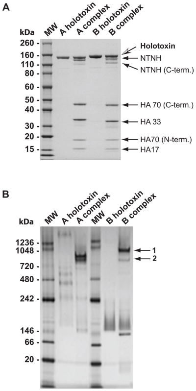
Figure 1.
Protein size and content comparisons of BoNT/A and BoNT/B holotoxin and complexes. A. Purified BoNT/A and BoNT/B holotoxins and complexes were size-separated on a 4–12% SDS-PAGE gel and stained with Coomassie Blue. Individual protein components in the toxin complexes were identified by mass spectrometry (arrows). B. The sizes of purified BoNT/A and BoNT/B holotoxins and complexes were compared under native gel conditions. Two different sized BoNT/B complexes were denoted with arrows. Gel bands 1 and 2 were identified by mass spectrometry to contain BoNT/B holotoxin, NTNH, HA70, HA33 and HA17. MW: molecular weight marker (sizes in kDa).

