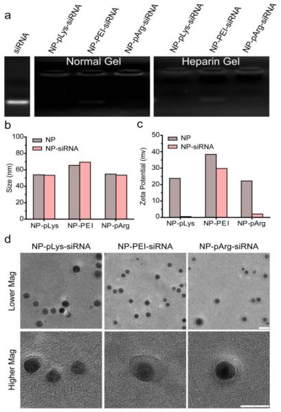
Figure 3.
Characterization of constructed nanovectors. (a) Gel retardation assay evaluating covalent attachment of siRNA to NPs under normal electrophoresis conditions and under heparin treatment to disrupt electrostatic interactions between cationic NPs and anionic siRNA. (b) Z-average hydrodynamic sizes of nanovectors before and after siRNA attachment. (c) Zeta potentials of nanovectors before and after siRNA attachment. (d) TEM images of nanovectors (scale bars correspond to 10 nm).

