Abstract
Identification of individuals is a challenging task in forensic odontology. In circumstances where identification of an individual by fingerprint or dental record comparison is difficult, the palatal rugae may be considered as an alternative source. Palatal rugae have been shown to be highly individualistic and it maintains consistency in shape throughout life.
Aims and Objectives:
The present study is conducted to test the efficiency of computerized software in the identification of individuals after obtaining digital photographic images of the rugae.
Materials and Methods:
The intra oral photographs of 100 individuals were taken using a SLR digital camera. The custom made external attachment was attached to the camera to standardize all the photographs. A special software was designed called the Palatal Rugae Comparison Software (PRCS Version 2.0) to match the clinical photographs. Five evaluators including 3 dentists, 1 computer professional, and 1 general surgeon were asked to match the rugae pattern using the software. The results were recorded along with time taken by each operator to match all the photos using software.
Results:
The software recorded an accuracy of 99% in identification of individuals.
Conclusion:
The present study supports the fact of individuality of the rugae. Computerized method has given very good results to support the individualization of rugae. Through our study, we feel that palatal rugae patterns will be of great use in the future of forensic odontology.
Keywords: Forensic odontology, human identification, palatal rugae
Introduction
One of the main focuses of the forensic odontologist is identification of an individual. Dental identification can be used as the sole method of identifying a deceased person. Dental identification is based on the comparison of antemortem and postmortem records. The records collected to identify a decedent should be accurate and totally inclusive of objective findings.[1]
Palatoscopy or palatal rugoscopy, is the name given to the study of palatal rugae.[2] Palatal rugae are present in the maxillary portion of the oral cavity. Palatal rugae, also called plicae palatinae transversae and rugae palatina, refer to the ridges on the anterior part of the palatal mucosa, each side of the median palatal raphe and behind the incisive papilla, which is just behind the maxillary central incisor teeth. As an entity they form the rugae pattern. Anatomically, the rugae consist of about 3 to 7 rigid and oblique ridges that radiate out tangentially from the incisive papilla.[3]
Rugae patterns have been studied for various purposes, with reports being published mainly in the fields of anthropology, comparative anatomy,[4] genetics, forensic odontology,[5] prosthodontics, and orthodontics.[6–8]
The anatomical position of the palatal rugae inside the oral cavity is surrounded by cheek, lips, tongue, teeth, and a buccal pad of fat. All these afford some protection in case of fire and high impact trauma. Rugae are amongst the best protected, morphologically individualizing soft tissue structures in the body, which are preserved after death and also accessible during life.[9] There are several reports available in the literature supporting the use of palatal rugae in individual identification.[10–14] Attempts have also been made to study the lip prints (cheiloscopy) along with palatoscopy for the identification of individuals.[15,16]
The scope for the study of rugae pattern in individuals prompted us to conduct this study, with the aim to test the efficiency of computerized software in the identification of individuals after obtaining their digital photographic image of the rugae.
Materials and Methods
One hundred healthy adult individuals formed the study group. For this, a written consent form and a questionnaire having questions regarding the orthodontic treatment, palatal lesions etc. was given to the medical and dental students of Yenepoya dental and medical college who volunteered to participate in the study [Table 1].
Table 1.
QUESTIONAIRE
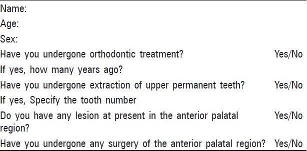
Individuals with cleft palate, below 18 years age, those who had undergone surgery in the anterior palatal region, those undergoing orthodontic treatment, those who had undergone extraction of upper permanent teeth, and individuals with palatal lesions, were excluded.
Intra oral photographs of all these 100 individuals were taken using a SLR digital camera (Canon EOS 300D). The custom made external attachment was attached to the camera to standardize all the photographs. The images were transferred to computer as a .jpeg image file [Figure 1a–b].
Figure 1.
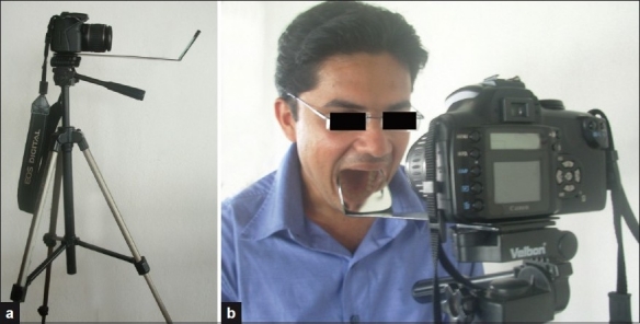
(a) SLR digital camera (Canon EOS 300D) along with the custom made external attachment; (b) Taking clinical photographs using SLR digital camera (Canon EOS 300D) along with the custom made external attachment
Identification of individuals using computer technique
For this purpose, a special software was designed called Palatal Rugae Comparison Software (PRCS Version 2.0). The initiation and termination points of rugae were marked on all 100 images of clinical photographs using MS paint version 5.1 software. A strict protocol was undertaken for the order in which points were plotted - tip and base of incisive papilla, then each ruga was plotted at the medial and lateral ends, working from anterior to posterior. Left sided rugae were plotted before right. These plotted points were processed by the software and the information sequentially stored corresponding to pixel position.
All the 100 photographs were stored in the software. Later same photographs were loaded to the software one by one. After marking the points in second set of photos the match command is given in the software. The software will search for its match from previously loaded photographs if it is already loaded in the computer.
Five evaluators including three dentists, one computer professional, and one general surgeon were asked to match the rugae pattern using the software. The results were recorded along with time taken by each operator to match all the photos using software.
Step-wise use of palatal rugae comparison software version 2.0
Click PRCS icon in the computer to open the software.
Load the photographs by pressing ANTEMORTEM button in the software [Figure 2a].
Press OPEN button to open the selected file. [Figure 2b] The photograph will open in a different window [Figure 2c].
Press MARK POINT buttons, [Figure 2b] mark the starting and end point of rugae using cursor [Figure 2d].
Press SAVE button to save the file. [Figure 2b] Enter the file name and person's name while saving. The software will save the file as shown in the Figure 2e.
Once all the photos are loaded then press POSTMORTEM button [Figure 2f].
Select the file to be matched and press OPEN button [Figure 2f].
Again mark the points on the photographs by pressing MARK POINT button [Figure 2f].
Save the file by pressing SAVE button [Figure 2f].
Press MATCH button to find the correct match, which was previously loaded in the software [Figure 2f].
If software does not find any match the result will come as NO MATCH [Figure 2g].
If it finds any match the result will come as MATCH SUCCESS along with the person's name [Figure 2h].
Figure 2.
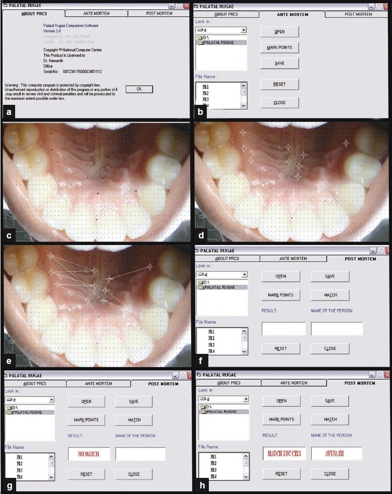
(a, b, f) Palatal Rugae Comparison Software (PRCS); (c) Initiation and termination points of rugae were marked on clinical photographs using MS paint version 5.1 software; (d) Initiation and termination points of rugae were marked on clinical photographs by using PRCS; (e) Showing photographs saved in the PRCS software; (g, h) PRCS showing the result
Results
When the results were analyzed it was found that the 3 evaluators got 100% results and 2 evaluators got 99% results [Figure 3].
Figure 3.
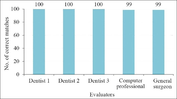
Correct matches in computer method by different evaluator
The time taken to match palatal rugae by different evaluators is as follows: The first dentist took 40 minutes, second dentist took 60 minutes and the third dentist took 55 minutes. The computer professional took 50 minutes, and general surgeon took 75 minutes [Figure 4].
Figure 4.
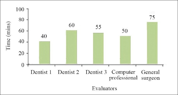
Time taken in minutes in computer method by different evaluators
Discussion
Palatal rugae have been shown to be highly individualistic and consistent in shape throughout the life. It is well-established fact that the palatal rugae pattern is unique to human being, as his fingerprints.
Once formed, they do not undergo any change except in length, due to normal growth,[17] and remain in the same position throughout a person's life time. Diseases, chemical aggression, or trauma do not seem to be able to change the palatal rugae form.[18]
It is also concluded that changes that occur from orthodontic movement, extraction, aging, and palatal expansion do not modify the rugae enough to hamper identification.[1]
Palatal rugae are used in human identification not only due to their singularity and unchangeable nature, but also due to other advantages, namely their low utilization costs.[19]
In a study conducted by Limson K.S. and Julian R[20] (2004), the comprehensive computerized antemortem records were constructed for 250 subjects and a comparison matching process performed using both recorded and unrecorded samples. The efficiency of the computer-based identification method was then assessed. The program proved to have an average sensitivity of 0.93 and specificity of 1 and had a success rate of 92–97% in matches with digitized rugae pattern samples.
Comparing the results of our study, we observed that the error rate observed is substantially less in our study. In our study, two evaluators had the error rate of 2%. This may be due to errors in the delineation of rugae or incorrect sequential plotting of characteristic points on the rugae pattern image while using the software. The error rate may be reduced by development of an intraoral scanning device to capture palatal rugae pattern, with image transferred directly to a computer, with appropriate software, as is presently available for fingerprints. This would eliminate the manual errors and time involved in the process of digitization of rugae pattern samples.
Limitations of the study
The computer software that is used in this method requires manual plotting of the points on the rugae before matching. This may lead to human errors in plotting the points. There is a scope for the development of new software, which can match the photographs of rugae without plotting any points on the image. The fully automation of the software will nullify the human errors, which may occur during manual plotting of the points.
The time required to match can be reduced by making a team effort while matching. Similar conclusion was drawn by English WR et al. in their study.[9] Further, with the use of interconnected computer networks it would be possible to store a large amount of data, facilitating quick retrieval of information, and fast and effective identification. The study can also be done by matching the photographs of the rugae manually. This can be done by using either clinical photographs or the photographs of the casts.
Conclusion
The present study supports the individuality of the rugae. Computerized method has given a very good result to support the individualization of rugae. The results can be improved furthermore by using advanced software.
Footnotes
Source of Support: Nil
Conflict of Interest: None declared.
References
- 1.Stuart LS, Leonard G. Missouri: The forensic examiner Spring; 2005. Forensic application of palatal rugae in dental identification; pp. 44–7. [Google Scholar]
- 2.Kapali S, Townsend G, Richards L, Parish T. Palatal rugae patterns in Australian aborigines and Caucasians. Aust Dent J. 1997;42:129–33. doi: 10.1111/j.1834-7819.1997.tb00110.x. [DOI] [PubMed] [Google Scholar]
- 3.Thomas CJ, Kotze TJ. The palatal rugae: A new classification. J Dent Assoc S Afr. 1983;38:153–7. [PubMed] [Google Scholar]
- 4.Hauser G, Daponte A, Roberts MJ. Palatine rugae. J Anat. 1989;165:237–49. [PMC free article] [PubMed] [Google Scholar]
- 5.Virdi M, Singh Y, Kumar A. Role of palatal rugae in forensic identification of the pediatric population. Internet J Forensic Sci. 2010;4 [Google Scholar]
- 6.Whittakar DK. Introduction to forensic dentistry. Quintessence Int. 1994;25:723–30. [PubMed] [Google Scholar]
- 7.Paliwal A, Wanjari S, Parwani R. Palatal rugoscopy: Establishing identity. J Forensic Dent Sci. 2010;2:27–31. doi: 10.4103/0974-2948.71054. [DOI] [PMC free article] [PubMed] [Google Scholar]
- 8.Damstra J, Mistry D, Cruz C, Ren Y. Antero-posterior and transverse changes in the positions of palatal rugae after rapid maxillary expansion. Eur J Orthod. 2009;31:327–32. doi: 10.1093/ejo/cjn113. [DOI] [PubMed] [Google Scholar]
- 9.English WR, Robison SF, Summitt JB, Oesterle LJ, Brannon RB, Morlang WM. Individuality of human palatal rugae. J Forensic Sci. 1988;33:718–26. [PubMed] [Google Scholar]
- 10.Hermosilla VV, San pedro VJ, Cantín LM, Suazo GI. C Palatal rugae: Systematic analysis of its shape and dimensions for use in human identification. Int J Morphol. 2009;27:819–25. [Google Scholar]
- 11.Patil MS, Patil SB, Acharya AB. Palatine rugae and their significance in clinical dentistry: A review of the literature. J Am Dent Assoc. 2008;139:1471–8. doi: 10.14219/jada.archive.2008.0072. [DOI] [PubMed] [Google Scholar]
- 12.Shetty SK, Kalia S, Patil K, Mahima VG. Palatal rugae pattern in Mysorean and Tibetan populations. Indian J Dent Res. 2005;16:51–5. [PubMed] [Google Scholar]
- 13.Vishlesh A, Anjana B, Vaishali K, Arvind S. Comparison of palatal rugae pattern in two populations of India. Int J Med Toxicol Leg Med. 2008;10:2. [Google Scholar]
- 14.Bansode SC, Kulkarni MM. Importance of palatal rugae in individual identification. J Forensic Dent Sci. 2009;1:2. [Google Scholar]
- 15.Caldas IM, Magalhães T, Afonso A. Establishing identity through cheloscopy and palatoscopy. Forensic Sci Int. 2007;165:1–9. doi: 10.1016/j.forsciint.2006.04.010. [DOI] [PubMed] [Google Scholar]
- 16.Sharma P, Saxena S, Rathod V. Comparative reliability of cheloscopy and palatoscopy in human identification. India J Dent Res. 2009;20:453–7. doi: 10.4103/0970-9290.59451. [DOI] [PubMed] [Google Scholar]
- 17.Campos ML. Rugoscopia palatina. [last accessed on 2007 July 27]. Available from: http://www.pericias-forenses.com.br .
- 18.Almeida MA, Phillips C, Kula K, Tulloch C. Stability of the palatal rugae as landmarks for analysis of dental casts. Angle Orthod. 1995;65:43–8. doi: 10.1043/0003-3219(1995)065<0043:SOTPRA>2.0.CO;2. [DOI] [PubMed] [Google Scholar]
- 19.Thomas CJ, van Wyk CW. The palatal rugae in identification. J Forensic Odontostomatol. 1988;6:21–5. [PubMed] [Google Scholar]
- 20.Limson KS, Julian R. Computerized recording of the palatal rugae pattern and an evaluation of its application in forensic identification. J Forensic Odontostomatol. 2004;22:1–4. [PubMed] [Google Scholar]


