Abstract
Current models and concepts of motor control represent the limb as a neuro-musculoskeletal system and rarely include other potentially important supporting tissues such as fascia and adipose tissue. It is possible that a normal complement of adipose tissue could contribute to the viscoelastic properties of supporting limbs and enhance stability during locomotion. The purpose of this study was to determine if the popliteal fat pad plays a role in locomotion in the cat. It is hypothesized that the fat pad limits flexion and reduces angular acceleration of the included hip, knee and ankle joints in the sagittal plane throughout the step cycle. 3D kinematics from 3 spontaneously locomoting decerebrate cats both before and after lipectomy were recorded during treadmill walking. Four time points throughout the step cycle were chosen for angular acceleration analysis: mid-stance, paw off, mid-swing and peak deceleration at the end of the re-extension of the knee. Significant increases in maximum angular acceleration for the hip, knee and ankle joints at these time points were observed. No significant increase in range of motion was found across all 3 included angles after lipectomy. Therefore, the hypothesis that the popliteal fat pad acts to decrease the angular acceleration is supported by these findings. The data indicate that the popliteal fat pad contributes to the damping component of the viscoelastic properties of the limb. These results may be applied to models of the hindlimb and knowledge of the effects of obesity on movement.
Key Words: Popliteal fat pad, Locomotion
Introduction
Animals produce a number of propulsive movements, such as running and walking, that engage the inertial properties of the body segments. The motor apparatus is also endowed with viscoelastic properties that provide for the storage and dissipation of energy and contribute to the stability of the body during locomotion. Muscles and limbs have been modeled as mass-spring-damper systems with the damping components often associated with the properties of muscle [McMahon, 1984; Lin and Rymer, 1993]. Current locomotion experiments in humans have investigated the potential for adipose tissue to influence the dynamics of movement [Devita and Hortobagyi, 2003]. The authors concluded that obese individuals have a decreased range of motion in the leg [Devita and Hortobagyi, 2003] and increased energy expenditure [Browning et al., 2009]. The purpose of this study was to test whether normal fat, in particular the popliteal fat pad, could influence movement parameters during locomotion in non-obese animals.
The popliteal fat pad in the cat is located between the medial and lateral hamstring muscles and covers the proximal attachments of the gastrocnemius muscles [Crouch, 1969]. The arrangement of these muscles forms a pocket posterior to the knee joint, and the fat pad is located in this pocket, beneath the crural fascia. Therefore, the popliteal fat pad is an incompressible and viscoelastic structure [Geerling et al., 2008] with limited room to expand. Other fat pads have been shown to have similar properties. For example, a study performed by Cirovic et al. [2005] described the orbital fat surrounding the eye globe as an incompressible and viscoelastic structure, which allows it to limit the acceleration of the eye globe through damping of the extraocular muscles on the eye globe. This damping increases the stability of the eye during movement.
We evaluated the effects of removing the popliteal fat on the kinematics of stepping in the feline hindlimb and asked whether removal of popliteal fat would enhance angular acceleration and increase the range of motion of the joints of the hindlimb. The spontaneously locomoting decerebrate cat was selected for the investigations into the role of the popliteal fat pad during locomotion, since this preparation allows for the evaluation of acute results without the influence of long-term neural or musculoskeletal adaptations that could develop even during surgical recovery in a chronic experiment. This preparation has been utilized extensively in studies in the field of motor control [Whalen, 1996]. We found that angular accelerations of the hip, knee and ankle joints did increase after lipectomy, supporting our hypothesis that popliteal fat contributes to damping in the hindlimb.
Materials and Methods
The experimental protocol used to determine the role of the popliteal fat pad in level treadmill walking was approved by the Emory University and Georgia Institute of Technology Animal Care and Use Committees. This protocol was conducted on 3 male cats <3 years old and weighing between 8 and 12 pounds.
Surgical Preparation
Each cat was initially anesthetized using isoflurane gas, and a tracheotomy was performed to control levels of anesthesia. An intravenous catheter was inserted into the external jugular vein to supply fluids and allow for the administration of concentrated pentobarbital upon the termination of the experiment. While the animal was under anesthesia, the skin on the right leg was split longitudinally from the popliteal fat pad behind the knee to within 1 cm of the calcaneus. The skin was blunt dissected off the fascia and muscle below and then closed with a flat-edged alligator clip. The cat was then supported atop a variable-speed treadmill in a natural stance with a stereotaxic frame fixing the head in place and a clamp supporting the base of the tail.
While still under anesthesia and in the stereotaxic frame, a premammillary decerebration was performed. The brainstem was transected at a 45° angle, beginning rostral to the superior colliculi and ending rostral to the mammillary bodies. The brain matter rostral and lateral to the transection was removed, preserving the subthalamic nucleus. The anesthetic was slowly titrated until eliminated. After the anesthesia had worn off, spontaneous stepping was observed with the onset of movement of the treadmill belt. Occasional manual stimulation at the base of the tail or stepping manipulation of the forelimbs was used to elicit consistent hindlimb stepping.
Once control trials were recorded, a lipectomy was performed to remove the popliteal fat pad. This was achieved by slightly extending the skin incision made under anesthesia on the distal limb. Blunt dissection was used to separate the fat pad from the surrounding muscles and tissues, and then the popliteal fat pad was manually pulled out using gauze to prevent damage to surrounding nerves or blood vessels. This procedure was conducted in such a way as to minimally disrupt the crural fascia.
Data Collection and Statistical Analysis
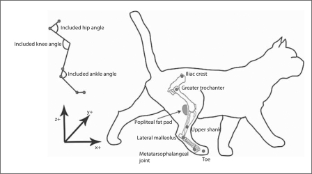
There were two conditions under which locomotion trials were collected: (1) control trials with an intact popliteal fat pad, and (2) after lipectomy. Kinematic data were collected for a minimum of two trials under each condition, where a trial consists of at least 30 s of continuous stepping and a minimum of 10 steps per trial at a treadmill speed of 0.7 m/s. Reflective markers to capture kinematics were placed at the following positions on the right leg: the iliac crest, greater trochanter, upper leg, lateral malleolus, metatarsophalangeal joint and toe (fig. 1). Data were collected using 6 Vicon cameras to record 3D marker locations from the cat at either 250 or 125 Hz to filter background noise. Video analysis of the experiment was used to manually demark events of paw contact and paw off. The location of the knee joint was calculated by projecting a vector from the lateral malleolus marker through the upper leg to the knee with the magnitude of the premeasured leg length. The marker trajectories were processed post-hoc in Matlab (Mathworks, Inc.) and with a low-pass 4th-order Butterworth filter at 6 Hz. The included angles for the hip, knee and ankle joints in the sagittal plane were calculated using custom-written Matlab code and oriented as shown in figure 1.
Fig. 1.
Anatomy of cat hindlimb and kinematic marker placement. The diagram on the right shows the placement of the popliteal fat pad as well as the locations of the kinematic markers. The axes demark the orientation of the markers in 3D space. The diagram on the top left depicts the included angles in relation to the placement of the kinematic makers.
The mean trajectories of the included hip, knee and ankle angles and angular accelerations were evaluated by averaging the first 10 steps of each trial in each condition (before or after lipectomy) together. The data points in each step were averaged into 50 bins (or 2% of the step cycle), which removed the length variability across steps but retained the variability of the trajectory in each individual step. Angular acceleration and velocity were calculated by taking the first and second time derivatives of the included angle, respectively. Specific time points throughout the step cycle were chosen for analysis: mid-stance (point 1), paw off (point 2), mid-swing (point 3) and peak deceleration at the end of the re-extension of the knee (point 4).
A two-way ANOVA test and a post-hoc Tukey test were performed using Statistica™ to determine if there was a significant change in the included angles and angular accelerations at the 4 chosen time points across all experiments. The two-way ANOVA was selected in order to test for a main effect of the disruption between animals.
Results
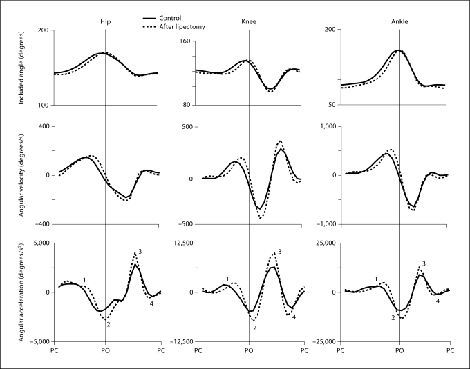
As is the case for the intact animal, the step cycle consisted of two distinct phases, based on the contact of the paw on the ground. The stance phase began immediately after paw contact and continued as the leg extended posteriorly and the paw was lifted from the ground (paw off). The swing phase began at paw off, usually when the limb was fully extended, and continued as the limb flexed and traveled forward until the paw contacted the ground again (paw contact). The included hip and ankle angles increased (extension) throughout stance, culminating in a maximum around paw off (fig. 2). In these experiments, ankle yield was minimal or absent due to the partial weight support offered by the tail clamp. The included angles at the hip and ankle then decreased (flexion) from paw off throughout swing, reaching a minimum around paw contact. The knee angle increased during stance, decreased during the first half of swing and then re-extended to paw contact. After removal of the popliteal fat pad, the trajectories of the included angles of the hip, knee and ankle did not show any substantial changes in range of motion (fig. 2). Further quantitative analysis of joint angles at paw contact and paw off did not reveal any significant changes following lipectomy, although the velocities and accelerations associated with these motions did show significant changes.
Fig. 2.
Included angles, velocities and angular accelerations recorded during level walking. The graphs show results from a single step of 1 intact limb cat, beginning and ending with paw contact (PC) and before and after lipectomy. PO = Paw off.
Included Angular Accelerations
Angular acceleration was quantified before and after lipectomy in order to evaluate damping properties of the popliteal fat. Angular velocities and accelerations for the hip, knee and ankle joints increased and decreased substantially throughout the step cycle, as shown in figure 2. All 3 joints accelerated in the extension direction during stance, achieved peak velocity at mid-stance and then decelerated toward paw off where maximum extension was achieved. After the reversal in direction from extension to flexion at paw off, the joint angles re-accelerated in the flexion direction and finally decelerated toward paw contact. For the knee, angular position exceeded the position at paw contact during swing and then re-extended toward paw contact. An additional peak of deceleration occurred at the knee at the end of this re-extension, and small corresponding peaks were observed for the hip and ankle. The maximum and minimum values of velocity and acceleration all increased following lipectomy, suggesting a decrease in damping of the system.
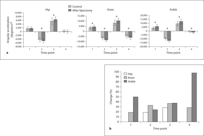
The peaks in the angular acceleration profiles occurred at approximately the same time before and after lipectomy. These peaks were measured in order to quantify and pool the results across animals, as shown in figure 3. The 4 time points corresponded approximately to mid-stance (point 1), paw off (point 2), mid-swing (point 3) and peak deceleration at the end of the re-extension of the knee (point 4). For all 3 joints, the largest accelerations occurred at time points 2 and 3, corresponding approximately to paw off and mid-swing. Following lipectomy, angular acceleration increased significantly for these time points. Substantial decelerations at time point 4 were observed for the knee alone, and lipectomy also had the effect of increasing this deceleration during the re-extension.
Fig. 3.
a Average (+SD) angular accelerations at the hip, knee and ankle joints, measured before and after lipectomy. Asterisks indicate significant differences. b Percentage changes in angular acceleration for joints and time points where the changes were significant.
The percentage changes in angular acceleration were also computed for each time point and joint where the changes were significant. The effects of lipectomy on angular acceleration were smaller for the hip than for the knee and ankle at time points 2 and 3. The average increases in angular acceleration for the 3 joints at these time points and at time point 4 for the knee ranged between approximately 20 and 40%.
Discussion
To determine the role of the popliteal fat pad in the hindlimb during level walking, the fat pad was removed between bouts of spontaneous stepping in the premammillary decerebrate cat. No significant changes were found in joint range of motion for the hip, knee and ankle joints. However, a significant increase in angular acceleration was observed at the included hip, knee and ankle angles, supporting the hypothesis that popliteal fat helps to control angular acceleration of the limb through damping.
Viscous Properties
The popliteal fat pad is a viscoelastic structure [Geerling et al., 2008] within the limb that can contribute to damping due to its mechanically parallel configuration with the muscular elements of the limb. The significant increases in angular acceleration of the included joint angles after lipectomy that were observed here constitute our evidence for contributions of popliteal fat to damping. Similarly, a study of the incompressible orbital fat surrounding the eye globe has shown that the limited room for expansion of the fat acts to decrease eye globe acceleration [Cirovic et al., 2005].
Intersegmental Effects
Accelerations of the joints during the swing phase may be particularly prone to interaction torques arising from the inertial properties of the limb segments. For example, extension of the knee may cause extension of the ankle [Prilutsky et al., 1996]. By providing damping to the muscular elements of the knee joint, the fat pad decreases the acceleration of the knee joint, consequently decreasing the interaction torque between the limb segments linked by the knee joint. This may be particularly important during the swing phase of the step cycle (time points 3 and 4) when the knee joint is flexed and the foot is not in contact with the ground, as inertial effects couple the actions for the knee joint and ankle joint. In fact, it was found that the greatest effect of the lipectomy to increase angular acceleration occurred at paw off and during swing.
Functional Considerations
It was noted in a recent report of a model of the feline hindlimb [Bunderson et al., 2008] that it was rather difficult to find muscular activation patterns that led to stable postures of the model limb, unless stiffness was increased by incorporating length feedback. The incorporation of additional damping might have also helped stabilize the limb. It is generally thought that muscles provide important sources of damping in the limb [Lin and Rymer, 2001]. The results from this study suggest that the popliteal fat pad also contributes to the damping of the limb through its ability to dissipate energy. Our results suggest that stability of model limbs may be improved by incorporating adipose tissue. In addition, stability might be improved further by the representation of fascia, a structure that provides mechanical coupling between joints [Stahl, 2010].
From these results, little increase in the range of motion of the hip, knee and ankle joints was observed following the removal of the popliteal fat pad. Due to the location of the incompressible fat behind the knee, the fat pad can only expand negligibly against the set of muscles behind the knee and slightly backwards towards the skin, thereby limiting the amount of deformation possible. This in turn may physically limit the extent to which the knee is able to flex. Therefore, although significant changes in range of movement during level walking were not observed, future studies will be important in determining the role of the fat pad in limiting the range of motion in situations requiring greater joint flexion, such as landing from jumps or crouching. Furthermore, the presence of excessive popliteal fat might lead to significant limitations of the range of knee motion.
Further understanding of the role of fat in the limb may contribute to the understanding of the effects of obesity on movement. The viscoelastic effect that the fat has on the knee decreases the propulsion provided to drive the limb through the step cycle. Consequently, an increased plantarflexion moment is seen at the ankle to increase propulsion in humans [Devita and Hortobagyi, 2003]. The popliteal fat pad in obese individuals results in overdamping of the limb, which results in greater energy dissipation when the fat pad is compressed. This increase in expenditure of energy was reported in a study comparing walking between obese and non-obese individuals [Browning et al., 2009]. The results showed that obese individuals pay a higher metabolic cost during walking.
Conclusion
The popliteal fat pad has a significant role in damping the angular accelerations of the hindlimb due to its anatomical location behind the knee and due to the viscoelastic properties of adipose tissue. The results from this experiment may be applied to models of the hindlimb and knowledge of the effects of obesity on movement. Further investigations should be done to examine the effects of popliteal fat in non-level walking and running.
Acknowledgements
The authors would like to thank Ramaldo Martin, Irrum Niazi and Chris Tuthill for their assistance during experiments and their input in results analysis. This project was funded by National Institutes of Health/National Institute of Child Health and Human Development grant 32571, and by financial support from the Georgia Institute of Technology.
References
- Browning R.C., McGowan C.P., Kram R. Obesity does not increase external mechanical work per kilogram body mass during walking. J Biomech. 2009;42:2273–2278. doi: 10.1016/j.jbiomech.2009.06.046. [DOI] [PMC free article] [PubMed] [Google Scholar]
- Bunderson N.E., Burkholder T.J., Ting L.H. Reduction of neuromuscular redundancy for postural force generation using an intrinsic stability criterion. J Biomech. 2008;41:1537–1544. doi: 10.1016/j.jbiomech.2008.02.004. [DOI] [PMC free article] [PubMed] [Google Scholar]
- Cirovic S., Bhola R.M., Hoses D.R., Howard I.C., Lawfords P.V., Parsons M.A. A computational study of the passive mechanisms of eye restraint during head impact trauma. Comput Methods Biomech Biomed Engin. 2005;8:1–6. doi: 10.1080/10255840500062989. [DOI] [PubMed] [Google Scholar]
- Crouch J.E. Text-Atlas of Cat Anatomy. Philadelphia: Lea and Febiger; 1969. [Google Scholar]
- Devita P., Hortobagyi T. Obesity is not associated with increased knee joint torque and power during level walking. J Biomech. 2003;36:1355–1362. doi: 10.1016/s0021-9290(03)00119-2. [DOI] [PubMed] [Google Scholar]
- Geerling M., Gerrit W.M.P., Ackerman P.A.J., Oomens C.W.J., Baajens F.P.T. Linear viscoelastic behavior of subcutaneous adipose tissue. Biorheology. 2008;45:677–688. [PubMed] [Google Scholar]
- Lin D.C., Rymer W.Z. Mechanical properties of cat soleus muscle elicited by sequential ramp stretches: implications for control of muscle. J Neurophysiol. 1993;70:997–1008. doi: 10.1152/jn.1993.70.3.997. [DOI] [PubMed] [Google Scholar]
- Lin D.C., Rymer W.Z. Damping actions of the neuromuscular system with inertial loads: human flexor pollicis longus muscle. J Neurophysiol. 2001;85:1059–1066. doi: 10.1152/jn.2001.85.3.1059. [DOI] [PubMed] [Google Scholar]
- McMahon T.A. Muscles, Reflexes, and Locomotion. Princeton: Princeton University Press; 1984. [Google Scholar]
- Prilutsky B.I., Herzog W., Leonard T. Transfer of mechanical energy between ankle and knee joints by gastrocnemius and plantaris muscles during cat locomotion. J Biomech. 1996;29:391–403. doi: 10.1016/0021-9290(95)00054-2. [DOI] [PubMed] [Google Scholar]
- Stahl, V.A. (2010) A Biomechanical Analysis of the Crural Fascia in the Cat Hindlimb; doctoral dissertation, Georgia Institute of Technology, Atlanta.
- Weisburg S.P., McCann D., Desai M., Rosenbaum M., Leibel R., Ferrante A.W. Obesity is associated with macrophage accumulation in adipose tissue. J Clin Invest. 2003;112:1796–1808. doi: 10.1172/JCI19246. [DOI] [PMC free article] [PubMed] [Google Scholar]
- Whalen P.J. Control of locomotion in the decerebrate cat. Prog Neurobiol. 1996;49:481–515. doi: 10.1016/0301-0082(96)00028-7. [DOI] [PubMed] [Google Scholar]


