Abstract
The US04 trial was a multicenter, open-label, single arm trial of deferasirox monotherapy (30–40 mg/kg/day) for 18 months. Cardiac iron response was bimodal with improvements observed in patients with mild to moderate initial somatic iron stores; relationship of cardiac response to labile plasma iron is now presented. Labile plasma iron was measured at baseline, six months, and 12 months. In patients having a favorable cardiac response at 18 months, initial labile plasma iron was elevated in only 31% of patients at baseline and no patient at six or 12 months. Cardiac non-responders had elevated labile plasma iron in 50% of patients at baseline, 50% patients at six months, and 38% of patients at 12 months. Risk of abnormal labile plasma iron and cardiac response increased with initial liver iron concentration. Persistently increased labile plasma iron predicts cardiac non-response to deferasirox but labile plasma iron suppression does not guarantee favorable cardiac outcome. Study registered at www.clinicaltrials.gov (NCT00447694).
Keywords: labile plasma iron, non transferrin bound iron, cardiac T2*, chelation, deferasirox, heart, MRI, siderosis, thalassemia
Introduction
Despite steady advances in iron chelation, iron-mediated cardiotoxicity remains a leading problem in thalassemia major. Cardiac iron loading occurs through non-specific transport of non-transferrin bound iron species.1 Labile plasma iron (LPI) represents a redox active form of non-transferrin bound iron that is chelatable, making it potentially available for transport into extrahepatic tissues. LPI can be accurately and reproducibly assayed by fluorescent methods.2,3 Chronic control of circulating LPI is an important goal for iron chelation therapy in order to prevent oxidative damage4,5 and to lower the risk of extrahepatic organ dysfunction6. Intuitively, serial LPI measurements may provide more information regarding response of extrahepatic iron stores than measurements of serum ferritin or liver iron,7 although LPI levels might more closely reflect efficacy of primary prevention than removal of established iron stores.
In April 2010, we reported the cardiac efficacy of deferasirox monotherapy for 18 months in heavily iron overloaded thalassemia major patients.8 In that study, initial liver iron concentration (LIC) and serum ferritin strongly predicted cardiac iron response to therapy. We suggested initial iron stores were most likely surrogates for drug compliance as well as surrogates for increased LPI. Here, we report labile plasma iron results from this trial (CICL670AUS04) and their relationship to cardiac and liver iron stores.
Design and Methods
The US04 trial was a multicenter, open-label, single arm trial of deferasirox monotherapy (30–40 mg/kg/day) for 18 months in 28 subjects (NCT00447694). The study was approved by the Institutional Review Board at all participating institutions. All patients were required to have MRI evidence of cardiac iron (T2* <20 ms) with normal cardiac function at baseline. Liver iron, cardiac T2*, and cardiac function were measured at baseline, six months, 12 months, and 18 months; serum ferritin was measured at each transfusion visit. Favorable cardiac response to iron chelation therapy was an improvement in cardiac T2* of more than 14.7% (the 95% confidence interval for a single MRI measurement) at 18 months; all patients who failed to complete the trial were scored as non-responders. Complete details of methods used may be found elsewhere.8
Serum labile plasma iron (LPI) was assessed at baseline, 25 weeks, and 49 weeks, using a central laboratory (Afferix Limited, Rehovot, Israel); studies were part of the original study design and not a retrospective analysis. Samples were drawn in the morning and iron chelation was held until after the blood draw. Upper limit of normal for the LPI assay was 0.5 umol/L, with a lower limit of detection of 0.1 umol/L. The ability of initial LIC to predict increased LPI and cardiac response was assessed by logistic regression and receiver operator characteristic (ROC) analysis. All statistics were performed in JMP5.1 (SAS, Cary, NC, USA).
Results and Discussion
The patient population was severely iron-loaded at baseline with a mean LIC of 20.3±3.0, serum ferritin 4417±669 ng/mL (394–16,249) and cardiac T2* (geometric mean) of 8.6±1.1 ms (1.8–16.1). Twenty-two patients completed the trial. Cardiac response to iron chelation was bimodal, determined primarily by initial iron burden. Responders had initial LICs of 10.5±5.8 mg/g dry weight compared with 29.2±4.5 mg/g dry weight in non-responders (P<0.0001).
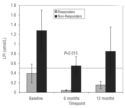
LPI was successfully obtained in 24 patients at baseline, 23 patients at six months, and 20 patients at 12 months. A summary of data is shown in Figure 1. In cardiac responders, LPI was 0.39±0.70 umol/L at baseline, declined to 0.04±0.12 umol/L at six months and remained low at 12 months; all responders had normal LPI levels (<0.5 umol/L) at six and 12 months. These changes were not statistically significant (P=0.09 and 0.58, respectively) because initial LPI was already in the normal range in 9 of 13 responders at baseline. In non-responders, baseline LPI was within the normal range in 6 of 12 subjects. Average LPI levels were higher than for responders at all three time points but only achieved statistical significance at six months (P=0.08, 0.015, and 0.21 respectively). However, LPI remained outside the normal range in 43% of non-responders at six and 12 months compared with 0% of the responders.
Figure 1.
Labile plasma iron at baseline, six months and 12 months of deferasirox therapy. Patients with favorable and unfavorable cardiac response at 18 months of therapy are displayed separately. Horizontal line at 0.5 umol/L represents the upper limit of normal for LPI.
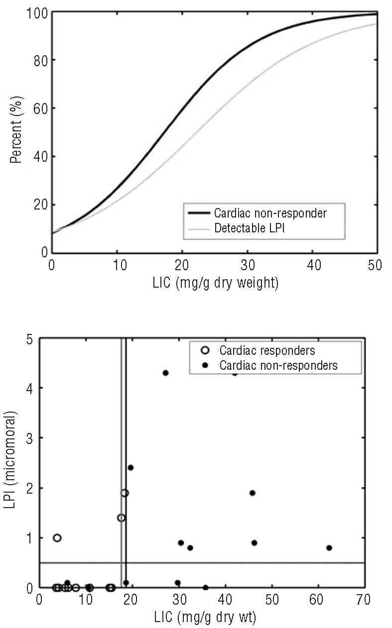
Using logistic regression, the risk of detectable LPI and poor cardiac response exhibited graded risk with LIC (Figure 2A), P<0.002, rising sharply as LIC exceeded 10 mg/g. Figure 2B shows LPI levels (vertical axis) and cardiac response (open and closed symbols) as functions of initial LIC. Vertical lines depict the discriminators calculated from ROC analysis (see below).
Figure 2.

Percentage of patients with increased LPI (LPI > 0.5 uMol, light line) or who were cardiac non-responders at 18 months (dark line) as a function of baseline liver iron concentration (LIC). Plot of labile plasma iron versus LIC. Solid symbols represent patients who did not respond or did not complete 18 months of deferasirox therapy. Open symbols represent patients whose cardiac T2* improved more than 14.7% from baseline after 18 months of deferasirox at 30–40 mg/kg/day. Horizontal line reflects LPI upper limit of normal (0.5 uMol). Vertical lines represent optimal cut offs generated by ROC analysis for detectable LPI (gray line) and cardiac response (black line).
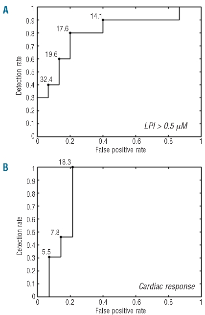
By receiver operator characteristic curve (ROC) analysis, initial LIC was a strong predictor of abnormal LPI and cardiac response, with an area under the ROC curve of 0.83 and 0.84, respectively (Figure 3). As shown in Figure 3A, the risk of detectable LPI increases sharply for LIC greater than 14.1 mg/g with an optimal LIC discriminator (best balance of sensitivity and specificity) of 17.6 mg/g. As shown in Figure 3B, all patients with liver iron greater than 18.3 did not reduce their cardiac iron over 18 months. One patient with an LIC less than 18.3 also failed to respond and 2 others did not complete the trial, accounting for the imperfect specificity of LIC in predicting response. To put this in perspective, the AUROCs for change in liver iron or ferritin over 18 months were 0.87 and 0.85, respectively, but the 5 patients who failed to complete the trial were excluded (improving prediction). Similarly, using 18-month LIC as a predictor yielded an AUROC of 0.94 with an optimal cut off of 17 mg/g dry weight.
Figure 3.
Receiver Operator Characteristic (ROC) (A) curve for prediction of elevated LPI by liver iron concentration (LIC). Curve represents the trade off between sensitivity and 1-specificity; numbers alongside the curve represent LIC values at critical points. (B) Same representation for the probability of favorable 18 month cardiac response to deferasirox.
Since extrahepatic iron loading occurs through unregulated transport of non-transferrin bound iron species, labile plasma iron measurements are potentially attractive metrics for iron chelation therapy.1,5,7,9 In our study, elevated LPI levels at six and twelve months were only observed in non-responders (43% of non-responders) making them a reasonable negative prognostic marker. However, LPI suppression did not guarantee cardiac response. In fact, 40% of the patients who had clinically significant cardiac T2* worsening on deferasirox had undetectable LPI for all three measurements, making it dangerous to rely exclusively on these measurements. The most likely explanation is that LPI measurements are short-term surrogates of chelation compliance and cardiac risk, having a time-scale of days. Thus even a generally non-compliant patient may have acceptable serum LPI values during check-ups if he or she remembers to take their deferasirox the day before the medical appointment. In contrast, LIC measurements reflect a longer horizon of chelation compliance. They also convey risk for elevated LPI for all the other days of the year when a chelator is not being taken.7,10–13
LPI measurements also have several other limitations. Patients must withhold chelation on the morning of the exam; this is not always easy to achieve in a busy clinical practice. Plasma must be quickly separated and shipped on dry ice to Israel. There is also no universal agreement among techniques to assay labile iron and no “optimal” method.14 However, promising new methods are under development that may overcome some of these challenges.
Although others have described the sharp increase in LPI with LIC, we can only speculate about the mechanisms.7,12 The liver is the predominant sink for circulating LPI and the source of the iron counter regulatory hormone, hepcidin. Liver damage, including increased transaminases and fibrosis, increase sharply for hepatic iron levels in the 15–20 mg/g range.15 We hypothesize that decreased hepatic scavenging of LPI and impaired hepcidin production, produced by iron toxicity to hepatocytes, may represent the causal link between cardiac risk and severe hepatic iron overload.11,13,16 However, two caveats must be attached to this model. First, significant LPI was present in a few patients having lower LICs, consistent with development of cardiac iron overload in some patients.17,18 Secondly, development of cardiac iron represents a combination of intrinsic risk and chelation use,11,19 making compliance critically important.19 Carefully controlled studies will need to be performed to test these hypotheses.
In summary, chelation compliance is the strongest predictor of cardiac response to iron chelation,19 but penalties for non-compliance appear to increase in patients with severe hepatic siderosis11,12 through chronic exposure to increased labile plasma iron. The relationships between increased LIC, LPI, and cardiac risk are graded, but appear to increase dramatically as liver iron exceeds 10 mg/g, consistent with prior observations.11,13,16 Persistent elevations of LIC and/or LPI appear to have negative prognostic value but neither a declining LIC nor suppression of LPI guarantees a favorable cardiac response to chelation therapy.8,17 Hence, vigilance for extrahepatic iron deposition is necessary, even in patients with apparently adequate control of somatic iron stores.
Acknowledgments
we are indebted to Anne Nord, RN, Susan Carson, PNP, Kelly Verel, Dena Fasheh, Elizabeth Evans, Jacqueline Madden, PNP, Matthew Cham, MD, and Matthew Herz for their help in patient recruitment and management. We also appreciate the help of Kevin Mennitt, MD, Barbara Bettencourt, and Cindy Rigsby, MD, for their assistance in MRI measurements as well as Ghulam Warsi, PhD, for his statistical assistance.
Footnotes
Funding: this study was supported by research funding from Novartis Pharma AG, Basel, Switzerland.
Authorship and Disclosures
The information provided by the authors about contributions from persons listed as authors and in acknowledgments is available with the full text of this paper at www.haematologica.org.
Financial and other disclosures provided by the authors using the ICMJE (www.icmje.org) Uniform Format for Disclosure of Competing Interests are also available at www.haematologica.org.
References
- 1.Oudit GY, Trivieri MG, Khaper N, Liu PP, Backx PH. Role of L-type Ca2+ channels in iron transport and iron-overload cardiomyopathy. J Mol Med. 2006;84(5):349–64. doi: 10.1007/s00109-005-0029-x. [DOI] [PMC free article] [PubMed] [Google Scholar]
- 2.Esposito BP, Breuer W, Sirankapracha P, Pootrakul P, Hershko C, Cabantchik ZI. Labile plasma iron in iron overload: redox activity and susceptibility to chelation. Blood. 2003;102(7):2670–7. doi: 10.1182/blood-2003-03-0807. [DOI] [PubMed] [Google Scholar]
- 3.Zanninelli G, Breuer W, Cabantchik ZI. Daily labile plasma iron as an indicator of chelator activity in Thalassaemia major patients. Br J Haematol. 2009;147(5):744–51. doi: 10.1111/j.1365-2141.2009.07907.x. [DOI] [PubMed] [Google Scholar]
- 4.Ghoti H, Fibach E, Merkel D, Perez-Avraham G, Grisariu S, Rachmilewitz EA. Changes in parameters of oxidative stress and free iron biomarkers during treatment with deferasirox in iron-overloaded patients with myelodysplastic syndromes. Haematologica. 2010;95(8):1433–4. doi: 10.3324/haematol.2010.024992. [DOI] [PMC free article] [PubMed] [Google Scholar]
- 5.Cabantchik ZI, Breuer W, Zanninelli G, Cianciulli P. LPI-labile plasma iron in iron overload. Best Pract Res Clin Haematol. 2005;18(2):277–87. doi: 10.1016/j.beha.2004.10.003. [DOI] [PubMed] [Google Scholar]
- 6.Koren A, Fink D, Admoni O, Tennenbaum-Rakover Y, Levin C. Non-transferrin-bound labile plasma iron and iron overload in sickle-cell disease: a comparative study between sickle-cell disease and beta-thalassemic patients. Eur J Haematol. 2010;84 (1):72–8. doi: 10.1111/j.1600-0609.2009.01342.x. [DOI] [PubMed] [Google Scholar]
- 7.Daar S, Pathare A, Nick H, Kriemler-Krahn U, Hmissi A, Habr D, et al. Reduction in labile plasma iron during treatment with deferasirox, a once-daily oral iron chelator, in heavily iron-overloaded patients with beta-thalassaemia. Eur J Haematol. 2009;82 (6):454–7. doi: 10.1111/j.1600-0609.2008.01204.x. [DOI] [PMC free article] [PubMed] [Google Scholar]
- 8.Wood JC, Kang BP, Thompson A, Giardina P, Harmatz P, Glynos T, et al. The effect of deferasirox on cardiac iron in thalassemia major: impact of total body iron stores. Blood. 2010;116(4):537–43. doi: 10.1182/blood-2009-11-250308. [DOI] [PubMed] [Google Scholar]
- 9.Piga A, Longo F, Duca L, Roggero S, Vinciguerra T, Calabrese R, et al. High non-transferrin bound iron levels and heart disease in thalassemia major. Am J Hematol. 2009;84(1):29–33. doi: 10.1002/ajh.21317. [DOI] [PubMed] [Google Scholar]
- 10.Angelucci E, Brittenham GM, McLaren CE, Ripalti M, Baronciani D, Giardini C, et al. Hepatic iron concentration and total body iron stores in thalassemia major. N Engl J Med. 2000;343(5):327–31. doi: 10.1056/NEJM200008033430503. [DOI] [PubMed] [Google Scholar]
- 11.Brittenham GM, Griffith PM, Nienhuis AW, McLaren CE, Young NS, Tucker EE, et al. Efficacy of deferoxamine in preventing complications of iron overload in patients with thalassemia major. N Engl J Med. 1994;331(9):567–73. doi: 10.1056/NEJM199409013310902. [DOI] [PubMed] [Google Scholar]
- 12.Jensen PD, Jensen FT, Christensen T, Eiskjaer H, Baandrup U, Nielsen JL. Evaluation of myocardial iron by magnetic resonance imaging during iron chelation therapy with deferrioxamine: indication of close relation between myocardial iron content and chelatable iron pool. Blood. 2003;101(11):4632–9. doi: 10.1182/blood-2002-09-2754. [DOI] [PubMed] [Google Scholar]
- 13.Telfer PT, Prestcott E, Holden S, Walker M, Hoffbrand AV, Wonke B. Hepatic iron concentration combined with long-term monitoring of serum ferritin to predict complications of iron overload in thalassaemia major. Br J Haematol. 2000;110(4):971–7. doi: 10.1046/j.1365-2141.2000.02298.x. [DOI] [PubMed] [Google Scholar]
- 14.Jacobs EM, Hendriks JC, van Tits BL, Evans PJ, Breuer W, Liu DY, et al. Results of an international round robin for the quantification of serum non-transferrin-bound iron: Need for defining standardization and a clinically relevant isoform. Anal Biochem. 2005;341(2):241–50. doi: 10.1016/j.ab.2005.03.008. [DOI] [PubMed] [Google Scholar]
- 15.Angelucci E, Muretto P, Nicolucci A, Baronciani D, Erer B, Gaziev J, et al. Effects of iron overload and hepatitis C virus positivity in determining progression of liver fibrosis in thalassemia following bone mar-row transplantation. Blood. 2002;100(1):17–21. doi: 10.1182/blood.v100.1.17. [DOI] [PubMed] [Google Scholar]
- 16.Olivieri NF, Nathan DG, MacMillan JH, Wayne AS, Liu PP, McGee A, et al. Survival in medically treated patients with homozygous beta-thalassemia. N Engl J Med. 1994;331(9):574–8. doi: 10.1056/NEJM199409013310903. [DOI] [PubMed] [Google Scholar]
- 17.Noetzli LJ, Carson SM, Nord AS, Coates TD, Wood JC. Longitudinal analysis of heart and liver iron in thalassemia major. Blood. 2008;112(7):2973–8. doi: 10.1182/blood-2008-04-148767. [DOI] [PMC free article] [PubMed] [Google Scholar]
- 18.Anderson LJ, Westwood MA, Prescott E, Walker JM, Pennell DJ, Wonke B. Development of thalassaemic iron overload cardiomyopathy despite low liver iron levels and meticulous compliance to desferrioxamine. Acta Haematol. 2006;115(1–2):106–8. doi: 10.1159/000089475. [DOI] [PubMed] [Google Scholar]
- 19.Gabutti V, Piga A. Results of long-term iron-chelating therapy. Acta Haematol. 1996;95(1):26–36. doi: 10.1159/000203853. [DOI] [PubMed] [Google Scholar]


