Abstract
Post-transplantation lymphoproliferative disorder (PTLD) with plasmacellular differentiation has been reported as a rare subtype of monomorphic B-cell post-transplant lympho-proliferation with histological and immunophenotypical features of plasmacytoma in the non-transplant population. Here we present clinical, laboratory and histopathological features, treatment and outcome of 8 patients from the German prospective PTLD registry. Clinically, extranodal manifestations were common while osteolytic lesions were rare and none of the patients had bone marrow involvement. Immunohistochemistry showed light chain restriction and expression of CD138 without CD20 expression in all samples. An association with Epstein-Barr virus was found in 3 out of 8 cases. We suggest that the Ann Arbor classification is most useful for this disease entity and report a generally good response to treatment including reduction of immuno-suppression, surgery and irradiation in localized disease and systemic chemotherapy analogous to plasmacell myeloma in advanced disease.
Keywords: plasmacytoma, plasmacell lymphoma, post-transplant lymphoproliferative disorder, CD138
Introduction
Post-transplantation lymphoproliferative disorder (PTLD), a spectrum of lymphatic diseases associated with the use of potent immunosuppressive drugs after transplantation,1 represents one of the most common neoplastic diseases following solid organ transplantation.2 Patients with PTLD commonly present with stage III or IV disease of varying histology, frequently involving extranodal sites.3–10 Organ dysfunction, especially renal function impairment, is also a common feature at presentation. Initial therapy in PTLD is immuno-suppression reduction (IR). This can result in complete remission (CR)11,12 but overall response rates are low.13 Additionally, IR is known to be associated with a high risk of graft rejection and graft loss. A rejection rate of 37% has recently been reported in a first prospective trial systematically evaluating IR in PTLD.13 Common subsequent therapies in patients failing to respond to upfront IR are rituximab monotherapy in CD20-positive B-cell PTLD,6–8 CHOP-like chemotherapy14 and a combination of rituximab and CHOP, either synchronous or in sequence. Both options are highly effective in the treatment of PTLD after an initial failure of upfront IR. Overall response rates of about 40–60% for single agent rituximab6–9 and up to 90% for sequential treatment with 4 courses of rituximab followed by 4 cycles of CHOP have been reported9 leading to an improved overall survival. Thus, significant progress has been made in the treatment of CD20-positive B-cell PTLD. However, PTLD is not a homogeneous entity, neither in its clinical presentation nor in its pathological appearance. While the majority of monomorphic PTLD lesions resemble non-Hodgkin’s lymphoma, specifically diffuse large B-cell lymphoma, rare forms have been identified, one of which is PTLD with plasmacellular differentiation. Plasma cell rich infiltrates in transplant recipients can be found in a spectrum of lesions comprising acute rejection, early PTLD lesions also known as plasmacytic hyperplasia and plasma cell rich polymorphic lymphoproliferations.15 These plasma cells usually show neither light chain restriction nor clonal proliferation. In contrast, PTLD with plasmacellular differentiation has been reported as a rare monomorphic B-cell PTLD subtype with a histomorphology comparable to plasmacytoma in the non-transplant population, clonal immunoglobulin gene rearrangements and light chain restriction.16 So far, only case reports and one retrospective series of 4 cases have been published on this disease entity.17 Here, we present the first prospective case series, including clinicopathological features, treatment and treatment outcome.
Design and Methods
To assess the clinical features, treatment options and outcome of rare PTLD subtypes, a prospective PTLD registry was set up in Germany in 2006. Here we present our data on patients diagnosed with plasmacytoma-like monomorphic PTLD from 2006 to 2010. Follow-up data was reviewed for all patients up to December 2010. Tumor response to treatment was defined according to the World Health Organization criteria. In plasmacytoma-like PTLD definition of complete remission (CR) additionally required the absence of monoclonal gammopathy. Progression-free survival (PFS) was defined from start of therapy to disease progression or to death from any cause, while overall survival (OS) was defined from diagnosis of PTLD to death from any cause. Clinical data on the patients in the registry was collected before, during and at least at four weeks, six, 12 and 24 months after treatment. Additional data on patients’ characteristics were retrieved from databases at the different transplant centers. The responsible local ethics committee approved the trial and patients gave written informed consent for non-anonymous documentation according to the Declaration of Helsinki.
The diagnosis of PTLD was based on the examination of histological material, obtained either by open biopsy or core needle biopsy. Diagnostic tissue samples were subsequently reviewed by a single expert pathologist (IA) and classified morphologically according to the WHO classification (2008). An association with the Epstein-Barr virus was confirmed by immunohistochemical staining for the latent membrane protein 1, Epstein-Barr nuclear antigen 2, Epstein-Barr ZEBRA antigen and/or by in situ hybridization for EBER-transcript detection. The extent of existing disease was determined through a complete patient history, physical examination, laboratory investigations (including full blood count, lactate dehydrogenase [LDH, upper limit 240U/l]), renal and liver function tests, as well as determination of the EBV DNA load in peripheral blood), bone marrow biopsy and computed tomography (CT) scans of the chest, abdomen and pelvis. All patients diagnosed with plasmacytoma-like PTLD had further blood tests to detect monoclonal gammopathy and an osteo-CT or osteo-MRI scan to detect local bone destruction. Patients for whom IR had provided no benefit received subsequent treatment as chosen by the treating physician. Treatment protocols included local irradiation and surgery for localized disease and systemic chemotherapy for disseminated PTLD.
Results and Discussion
By the end of 2010, 182 patients were reported to the German PTLD registry D2006-2010 from which 8 (4%) had a diagnosis of monomorphic plasmacytoma-like PTLD (6 male, 2 female). They had previously undergone solid organ transplantation of kidney (n=3), lung (n=1), liver (n=1), heart (n=2) or small intestine (n=1). The median age at diagnosis was 55 years (range 25–73). All patients received immunosuppressive treatment at the time of their PTLD diagnosis (Table 1A). In accordance with previously published data,17 most cases were late-onset PTLD with a median time from transplant to diagnosis of PTLD of 8.3 years (range 3 months to 26 years). Only 2 cases were diagnosed within the first year after transplantation while 4 cases were diagnosed more than ten years after transplantation. There was no obvious association with an underlying disease (Table 1A).
Table 1A.
Baseline characteristics.
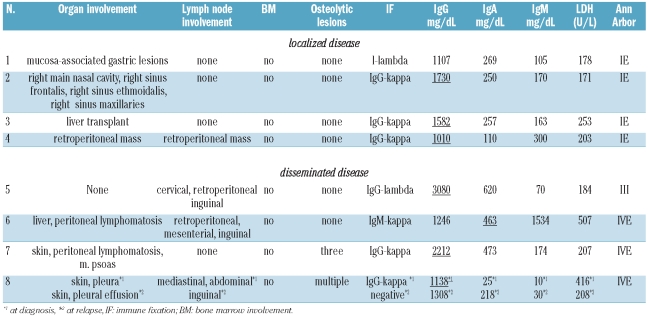
All 4 cases with localized disease at diagnosis presented with exclusively extranodal manifestations. Out of the 4 patients with disseminated disease, one had only extranodal and another only nodal manifestations, while 2 patients had both. Of note, osteolytic lesions were rare (2 of 8) and none of the patients had bone marrow involvement (Table 2). This is in contrast to the 3 patients diagnosed with monomorphic multiple myeloma-like PTLD described by Sun et al. who all presented with osteolytic lesions and bone marrow involvement but without nodal or extranodal disease.18
Table 2.
Clinical presentation.
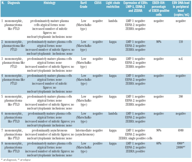
The neoplastic plasma cell population was well-differentiated (Marschalko-type) in 7 of 8 cases and showed either lambda (2 of 8) or kappa (6 of 8) light chain restriction and positivity for CD138 (8 of 8), while CD20 was negative in all and CD56 in 7 of 8 cases. A paraprotein could be detected in all cases, but overall serum immunoglobulin levels were low compared to plasmacell myeloma (Table 3). An association with latent EBV infection was verified by EBER-ISH in 3 of 8 cases (Table 1B). EBV-associated plasmacytoma-like PTLD showed no expression of LMP-1 or EBNA-2 proteins, while the ZEBRA protein indicative for a transition from the lytic to the latent infection cycle was expressed in 2 of 3 cases. All 3 cases of EBV-associated PTLD showed remarkably elevated EBV DNA loads in their peripheral blood, while non-EBV associated cases often had no detectable blood levels of EBV DNA (Table 1B).
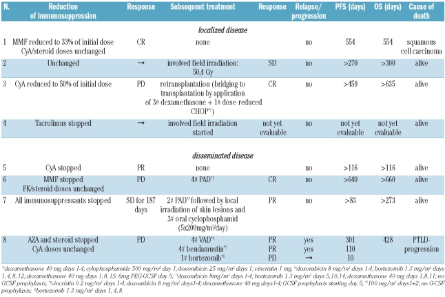
Table 3.
Treatment.
Table 1B.
Baseline characteristics.
Out of the 6 patients who received a reduction of immunosuppression as the initial therapeutic intervention, 2 responded while 4 patients showed progressive disease. All 3 evaluable patients with localized disease achieved sustained lymphoma control (including CR after IR and CR after surgery). The 3 patients with disseminated disease and PD after IR received systemic chemotherapy analogous to plasmacell myeloma (Table 3) which was remarkably well tolerated with supportive treatment including GCSF. All 3 responded to treatment (including one CR after PAD). None of the patients received rituximab, as staining for CD20 was negative in all cases.
The group of 8 adult patients presented here included different types of solid organ transplant recipients under immunosuppressive therapy. The small number of patients does not allow any conclusions to be drawn as to whether either transplantation of a particular organ or a specific immunosuppressive drug is linked to the development of plasmacytoma-like PTLD. It is likely that the total amount of immunosuppression plays an important role, as it does in other PTLD.19
Like other monomorphic PTLD subtypes, EBV-association is present in half of the lymphomas.9 This is in contrast to classical plasmacytoma in non-transplant patients in whom EBV is rarely present. The fact that LMP-1 and EBNA-2 expression was negative in all cases underscores the importance of EBER in situ hybridization. With positive PCR results for EBV DNA in peripheral blood in all patients with EBV-associated plasmacytoma-like PTLD, monitoring of EBV copy numbers might provide a potential further method for monitoring treatment in these patients.20–23 However, a careful interpretation of EBV loads is necessary.24
As bone marrow involvement and lytic bone lesions are rare, and most patients suffer from impaired renal function due to their underlying disease or the use of calcineurin inhibitors even before the diagnosis of plasmacytoma-like PTLD, neither the Durie and Salmon classification nor the ISS classification seem appropriate to predict prognosis. The presentation with mass lesions and successful therapy of localized disease with surgery and irradiation make the Ann Arbor classification appear most useful. In this respect, plasmacytoma-like PTLD behaves similarly to extramedullary plasmacytoma in immunocompetent patients.
In conclusion, patients with plasmacytoma-like PTLD in this case series had a relatively good treatment outcome, with only one patient dying from progressive disease. As in the case of conventional PTLD, reduction of immunosuppression was an effective and potentially curative therapy in 2 out of 7 patients11,12 and patients with localized disease responded well to localized treatment such as surgery and irradiation. In more advanced disease, anti-plasma cell chemotherapy including treatment with the proteasome inhibitor bortezomib could induce disease remission.
Footnotes
Funding: the PTLD D 2006-2012 registry is supported by grants from AMGEN, CLS Behring, Mundipharma GmbH and Roche Pharma AG. The German Study Group on PTLD (DPTLDSG) is a member of the German Competence Network Malignant Lymphomas (KML).
Authorship and Disclosures
The information provided by the authors about contributions from persons listed as authors and in acknowledgments is available with the full text of this paper at www.haematologica.org.
Financial and other disclosures provided by the authors using the ICMJE (www.icmje.org) Uniform Format for Disclosure of Competing Interests are also available at www.haematologica.org.
References
- 1.Harris NL, Ferry JA, Swerdlow SH. Posttransplant lymphoproliferative disorders: summary of Society for Hematopathology Workshop. Semin Diagn Pathol. 1997;14(1):8–14. [PubMed] [Google Scholar]
- 2.Penn I, Hammond W, Brettschneider L, Starzl TE. Malignant lymphomas in transplantation patients. Transplant Proc. 1969;1:106–12. [PMC free article] [PubMed] [Google Scholar]
- 3.Dodd GD, 3rd, Greenler DP, Confer SR. Thoracic and abdominal manifestations of lymphoma occurring in the immunocompromised patient. Radiol Clin North Am. 1992;30(3):597–610. [PubMed] [Google Scholar]
- 4.Pickhardt PJ, Siegel MJ. Posttransplantation lymphoproliferative disorder of the abdomen: CT evaluation in 51 patients. Radiology. 1999;213(1):73–8. doi: 10.1148/radiology.213.1.r99oc2173. [DOI] [PubMed] [Google Scholar]
- 5.Leblond V, Dhedin N, Mamzer Bruneel MF, Choquet S, Hermine O, Porcher R, et al. Identification of prognostic factors in 61 patients with posttransplantation lymphoproliferative disorders. J Clin Oncol. 2001;19(3):772–8. doi: 10.1200/JCO.2001.19.3.772. [DOI] [PubMed] [Google Scholar]
- 6.Oertel SH, Verschuuren E, Reinke P, Zeidler K, Papp-Vary M, Babel N, et al. Effect of Anti-CD 20 Antibody Rituximab in Patients with Post-Transplant Lymphoproliferative Disorder (PTLD) Am J Transplant. 2005;5 (12):2901–6. doi: 10.1111/j.1600-6143.2005.01098.x. [DOI] [PubMed] [Google Scholar]
- 7.Choquet S, Leblond V, Herbrecht R, Socie G, Stoppa AM, Vandenberghe P, et al. Efficacy and safety of rituximab in B-cell post-transplant lymphoproliferative disorders: results of a prospective multicentre phase II study. Blood. 2006;107:3053–7. doi: 10.1182/blood-2005-01-0377. [DOI] [PubMed] [Google Scholar]
- 8.Gonzalez-Barca E, Domingo-Domenech E, Capote FJ, Gomez-Codina J, Salar A, Bailen A, et al. Prospective phase II trial of extended treatment with rituximab in patients with B-cell post-transplant lymphoproliferative disease. Haematologica. 2007;92(11):1489–94. doi: 10.3324/haematol.11360. [DOI] [PubMed] [Google Scholar]
- 9.Trappe R, Choquet S, Oertel SHK, Leblond V, Dierickx D, Mollee P, et al. Sequential Treatment with Rituximab and CHOP Chemotherapy in B-Cell PTLD - Moving Forward to a First Standard of Care: Results From a Prospective International Multicenter Trial. Blood. 2009;114(22):100. [Google Scholar]
- 10.Evens AM, David KA, Helenowski I, Nelson B, Kaufman D, Kircher SM, et al. Multicenter analysis of 80 solid organ trans-plantation recipients with post-transplantation lymphoproliferative disease: outcomes and prognostic factors in the modern era. J Clin Oncol. 28(6):1038–46. doi: 10.1200/JCO.2009.25.4961. [DOI] [PMC free article] [PubMed] [Google Scholar]
- 11.Tsai DE, Hardy CL, Tomaszewski JE, Kotloff RM, Oltoff KM, Somer BG, et al. Reduction in immunosuppression as initial therapy for posttransplant lymphoproliferative disorder: analysis of prognostic variables and long-term follow-up of 42 adult patients. Transplantation. 2001;71(8):1076–88. doi: 10.1097/00007890-200104270-00012. [DOI] [PubMed] [Google Scholar]
- 12.Starzl TE, Nalesnik MA, Porter KA, Ho M, Iwatsuki S, Griffith BP, et al. Reversibility of lymphomas and lymphoproliferative lesions developing under cyclosporin-steroid therapy. Lancet. 1984;1(8377):583–7. doi: 10.1016/s0140-6736(84)90994-2. [DOI] [PMC free article] [PubMed] [Google Scholar]
- 13.Swinnen LJ, LeBlanc M, Grogan TM, Gordon LI, Stiff PJ, Miller AM, et al. Prospective study of sequential reduction in immunosuppression, interferon alpha-2B, and chemotherapy for posttransplantation lymphoproliferative disorder. Transplantation. 2008;86(2):215–22. doi: 10.1097/TP.0b013e3181761659. [DOI] [PMC free article] [PubMed] [Google Scholar]
- 14.Choquet S, Trappe R, Leblond V, Jager U, Davi F, Oertel S. CHOP-21 for the treatment of post-transplant lymphoproliferative disorders (PTLD) following solid organ transplantation. Haematologica. 2007;92 (2):273–4. doi: 10.3324/haematol.10595. [DOI] [PubMed] [Google Scholar]
- 15.Meehan SM, Domer P, Josephson M, Donoghue M, Sadhu A, Ho LT, et al. The clinical and pathologic implications of plasmacytic infiltrates in percutaneous renal allograft biopsies. Hum Pathol. 2001;32 (2):205–15. doi: 10.1053/hupa.2001.21574. [DOI] [PubMed] [Google Scholar]
- 16.Knowles DM, Cesarman E, Chadburn A, Frizzera G, Chen J, Rose EA, et al. Correlative morphologic and molecular genetic analysis demonstrates three distinct categories of posttransplantation lymphoproliferative disorders. Blood. 1995;85(2):552–65. [PubMed] [Google Scholar]
- 17.Richendollar BG, Hsi ED, Cook JR. Extramedullary plasmacytoma-like post-transplantation lymphoproliferative disorders: clinical and pathologic features. Am J Clin Pathol. 2009;132(4):581–8. doi: 10.1309/AJCPX70TIHETNBRL. [DOI] [PubMed] [Google Scholar]
- 18.Sun X, Peterson LC, Gong Y, Traynor AE, Nelson BP. Post-transplant plasma cell myeloma and polymorphic lymphoproliferative disorder with monoclonal serum protein occurring in solid organ transplant recipients. Mod Pathol. 2004;17(4):389–94. doi: 10.1038/modpathol.3800080. [DOI] [PubMed] [Google Scholar]
- 19.Gao SZ, Chaparro SV, Perlroth M, Montoya JG, Miller JL, DiMiceli S, et al. Post-transplantation lymphoproliferative disease in heart and heart-lung transplant recipients: 30-year experience at Stanford University. J Heart Lung Transplant. 2003;22(5):505–14. doi: 10.1016/s1053-2498(02)01229-9. [DOI] [PubMed] [Google Scholar]
- 20.Rowe DT, Webber S, Schauer EM, Reyes J, Green M. Epstein-Barr virus load monitoring: its role in the prevention and management of post-transplant lymphoproliferative disease. Transpl Infect Dis. 2001;3(2):79–87. doi: 10.1034/j.1399-3062.2001.003002079.x. [DOI] [PubMed] [Google Scholar]
- 21.Green M, Cacciarelli TV, Mazariegos GV, Sigurdsson L, Qu L, Rowe DT, et al. Serial measurement of Epstein-Barr viral load in peripheral blood in pediatric liver transplant recipients during treatment for post-transplant lymphoproliferative disease. Transplantation. 1998;66(12):1641–4. doi: 10.1097/00007890-199812270-00012. [DOI] [PubMed] [Google Scholar]
- 22.Riddler SA, Breinig MC, McKnight JL. Increased levels of circulating Epstein-Barr virus (EBV)-infected lymphocytes and decreased EBV nuclear antigen antibody responses are associated with the development of posttransplant lymphoproliferative disease in solid-organ transplant recipients. Blood. 1994;84(3):972–84. [PubMed] [Google Scholar]
- 23.Kenagy DN, Schlesinger Y, Weck K, Ritter JH, Gaudreault-Keener MM, Storch GA. Epstein-Barr virus DNA in peripheral blood leukocytes of patients with posttransplant lymphoproliferative disease. Transplantation. 1995;60(6):547–54. doi: 10.1097/00007890-199509270-00005. [DOI] [PubMed] [Google Scholar]
- 24.Ahya VN, Douglas LP, Andreadis C, Arnoldi S, Svoboda J, Kotloff RM, et al. Association between elevated whole blood Epstein-Barr virus (EBV)-encoded RNA EBV polymerase chain reaction and reduced incidence of acute lung allograft rejection. J Heart Lung Transplant. 2007;26 (8):839–44. doi: 10.1016/j.healun.2007.05.009. [DOI] [PubMed] [Google Scholar]


