Abstract
This report summarises the case of a 19-year-old male, with a history of gastro-oesophageal reflux disease, who presented to hospital with an acute chest pain. An electrocardiographic and biochemical diagnosis of ST elevation myocardial infarction was made; however, subsequent coronary angiography and echocardiography were both normal. In the week preceding the admission, the patient had consumed large quantities of a popular energy drink and the authors believe this may have implicated the development of his coronary event. This is an association that has been suggested previously and this report briefly summarises the evidence supporting the connection.
Background
Chest pain is a very common complaint on hospital presentation. A range of potential diagnoses must be considered; however, in the young, previously healthy individuals, a cardiac cause is not the most likely diagnosis. Here, we present the case of young gentleman who admitted with chest pain secondary to ST elevation myocardial infarction that occurred after consuming large quantities of an energy drink. Similar cases have been described previously and we believe, given their popular use, the association requires further investigation.
Case presentation
A 19-year-old male presented himself to the accident and emergency department of a district general hospital. He had been feeling generally unwell for several hours, but just prior to admission had developed sudden onset, central, dull chest pain radiating to his right arm. This was associated with the patient feeling cold, clammy and short of breath. The pain slightly improved on lying flat, although it was not fully relieved until he received sublingual glyceryl trinitrate and intravenous diamorphine within the emergency department. In total, the patient had chest pain for around 1.5 h.
Other than a 2-year history of gastro-oesophageal reflux disease, for which he had recently been using domperidone, the patient had no other past medical problems. He described the pain with which he had presented as very different from his reflux symptoms. There were no obvious risk factors for coronary heart disease; he was a lifelong non-smoker and drank alcohol only occasionally. He had recently started weight lifting, but denied any performance enhancing or illicit drug use. However, in the week prior to his admission, he had been drinking around two to three cans of the energy drink ‘Red Bull’ daily. There was no relevant family medical history.
On admission to the department, the patient was afebrile, haemodynamically stable, had a respiration rate of 16 breaths/min and oxygen saturations were at 98% on room air. Physical examination was unremarkable, aside from the obvious chest pain.
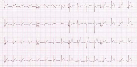
The admission ECG (figure 1) showed 2 mm ST segment elevation in leads I, II, aVL and V4 to V6, with 2 mm ST depression in leads V1 and V2; consistent with a diagnosis of posterolateral myocardial infarction. After appropriate initial resuscitation and treatment for acute coronary syndrome, the patient was transferred immediately by emergency ambulance to the local tertiary centre for primary percutaneous coronary intervention (figure 2).
Figure 1.
ECG recorded on admission. 2 mm ST segment elevation in leads I, II, aVL and V4 to V6, with 2 mm ST depression in leads V1 and V2; consistent with a diagnosis of posterolateral myocardial infarction.
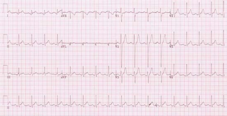
Figure 2.
ECG on repatriation to District General Hospital 24 h following percutaneous coronary angiography.
Investigations
Coronary angiography revealed entirely normal coronary arteries with very mild left ventricular systolic impairment. His 12-h troponin I was significantly elevated at 34.67 µg/ml (normal range <0.07); confirming definite myocardial infarction. d-Dimers were 375 ng/ml (normal range <500). Chest radiography and a subsequent thrombophilia screen were also negative.
Transthoracic echocardiography later revealed good left ventricular systolic function, with an ejection fraction of 62%. Left ventricular diameters were also within normal limits.
Differential diagnosis
Vasospastic causes of ST segment elevation ECG patterns with angiographically normal coronary vessels are well established. These include Prinzmetal’s angina and the use of illicit stimulants, most notably cocaine. However, these are not associated with dramatic rises in troponin and our patient strongly denied any illegal drug use.
Cardiomyopathies, such as hypertrophic obstructive cardiomyopathy or Tako–Tsubo syndrome, could also be considered. They would account for both the ECG and angiographic findings, but would be associated with characteristic echocardiographic abnormalities, so can be excluded in this patient.
Treatment
Following repatriation to the district general hospital, the patient was pain free during the 5-day observation period and was discharged home. He had been prescribed aspirin, clopidogrel, an ACE inhibitor and a β-blocker, all to continue on discharge.
Outcome and follow-up
The patient returned to the outpatient clinic for the routine follow-up at 2 months. He had been completely abstinent of energy drinks and had experienced no further episodes of chest pain.
Discussion
The case presented illustrates an example of electrocardiographically and biochemically proven myocardial infarction in an individual with structurally normal coronary vessels. We suspect the cardiac event experienced by this individual was secondary to his high intake of an energy drink in the days preceding his admission.
There has been apprehension for some time that the consumption of energy drinks in excessive volumes can have deleterious health effects.1 2 Concern has focused primarily on problems of caffeine intoxication and withdrawal. However, there is growing evidence that energy drinks may be associated with myocardial infarction and there is existing case report describing this.3 It is similar to the presented case in that it describes ST segment elevation myocardial infarction in a young male following excessive consumption of caffeine and taurine based energy drinks; both associated with significant troponin elevation and normal coronary angiography.
The energy drink consumed by our patient contained high concentrations of caffeine, taurine and glucoronate. It is not entirely clear which, if any, of these ingredients is responsible for inducing myocardial ischaemia, although a number of potential mechanisms, particularly implicating caffeine and taurine, have been suggested.
The cardiovascular effects of caffeine are perhaps the best defined. Its primary pharmacological effect is the competitive inhibition of adenosine receptors. However, it also induces catecholamine release and, as such, is positively inotropic. It also exerts a physiological effect upon intracellular calcium concentrations within vascular smooth muscle, potentially inducing a coronary vasospasm.
The cardiovascular action of taurine is less well established, although there is in vitro evidence that it has similar inotropic and vascular effects to caffeine. It is thought that taurine can also potentiate the physiological actions of caffeine.
There is also human evidence linking energy drink consumption with a potential mechanism of myocardial ischaemia. Healthy individuals showed a transient increase in both platelet aggregation and mean arterial pressure following consumption of 250 ml (one can) of an energy drink similar to that used by our patient. However, this study did not distinguish which component of the energy drink was responsible for these changes and the effects were only short-lived.4
We believe the case presented provides further evidence that there may be an association between energy drink consumption and myocardial infarction. A number of potential mechanisms do exist which, either individually or in combination, could support this hypothesis. Indeed, a potential link between energy drinks and myocardial infarction has been made previously3 and, given their popular and increasing use, it is certainly an association that warrants further investigation.
Learning points.
-
▶
There is growing evidence of a causative role of caffeinated energy drinks in the pathophysiology of myocardial infarction
-
▶
Myocardial infarction can occur in young patients and it should remain as a differential diagnosis in any patient with chest pain, irrespective of age
-
▶
A thorough clinical history is imperative in the diagnosis and management of any patient with chest pain. However, with young patients, in particular, the history is especially important. Non-conventional risk factors, including the use of caffeinated energy drinks and illicit drugs, should always be considered.
Footnotes
Competing interests None.
Patient consent Obtained.
References
- 1.Reissiga CJ, Straina EC, Griffiths RR. Caffeinated energy drinks – a growing problem. Drug Alcohol Depend 2009;99:1–10 [DOI] [PMC free article] [PubMed] [Google Scholar]
- 2.Birchard K. Irish concerned about health effects of stimulant soft drinks. Lancet 2000;356:1911. [DOI] [PubMed] [Google Scholar]
- 3.Berger AJ, Alford K. Cardiac arrest in a young man following excess consumption of caffeinated “energy drinks”. Med J Aust 2009;190:41–3 [DOI] [PubMed] [Google Scholar]
- 4.Worthley MI, Prabhu A, De Sciscio P, et al. Detrimental effects of energy drink consumption on platelet and endothelial function. Am J Med 2010;123:184–7 [DOI] [PubMed] [Google Scholar]


