

INTRODUCTION
The inner root sheath (IRS) is an important structure of the lower part of the hair follicle that surrounds and protects the growing hair. It lies between the hair in the center and the outer root sheath (ORS) peripherally, and all these three structures are derived from the matrical cells of the follicular bulb. The inner root sheath is made up of three distinct layers, namely, Henle's layer, Huxley's layer, and the cuticle of the IRS.
This article describes in brief the microanatomy, cornification patterns, and functions of the IRS and provides short biographical sketches of Jacob Henle and Thomas Huxley, who in the 19th century discovered and described the layers that make up the IRS of the human hair follicle.
THE INNER ROOT SHEATH
Origin, extent, and structure
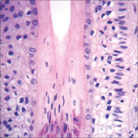
Like the hair (cortex, and medulla in terminal hairs, and the cuticle of the hair) the IRS and the ORS are also derived from the rapidly replicating matrical cells which form the bulk of the pyriform-shaped lower part of the follicular bulb. The centrally located matrical cells of the bulb give rise to the inner root sheath structures, which are from medial to lateral, the cuticle of the IRS, Huxley's layer, and Henle's layer[1] [Figure 1].
Figure 1.
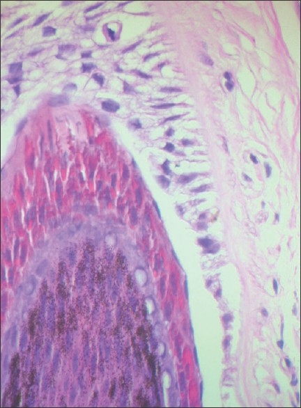
Follicular bulb medially, inner root sheath (IRS) with bright pink TH granules in the center and pale cells of outer root sheath laterally (H and E, ×400)
The IRS extends from the lower part of the follicular bulb to the lower part of the isthmus where the cornified cells of the inner root sheath desquamate and mark the end of the IRS and the stem of the follicle. It forms a rigid funnel like sleeve that covers and protects the growing hair within it.
Structurally the inner root sheath is made up of three distinct layers, namely, from inside out, the cuticle (single layer of cells), Huxley's layer (2-4 cells thick and containing numerous trichohyalin granules), and Henle's layer (a single layer thick) that abuts onto the cells comprising the outer root sheath. The interface between the ORS and Henle's layer is smooth and facilitates movement of the IRS against the ORS.
Trichohyalin is expressed in specialized epithelia that are unusually mechanically strong, such as the IRS cells of the hair follicle and is a multi-functional cross-bridging protein that functions in the IRS by conferring to and coordinating mechanical strength between the peripheral cell envelope barrier structures and the cytoplasmic keratin filament networks.[2]
Flugzellen[3,4] (winged cells) are specialized cells of Huxley's layer in the supra-bulbous portion of the follicle, which show elongated cytoplasmic pseudopods that traverse the rigid cornified impermeable Henle's layer and connect with the innermost cells of the ORS.
Cornification of the inner root sheath
The cornification of the cells of Huxley's and Henle's layers occurs through the formation of bright pink trichohyalin granules (the trichological counterpart of the keratohyaline granules of the epidermis and the follicular infundibulum).
Henle's layer is the first to cornify followed by the cuticle while the cells of Huxley's layer cornify last of all.
The process of cornification (full keratinization) is similar in the cells of both Huxley's and Henle's layers and begins with the development of large and numerous trichohyalin granules associated with numerous tonofilaments. As the cells transform and get fully keratinized the cytoplasm appears homogenous with filaments in a dense matrix. The trichohyalin granules and the nucleus disappear with scattered remnants of cytoplasmic organelles and nuclear ghosts.[4]
On light microscopy, the trichohyalin granules appear bright pink and are admixed with few nuclei of inner root sheath cells, while the fully cornified cells are devoid of nuclei and assume a blue-gray color.
Based on cornification patterns of the IRS, the lower part of the follicle (bulb to isthmus) has been divided into four areas[4] [Figures 2–5]:
Figure 2.
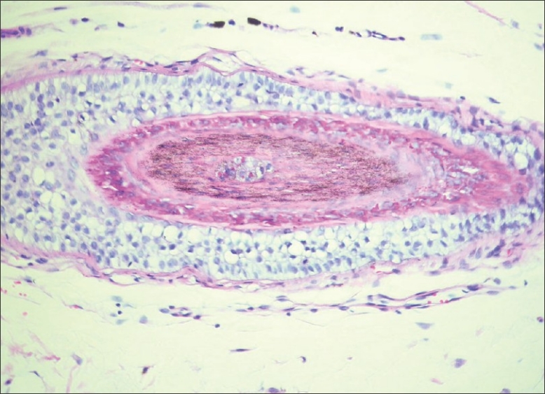
T.S at level of upper bulb, showing nucleated hair shaft, TH granules in Huxley's layer and partial keratinization of cuticle and Henle's layer and prominent pale outer root sheath (H and E, ×400)
Figure 5.
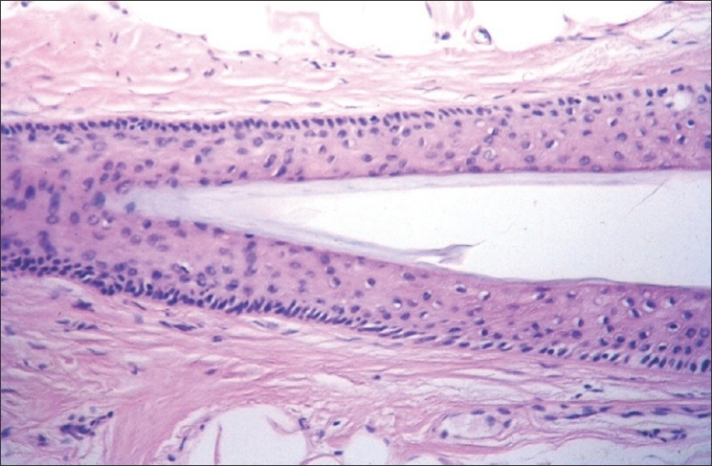
T.S at level of upper stem just beneath the isthmus, with complete keratinization of IRS which appears as blue-gray compactly arranged corneocytes (H and E, ×400)
Lower bulb: has no trichohyaline granules, and extends from the lowest part of the follicle to the level where trichohyaline granules are first seen in Henle's layer.
Upper bulb: extends upwards till the point of full keratinization of Henle's layer and this area usually corresponds with the margin of the keratogenous zone (Adamson's fringe) of the hair shaft where the hair shaft gets fully keratinized and loses its nuclei. (the cuticle too acquires few trichohyalin granules and gets fully keratinized in this portion of the follicle)
Suprabulbous portion: this extends till the level of complete keratinization of Huxley's layer, i.e., there is complete keratinization of the entire IRS.
Sub-isthmic part: the portion just below the isthmus where the IRS is fully keratinized and eventually desquamates completely and disappears.
Figure 3.
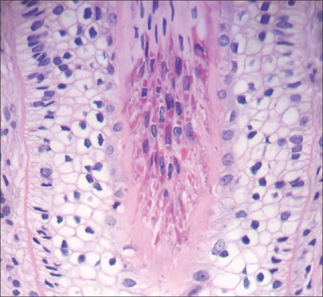
V.S at level just beneath Adamson's fringe, with cornified Henle's layer, Huxley's layer shows TH granules admixed with nuclei of cells of Huxley's layer (H and E, ×400)
Figure 4.
V.S at level straddling Adamson's fringe and just above with the lower part showing few TH granules and nuclei while the upper part shows full keratinization of the IRS with absence of nuclei and TH granules (H and E, ×400)
In sum, therefore, the cornification of the inner root sheath begins soon after its development from the matrical cells of the follicular bulb. It begins first in Henle's layer, which is completely keratinized at the level of Adamson's fringe. The cuticle too is fully keratinized at this level and its cells are intimately interlocked with the cells of the cuticle of the hair shaft. The cells of Huxley's layer begin keratinization just below Adamson's fringe and are fully keratinized mid way in the stem of the follicle. The upper part of the IRS in the stem (sub-isthmic portion) is fully keratinized and made up of only blue-gray corneocytes, which are completely desquamated and the IRS withers away at the level of the beginning of the isthmus of the follicle.
Functions of the inner root sheath
Functionally the IRS serves as a rigid funnel-shaped protective covering around the hair and the flattened corneocytes of the cuticle of the IRS overlap and interlock intimately with the cells of the cuticle of the hair ensuring that the IRS and the growing hair that it protects, ascend upwards in the follicle together.[1]
The IRS is essential for the proper moulding, adherence, and keratinization of the growing hair. The Flugzellen of Huxley's layer are postulated to function as nutritive canals between the not yet keratinized Huxley's layer and the outer root sheath, bypassing the fully cornified and impermeable Henle's layer.[4,6,7]
From a clinical perspective, in the condition described as loose anagen hair of childhood[5] premature keratinization of the inner root sheath has been implicated in the loss of the anchoring function and subsequent deformation of the hair shaft.
Friedrich Gustav Jacob Henle (1809-1885)
Jacob Henle was born to Jewish parents in Furth in Bavaria on July 9, 1809 [Figure 6]. His father was a prosperous merchant and his work required the family to move frequently throughout Germany.
Figure 6.
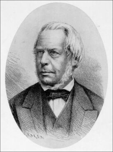
Jacob Henle (Accessed from Wikipedia, original source http://clendening.kumc.edu/dc/pc/h.htm)
Henle studied medicine at Heidelburg and Bonn where he received his doctor's degree in 1832. In Bonn, Henle was strongly influenced by Johannes Muller who graduated from Bonn but later was Professor of Anatomy in the Humboldt University in Berlin.
Henle joined Muller in Berlin, as Prosector in Anatomy, and during the six years he spent in that position he published a large amount of work. Muller founded the Archives of Anatomy, Physiology, and Scientific Medicine with Henle as managing editor. From 1837 to 1840, Henle contributed greatly to the modern understanding of the epithelial tissues of the body. He was the first to describe the epithelium of the skin and intestines, define columnar and ciliated epithelium, and point out that this tissue constitutes the true lining of all the free tissues of the body and the inner lining of its tubes and cavities.
Henle was a histologist at heart and continued working in that discipline all his life at various universities. Numerous structures in the human body have been eponymically named after him and include in the eye, the trachoma glands of Henle, the lamina basalis (Henle's membrane), and Henle's fibrous layers in the macula lutea. Other eponyms followed his discoveries in the urogenital field, namely, the expanded outer half of the Fallopian tube (Henle's ampulla), a portion of the uriniferous tubule (the canal of Henle), Henle's internal cremaster, and Henle's loop of the uriniferous tubule.
Henle's original description of the IRS layer of the hair follicle named after him has been narrated by Steffen6 from a contemporary French translation,[8] “At the base of the follicle and external to the hair lies a distinct structure that I shall call the internal root sheath. This clear layer has the appearance of an oily liquid, fits between the external root sheath and the hair itself and must be distinguished from the external root sheath laterally and the cortex of the hair medially. The internal root sheath is very thin, but quite apparent. This narrow space seems to hold the hair in its grip. I was not able to determine its exact width. The internal root sheath extends from base of the follicle and merges directly with the epidermis. I cannot say if the internal root sheath arises from the epidermis at the raised point where the hair protrudes. However, it desquamates and undergoes structural changes in a unique fashion thus proving that it is a distinct part of the follicle”.
Steffen[6] commented that “Although Henle described only one layer of the internal root sheath, in fact, it contains three of these (cuticle, Huxley's and Henle's). Henle did not notice Huxley's layer or the cuticle for evidently, he must have examined Henle's layer at a level where cornification had already taken place, because he thought that the layer was anucleated. He also erred when he wrote that “his” layer extended to the epidermis”.
In 1840 after a brief disagreeable incident in Berlin, Henle eagerly accepted the professorship at Zurich. In Zurich, Henle and Carl Pfeufer founded the School of Rational Medicine and its mouthpiece The Journal of Rational Medicine. In 1841, Henle published his text, Allgemeine Anatomie, which was the first treatise on microscopic histology. In 1844, Henle and Pfeufer left Zurich to become members of the Heidelberg faculty. Unhappy at Heidelberg, Henle in 1852 accepted the professorship of anatomy at Gottingen, a town which he loved and where he remained until his death on May 13, 1885. In Gottingen, he published his Handbook of Systemic Human Anatomy, which was remarkable not only for the fullness and minuteness of the anatomical descriptions, but also for the number and excellence of the illustrations with which they were elucidated.
Thomas Henry Huxley (1825-1895)
Thomas Huxley was a famous English biologist, anatomist, and a strong supporter of Darwin's theory of natural selection [Figure 7].[9,10] He was commonly referred to as ‘Darwin's bulldog’ a self-designated aphorism to indicate his strong support for Darwin's theory of natural selection and evolution. As part of his support for the theory of evolution he participated in the 1860 Oxford evolution debate which took place at the Oxford University Museum on 30 June 1860, seven months after the publication of Charles Darwin's On the Origin of Species.
Figure 7.
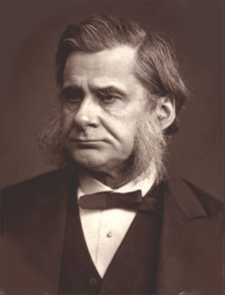
Thomas Huxley at about age 55. (Accessed from Wikipedia, original print by Samuel R. Lock and George C. Whitfield, London.)
Several prominent British scientists and philosophers participated, including Thomas Henry Huxley, Bishop Samuel Wilberforce, Benjamin Brodie, Joseph Dalton Hooker and Robert FitzRoy. The debate is best remembered today for a heated exchange in which Bishop Wilberforce supposedly asked Huxley whether it was through his grandfather or his grandmother that he claimed his descent from a monkey. Huxley is said to have replied that he would not be ashamed to have a monkey for his ancestor, but he would be ashamed to be connected with a man who used his great gifts to obscure the truth. However, no verbatim account of the debate exists, and there is considerable uncertainty regarding what Huxley and Wilberforce actually may have said, however, the general view was that Huxley got much the better of the exchange, and more importantly paved the way for development of scientific thought without interference from the clergy of that period. It also made Huxley a believer in the power of public debate.[11]
He was member of several scientific societies, served as President of the Royal Society and was an active writer, debater and champion of educational reforms and universal education in Victorian England. He was a strong supporter of scientific enquiry, agnosticism (a term coined by him) and was against irrational blind faith and dogma.
His achievements and honors are too numerous to be detailed in this article which will focus on his early life and what is relevant to us, namely, his discovery of the layer of the inner root sheath that bears his name eponymically.
Huxley was born on May 4, 1825, in the village of Ealing, county of Middlesex, England.
His father George Huxley, was an impoverished school teacher and the family fell on hard times due to closing of the local school where the senior Huxley was employed. Thomas Huxley therefore had only two years of formal education and had to leave school at age ten, but was determined to self-educate himself, which he did with panache, becoming fluent in German and Latin and knew enough of the Greek language to read Aristotle in the original. Not only did he dwell on the classics and languages but was also well read in the emerging science of that age. He was an expert in comparative anatomy, a superb illustrator, and in later life was a keen debator and writer on matters pertaining to science, education and theology, and was considered to be one of the most knowledgeable men in Britain.
Huxley had keen interest in mechanical engineering but being impoverished had to accept apprenticeship with medical practitioners in London, who happened to be his brothers in law. At age sixteen, he joined Sydenham College with very good results and a year later won the silver medal in the Apothecaries’ annual competition. He was admitted to Charing Cross Hospital with a small scholarship.
At Charing Cross, he had the fortune to be taught by Thomas Wharton Jones, who was a fine teacher, physiologist, and ophthalmic surgeon, and who encouraged Huxley to pursue his natural desire for scientific knowledge. At age 19, in 1845, Huxley under Wharton Jones’ guidance published his first scientific paper demonstrating the existence of a previously unrecognized layer in the inner root sheath of the hair follicle, a layer since then known as Huxley's layer.[12] Readers will remember that a few years earlier Jacob Henle had in Germany described Henle's layer of the inner root sheath but had failed to recognize the medially located 2-4 cell thick layer that Huxley described later.
At age twenty, he passed his first M. B. examination of University of London, winning the gold medal for anatomy and physiology. He, however, failed to appear for the final M. B exams and did not thus qualify with a university degree.
At age 20, deep in debt, too young to apply to the Royal College of Surgeons for a license to practice, he applied for and was accepted into the Royal Navy as Assistant Surgeon. He joined HMS Rattlesnake a British ship, which undertook a voyage of discovery and surveying to the southern hemisphere. On board the HMS Rattlesnake, Huxley left England on December 3, 1846, and devoted his time to the study of marine invertebrates, a field in which he was to become an authority in the coming years. The value of Huxley's work was recognized and on returning to England in 1850, he was elected a Fellow of the Royal Society.
In the following year, at the age of twenty-six, he not only received the Royal Society Medal but was also elected to the Council. This marked a change of course in life for the young Huxley, who always uninterested in the practice of medicine devoted his further life to comparative anatomy and Natural History. He was appointed in 1854 as the Professor of Natural History at the Royal School of Mines, where he remained for 31 years till he retired in 1885, aged 60, after suffering from a prolonged bout of depressive illness, a malady that he had been prone to as a youth. He received numerous awards, medals, several Royal Commissions, and even recognition from the King of Sweden for his meritorious work in natural history and public work. He died in 1895 after an attack of influenza and pneumonia and is buried in North London.
COMMENT
It is relevant here to compare and contrast the two men who described the layers of the IRS.
Both Henle and Huxley were astute and diligent microscopists and anatomists who contributed immensely to scientific knowledge. Both had dislike and disdain for the commercial practice of medicine as is evident from Henle's reasons for pursuing scientific investigation rather than medical practice inunciated in the early 19 th century but which nevertheless remain relevant for the medical student of today:[6] ‘Without laying a scientific foundation, everyone rushes into practice. The machine-like prescription writing is certainly not our highest goal, and whatever such a mechanical man appropriates through many years of experience can soon be equaled if we are willing to seek reason and ground. This is the only way by which we will not be compelled to follow blindly what others tell us from their experience. The practitioner who is merely concerned about practice may through some luck and savoir faire, attain a decent income, but he will never attain anything for his science, and I do not call him fortunate, who is satisfied with that.’
Henle continued his work in the setting of Universities all over Germany and Zurich in Switzerland and continued with his passion of anatomy and histology, while Huxley relinquished medicine and made a mark in Natural history, zoology and public service.
Even today, with the latest in optics of modern microscopes it is difficult for a histopathologist to clearly delineate the layers of the IRS and it is a testimony to the amazing powers of observation of these men, that Henle and Huxley described these structures of the IRS more than a century and a half ago.
Footnotes
Source of Support: Nil
Conflict of Interest: None declared.
REFERENCES
- 1.Ackerman AB, Boer A, Bennin B, Gottlieb GJ. Embryonic, histologic, and anatomic aspects. In: Histologic diagnosis of inflammatory skin diseases. 3rd Ed. New York: Ardor Scribendi; 2005. pp. 26–3. [Google Scholar]
- 2.Steinert PM, Parry DA, Marekov LN. Trichohyalin mechanically strengthens the hair follicle: multiple cross-bridging roles in the inner root sheath. J Biol Chem. 2003;278:41409–19. doi: 10.1074/jbc.M302037200. [DOI] [PubMed] [Google Scholar]
- 3.Hoepke H. Die Haare. In: Mullendorf W, editor. Handbuch der mikroskopischen Anatomie des Menschen. 3rd Ed. Berlin: Spinger Verlag; 1927. pp. 66–8. [Google Scholar]
- 4.Clemmensen OJ, Hainau B, Hanstead B. The ultrastructure of the transition zone between specialized cells (Flugelzellen) of Huxley's layer of the inner root sheath and cells of the outer root sheath of the human hair follicle. Am J Dermatopathol. 1991;13:264–70. doi: 10.1097/00000372-199106000-00008. [DOI] [PubMed] [Google Scholar]
- 5.Hamn H, Traupe H. Loose Anagen hair of childhood: The phenomenon of easily pluckable hair. J Am Acad Dermatol. 1989;20:242–8. doi: 10.1016/s0190-9622(89)70029-3. [DOI] [PubMed] [Google Scholar]
- 6.Steffen C. The Man Behind the Eponym: Jacob Henle – Henle's layer of the Innternal Root Sheath. Am J Dermatopathol. 2001;23:549–51. doi: 10.1097/00000372-200112000-00011. [DOI] [PubMed] [Google Scholar]
- 7.Jacob Henle. [Last accessed on 2011, March 02]. Available from: http://whonamedit.com/doctor.cfm/710.html .
- 8.Henle J. In: Traite d’anatomie generale ou histoire des tissue et de la composition chimique de corps human. Jordan A. J. L, editor. Paris: J B Bailliere; 1843. p. 318. [Google Scholar]
- 9.Steffen C. The Man behind the Eponym: Thomas Henry Huxley- Huxley's Layer of the Inner Root Sheath. Am J Dermatopathol. 2002;24:82–4. doi: 10.1097/00000372-200202000-00017. [DOI] [PubMed] [Google Scholar]
- 10.Thomas Huxley. [last Accessed on 2011, March 02]. Available from: http://en.wikipedia.org/wiki/Thomas_Henry_Huxley .
- 11.Lucas JR. Wilberforce and Huxley: A legendary Encounter. Hist J. 1979;22:313–30. doi: 10.1017/s0018246x00016848. [DOI] [PubMed] [Google Scholar]
- 12.Huxley TH. On a hitherto undescribed structure in the human hair sheath. London Med Gazette. 1845;38:1340–1. [Google Scholar]