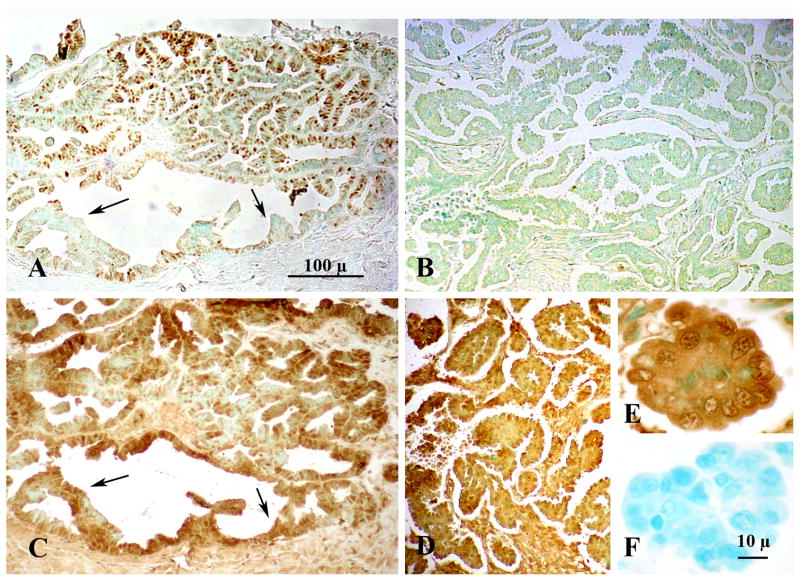
Figure 1.
Immunohistochemical localization of Progesterone Receptor (PGR) (A,B) and Progesterone Receptor Membrane Component-1 (PGRMC1) (C,D) in Stage IIIc Grade 2 (A,C) and Stage IIIc Grade 3 (B,D,E) ovarian tumors. Images from panels A and C and B and D are taken from adjacent sections from respective tumors. Panels A, B C and D are shown at the same magnification. The arrows in panels A and C mark the location of ovarian cancer cells that do not express PGR but do express PGRMC1. The image in panel E is a higher magnification of the image shown in panel D. Panel F is a negative control. Both panel E and F are shown at the same magnification. Data taken from Peluso et al [20].

