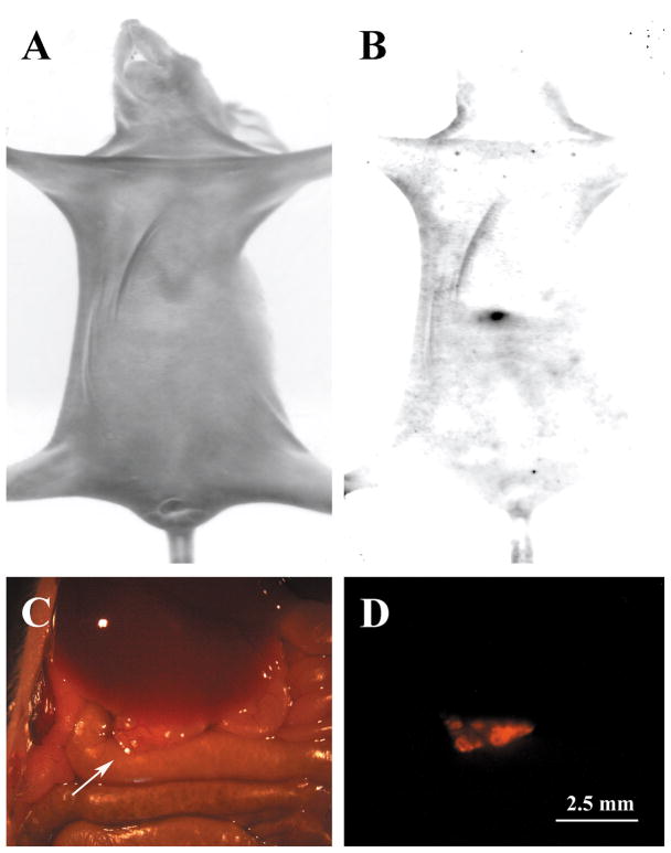
Figure 3.
Detection of ovarian tumors within intact living nude mice. The mouse shown in these images was injected with 10 million dsRed SKOV-3 cells and the tumors were allowed to develop for 5 weeks. In panel A, a bright field image of a nude mouse is shown. When observed under dsRed fluorescent filters (B), a tumor was observed (black arrow). This animal was dissected to expose the peritoneum and a tumor (arrow) was observed under bright field (C) and fluorescent optics (D).

