Abstract
Early studies suggest that TR4 nuclear receptor is a key transcriptional factor regulating various biological activities, including reproduction, cerebella development, and metabolism. Here we report that mice lacking TR4 (TR4−/−) exhibited increasing genome instability and defective oxidative stress defense, which are associated with premature aging phenotypes. At the cellular level, we observed rapid cellular growth arrest and less resistance to oxidative stress and DNA damage in TR4−/− mouse embryonic fibroblasts (MEFs) in vitro. Restoring TR4 or supplying the antioxidant N-acetyl-l-cysteine (NAC) to TR4−/− MEFs reduced the DNA damage and slowed down cellular growth arrest. Focused qPCR array revealed alteration of gene profiles in the DNA damage response (DDR) and anti-reactive oxygen species (ROS) pathways in TR4−/− MEFs, which further supports the hypothesis that the premature aging in TR4−/− mice might stem from oxidative DNA damage caused by increased oxidative stress or compromised genome integrity. Together, our finding identifies a novel role of TR4 in mediating the interplay between oxidative stress defense and aging.
Keywords: testicular nuclear receptor 4, senescence
testicular nuclear receptor 4 (TR4) belongs to the nuclear receptor superfamily (4) that regulates various genes via binding to AGGTCA-like direct-repeat motifs (23). Regulation of various genes in diverse pathways, for example, apoE (21), Gata1 (42), PEPCK (25), and CD36 (47), indicates that TR4 plays vital roles in many important biological events. Knockout of TR4 gene in mice (TR4−/−) results in abnormal maternal behavior (8), impairment in male (32) and female (5) fertility, hypoglycemia (25), and neurological abnormalities (6), indicating TR4's critical role in maintaining tissue homeostasis. Hence, loss of TR4 might impair mouse physiological function in general, which might gradually lead to systematic declines over time. Indeed, TR4−/− mice suffered progeriod syndrome as demonstrated by shorter life span and signs of accelerating aging, including cachexia, graying hair, osteoporosis, and kyphosis, at a much younger age than their wild-type littermates.
Aging is characterized by the deterioration of physiological functions caused by the accumulation of stochastic damage to cellular macromolecules (26). Organisms are constantly bombarded by endogenous and environmental genotoxic insults such as reactive oxygen species (ROS), which cause damage to macromolecules, including DNA, and finally lead to genome instability. A number of theories have been put forth to explain the cause and mechanism of aging, and yet no single mechanism is sufficient to account for age-related frailty, disability, or diseases. In general, elevated oxidative stress, defective stress responses, and increased genomic instability are among the major causes that lead to accelerated aging (15).
Reducing stress tolerance is one characteristic of organism aging (33), during which cells become less resistant to stresses, and thus increased cellular senescence was triggered by the accumulated DNA damage to deplete the damaged cells (40). Remarkably, transgenic mice that overexpress mitochondrial catalase show a prolonged life span that is correlated with an increased ROS-scavenging activity (38). Also, loss of genome stability due to malfunctions in DNA repair machineries can have catastrophic consequences, including premature aging. This is supported by studies in premature aging mouse models with disruptions of genes involved in maintaining DNA integrity, such as DNA-PKcs (11), Ku 80 (45), and XPD (10).
In our previous study, we have demonstrated that oxidative stress stimulates TR4 expression via the stress responder FOXO3a (24), which has been associated with longevity in various organisms (19, 46). Furthermore, we also found that TR4 expression level can be stimulated upon other various genotoxic stresses, including UV and gamma irradiation (Yan SJ, Lee YF, Ting HJ, Liu NC, Liu S, Yeh SD, and Chang C, unpublished observations). Thus, our findings have established TR4's role in the regulation of the cellular response to multi-stresses. In this report, we demonstrate further that elevated ROS and increased genome instability contribute to the premature aging phenotypes in TR4−/− mice. Furthermore, we showed that the application of the antioxidant N-acetyl-l-cysteine (NAC) to TR4−/− mouse embryonic fibroblasts (MEFs) reduced the DNA damage and slowed down cellular growth arrest. Therefore, our report provides a novel mechanism that links TR4, stress defense, and aging.
EXPERIMENTAL PROCEDURES
Mice information generation, breeding, and genotyping of TR4−/− mice were described previously (8). The original TR4−/− mice were of C57/BL6/129 mixed background. TR4−/− mice were back-crossed to C57/BL6 mice for five generations and used in this study. Continuous back-cross to C57/BL6 background over six to seven generations caused embryonic lethality in TR4−/− mice.
Tissue preparation and histology.
Mice were anesthetized with an overdose of pentobarbital sodium, and tissues were removed and fixed by immersion in 10% neutral buffered formalin. Tissues were cut into 5-μm sections, deparaffinized, and stained with haematoxylin and eosin by standard procedures.
Bone analyses.
Mice were subjected to whole body X-ray in situ under anesthesia. Bone mineral density was quantified by dual-energy X-ray absorptiomentry (DEXA) scanning.
Immunofluorescence staining of 8-oxodeoxyguanosine.
The assessment of 8-oxodeoxyguanosine (8-OxodG) followed the protocol with small revision (41). Liver tissues were prefixed by the addition of 2% (wt/vol) paraformaldehyde (in PBS at pH 7.4) for 15 min after the tissues were washed with PBS, fixed and permeabilized with ice-cold methanol for 15 min, and rehydrated in PBS before blocking with PBS containing 10% (wt/vol) normal goat serum (NGS). The blocking solution was washed off with PBS containing 0.2% (wt/vol) NGS. DNA damage was visualized with avidin-conjugated FITC (1:200 in PBS for 1 h) under fluorescence microscopy. The percentage of the 8-OxodG-containing cells per 500 cells was counted, and the average percentage was achieved from a total of 2,000 cells for each sample.
Generation of MEF cells.
We removed the heads and all of the internal organs from E14.5 embryos and rinsed with PBS, added 5 ml of DMEM, passed through a 22-gauge needle a few times to mince tissues, and then allowed the fibroblasts to attach to the culture flask for 24 h and changed the medium to remove unattached cells and debris. MEFs at passage 0 (P0) would form a confluent monolayer after 2–3 days. Cells were then trypsinized and subcultured for genotyping and experiments. All of the experiments were finished before passage 4 (P4).
Protective effect of cellular protein on pUC19 DNA damage.
To determine the protective effects of cellular proteins on plasmid DNA, 5 μl of 0.25 μg/μl pUC19 DNA was incubated with 5 μg of cellular proteins from TR4−/− or TR4+/+ MEFs (PBS was used as control). Then, 1 μl of 6 mmol/l H2O2 and 1 μl of 0.4 mmol/l FeSO4 were added and incubated at 37°C for 60 min. The reaction was electrophoresed on an agarose gel, and DNA damage evaluations were based on the loss of supercoiled (SC) monomer.
Cell cycle profiling.
MEFs from TR4−/− or TR4+/+ mice were collected and fixed with 70–75% EtOH at 4°C for ≥12 h. Cells were then centrifuged at 1,000 rpm for 7 min at 4°C, the supernatant was decanted, and 1 ml of RNase (1 mg/ml in 1× PBS) was added for 30 min. Cells were then incubated with 500–1,000 μl of propidium iodide (20 μg/ml) and analyzed by flow cytometry.
Growth assay (MTT).
We seeded 2,000 cells in 96-well plates and waited ≥12 h or overnight for attachment. We then infected with retrovirus (vector or TR4 virus) for 24 h, washed with culture medium, and replaced with 200 μM H2O2 containing medium for 2 h. Cells without H2O2 treatment were recorded as day 0. After 2 h H2O2 treatment, we washed cells with culture medium and replaced with the fresh medium and harvested cells on days 1, 3, and 5 for 3-[4,5-dimethylthiazole-2-yl]-2,5-diphenyltetrazolium bromide (MTT) assay. To determine the cells' sensitivity to stress, MEFs from TR4−/− and TR4+/+ mice were seeded and treated with various doses of γ-irradiation or 2 h of H2O2. We harvested the cells on day 3 to determine cell growth by MTT assay. The survival rate was determined as the ratio between treated and nontreated groups.
Measurement of DNA single-stranded breaks.
A DNA precipitation assay was used for DNA strand breaks detection. Confluent MEFs from TR4−/− and TR4+/+ mice were labeled with 0.25 μCi/ml [3H]methylthymidine for 24 h, and then the cells were washed thoroughly with PBS and supplied with serum-free medium in the presence and absence of 250 μM H2O2 for another 30 min. The cells were washed and lysed with lysis buffer (10 mM Tris·HCl, 10 mM EDTA, 50 mM NaOH, and 2% SDS, pH 12.4), followed by addition of KCl (12 mM) for 10 min at 65°C, followed by a 5-min cooling and precipitation period on ice. A DNA protein K-SDS precipitate was formed under these conditions, from which low-molecular-mass broken DNA was released. DNA fragments were recovered in the supernatant from a 10-min centrifugation at 200 g at 10°C and transferred into a liquid scintillation vial containing 1 ml of 50 mM HCl. The precipitated pellet (intact double-stranded DNA) was solubilized with water at 65°C. Radioactivity was determined by scintillation counter. The amount of double-stranded DNA remaining was calculated for each sample by dividing the dpm value of the pellet by the total dpm value of the pellet plus supernatant and multiplying by 100. In control cells (cells incubated in Ca2+-containing or Ca2+-free EGTA), the level of total double-stranded DNA was ∼75%. Pretreatment with the various chelators did not affect this level.
Measurement of intracellular ROS by flow cytometry.
The production of intracellular ROS was detected by flow cytometry using dichlorofluorescein diacetate (DCFH-DA). The MEFs from TR4−/− and TR4+/+ mice were cultured in 35-mm tissue culture dishes. When cells reached 80% confluence, they were incubated with 10 μM DCFH-DA together with 250 μM H2O2 for 30 min at 37°C in the dark, washed once with PBS, detached by trypsinization, collected by centrifugation, and suspended in PBS prior to flow cytometry. For the control group, the medium was changed to serum-free medium and incubated with 10 μM DCFH-DA and followed by flow cytometric analyses, and the amount of cellular ROS levels was quantified by the fluorescence of dichlorofluorescein density.
Senescence-associated β-galactosidase assay.
Briefly, cells were washed in phosphate-buffered saline and fixed in 2% formaldehyde-0.2% glutaraldehyde. Then the cells were washed and incubated at 37°C overnight with fresh senescence-associated β-galactosidase (SA-β-Gal) stain solution [1 mg/ml 5-bromo-4-chloro-3-indolyl-β-d-galactopyranoside (X-Gal), 40 mM citric acid-sodium phosphate (pH 6.0), 150 mM NaCl, 2 mM MgCl2, 5 mM potassium ferrocyanide, 5 mM potassium ferricyanide]. All visualization and photography of cells were performed on a Leica microscope with Sony digital imaging.
Pathway-focused RT2 Profiler PCR Array.
The relative expression of 84 DNA damage signaling-related genes and 84 oxidative stress defense-related genes was evaluated using the mouse DNA damage signaling pathway and oxidative stress and antioxidant defense signaling pathway RT2 Profiler PCR Array system (SuperArray Bioscience) according to the manufacturer's instructions. DNase-treated total RNA was purified from cultured MEF cells, and cDNA was generated by reverse transcription from 1 μg of total RNA from each sample using the RT2 First Strand kit and then combined with the RT2 qPCR Master Mix and added to lyophilized primer pairs in the 96-well arrays. Thermal cycling was performed in a Bio-Rad iCycler. Relative gene expression levels were calculated using the ΔΔCT method with normalization to the average expression level of five common genes (ACTB, B2M, GAPDH, HPRT, and RPL13A).
RESULTS
TR4−/− mice display premature aging phenotypes.
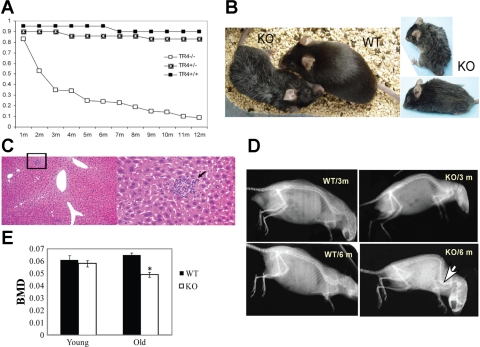
We first found that almost all TR4−/− mice developed cachexia by 1 mo, characterized by severe weight loss, prominent reduction of fat tissue, and weakness (8). Cachexia in TR4−/− mice was progressive, heterogeneous in severity, and followed by premature death. Generally, TR4−/− mice had much shorter life span compared with TR4 wild-type (TR4+/+) and heterozygote (TR4+/−) mice. Seventy percent of TR4−/− mice died before 4 mo, and >90% of the TR4−/− mice (72 out of 77 TR4−/− mice) could not live >1 yr (Fig. 1A). More than 90% of TR4+/+ and TR4+/− mice survived over this period of study, and the average life span of C57BL/6J mice exceeds 2 yr (13).
Fig. 1.
Premature aging phenotypes in testicular nuclear receptor 4 (TR4)−/− mice. A: 1-yr survival rate between TR4+/+, TR4+/−, and TR4−/− mice (n = 74 for TR4+/+ and TR4+/− mice, n = 77 for TR4−/− mice). B: general appearance of TR4−/− [knockout (KO)] mice. Gray hair and hunchback were seen in 6-mo-old TR4−/− female mice compared with age-matched TR4+/+ [wild-type (WT)] mice. C: extramedullary hematopoiesis in 6-mo-old TR4−/− mice liver. D: radiograph of 3- and 6-mo-old TR4−/− and TR4+/+ mice showed skeletal abnormalities in aging TR4−/− mice. Six-month-old TR4−/− mice display curvature of the spinal column (kyphosis). Pictures from B–D are the representative pictures from ≥3 pairs of WT and KO mice. E: dual-energy X-ray absorptiometry scan analyses of young (2–4 mo; n = 4) and old male TR4−/− mice (n = 5) at the age of 6–7 mo compared with age/sex-matched TR4+/+ mice. Differences in bone mineral density (BMD) of a particular sex and genotypes were analyzed by Student's t-test *P < 0.05.
Besides the shorter life span, TR4−/− mice also acquired an aged appearance at an earlier stage. By 6 mo, most TR4−/− mice have gray hair (Fig. 1B) and an extramedullary hematopoiesis in the liver (Fig. 1C) that is often found in aging mice and the PolgA mutant premature aging mouse model (43).
Furthermore, bone condition was examined for age-associated changes in TR4+/+ and TR4−/− mice. Although 2- to 3-mo-old TR4−/− mice showed no significant skeletal abnormalities (Fig. 1D) and had similar bone mineral density (BMD) compared with their littermate TR4+/+ mice (Fig. 1E), radiographs of 6-mo-old TR4−/− mice revealed a severe kyphosis (curvature of the spine; Fig. 1D), a landmark for aging bone. A reduction of BMD in 6- to 7-mo-old TR4−/− mice spine was revealed by DEXA scanning (Fig. 1E), although there was no difference of BMD in the skull between TR4−/− and TR4+/+ (data not shown).
Taken together, Fig. 1, A–E, demonstrated that TR4−/− mice developed a segmental progeroid syndrome characterized by the early onset of premature aging phenotypes and shorter life span.
Increasing genome instability and ROS in TR4−/− mice.
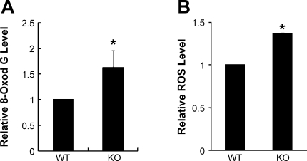
Mammalian aging is characterized by the functional decline caused by accelerated accumulation of somatic damage to macromolecules. To examine the level of macromolecular damage in TR4−/− mice, the 8-OxodG level was assayed. 8-OxodG is a major form of oxidatively modified DNA that increases with age and can lead to a variety of diseases and the age-associated decline of physiological functions (29). The level of 8-OxodG in the liver tissue of old TR4−/− mice was 50% higher than that in the littermate TR4+/+ mice (Fig. 2A), suggesting that TR4 is involved in the regulation of redox homeostasis or the defense against ROS. This is consistent with our previous finding that TR4 mRNA was induced directly by FOXO3a under hydrogen peroxide treatment (24).
Fig. 2.
Increased reactive oxygen species (ROS) and oxidative damage in TR4−/− tissues. A: 8-oxodeoxyguanosine (8-OxodG) staining was carried out in old (>12 mo) WT and KO mouse (>12 mo) liver tissues, as described in experimental procedures. The number of 8-OxodG-positive cells/500 cells was counted, and the percentage was calculated. A total of 2,000 cells were counted from each mouse. Shown are the average percentage ± SD (n = 4). B: tissue ROS levels were quantified in liver extracts from WT and KO mice by dichorofluorescein staining and flow cytometric analyses. Shown are the mean fluorescence intensity ± SD (n = 5). *P < 0.05 vs. WT.
Among all sources of somatic damage in organisms, ROS, the by-products of oxidative phosphorylation, are considered the main insult (37). To further investigate the possible implication of TR4 in ROS homeostasis, ROS levels were measured in liver extracts from TR4−/− and TR4+/+ mice. As shown in Fig. 2B, ROS level in the liver extracts from TR4−/− mice was significantly higher than that from their TR4+/+ littermates, indicating that TR4 plays essential roles in regulating redox homeostasis.
Early onset of senescence in TR4-deficient cells.
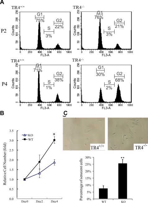
To better study the possible mechanisms leading to the progerorid phenotypic changes in TR4−/− mice at the cellular level, we examined the growth characteristics of primary MEFs from TR4−/− and TR4+/+ embryos. At early passages (P1–P3), both TR4−/− and TR4+/+ MEFs grew well, and their proliferation capacity did not show any noticeable difference (data not shown). Consistent with this, there was no difference between their cell cycle profiles at early passages (Fig. 3A). However, after P4, TR4−/− MEFs gradually slowed down their growth rate, whereas TR4+/+ MEFs kept up a steady growth before P8 (Fig. 3B), which was confirmed by their cell cycle characteristics in which two-thirds of TR4−/− cells were arrested at G2/M at P4, whereas less than one-third of TR4+/+ MEFs were in G2/M (Fig. 3A) at P4.
Fig. 3.
Growth arrest and early onset of cellular senescence in TR4−/− mouse embryonic fibroblasts (MEFs). A: cell cycle profile analysis of passages 2 (P2) and 4 (P4; MEFs from TR4−/− and TR4+/+ mice). TR4−/− display an early G2/M arrest in P4, whereas P4 TR4+/+ MEFs showed a normal cell cycle distribution. B: the growth rate of MEFs from TR4+/+ (WT) and TR4−/− (KO) was examined by 3-[4,5-dimethylthiazole-2-yl]-2,5-diphenyltetrazolium bromide (MTT) assay. The MEFs, from both genotypes at P5, were seeded and harvested at indicated days. The growth rate was calculated as the ratio to day 0. Three independent experiments were carried out, and the representative results are shown. Shown are means ± SD (*P < 0.05 vs. WT). C: P5 TR4+/+ and TR4−/− MEFs were subjected to senescence-associated β-galactosidase (SA-β-Gal) staining, as described in experimental procedures (top). Cellular senescence was indicated by the blue-colored staining. The percentage of SA-β-Gal staining positive cells was counted. Representative pictures from TR4+/+ and TR4−/− MEFs are shown (bottom). **P < 0.01 vs. WT.
We also observed an increased number of TR4−/− MEFs with flattened and enlarged morphology, a feature typically associated with senescence, after P4 (Fig. 3C, top). Senescence as a stress response to environmental insults, DNA damage, or telomere shortening (1) is a common feature among the in vitro cultured primary cells from individuals with progeroid syndromes (3). To determine whether TR4−/− MEFs underwent an early onset of senescence, we stained both TR4−/− and TR4+/+ MEFs for the senescence biomarker endogenous SA-β-gal and found a significantly increased number of TR4−/− MEFs with positive SA-β-gal staining (Fig. 3C, bottom). Together, our data suggest that TR4 deficiency leads to an early onset of senescence.
Higher cellular levels of ROS and increased DNA damage in TR4−/− MEFs could be rescued by restoration of TR4.
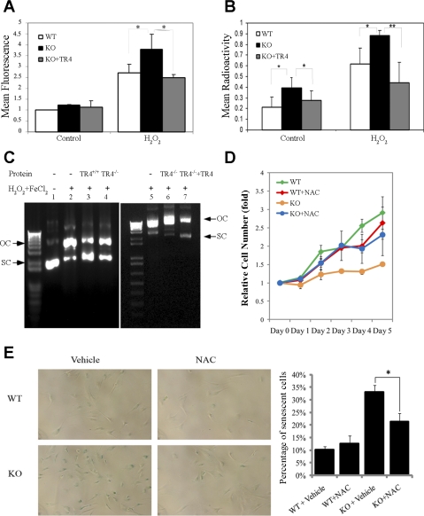
To investigate the cause of the early onset of senescence in TR4−/− MEFs, we assessed the levels of ROS and oxidative DNA damage in MEF cells. TR4−/− MEFs did display higher cellular ROS levels than TR4+/+ MEFs after H2O2 challenging (Fig. 4A), which suggests the involvement of TR4 in the ROS scavenging system. We then measured the single-stranded DNA breaks in MEFs with or without H2O2 treatment. As expected, the intrinsic as well as extrinsic (H2O2-induced) single-stranded DNA breaks were increased in TR4−/− MEFs compared with TR4+/+ MEFs (Fig. 4B), suggesting a protective role of TR4 against DNA damage. To validate the involvement of TR4 in cellular stress defense, we transfected functional TR4 into TR4−/− MEFs and found that restoring functional TR4 into TR4−/− MEFs reduced both endogenous and H2O2-induced ROS and single-stranded DNA breaks significantly (Fig. 4, A and B). These results suggested that TR4 is involved in the antioxidative stress system, one of the major defenses against genotoxic insults to maintain genome integrity, and this might be one of the mechanisms through which TR4 contributes to longevity.
Fig. 4.
Higher cellular levels of ROS, increased DNA single-stranded breaks, and growth arrest in TR4−/− MEFs could be rescued by TR4 or the antioxidant reagent N-acetyl-l-cysteine (NAC). A: TR4+/+ (WT) MEFs and TR4−/− (KO) MEFs transfected with vector or pBabe-TR4 MEFs were treated with 250 μM H2O2 or vehicle control and examined for cellular ROS levels by flow cytometry. Three independent experiments were carried out, and the representative results are shown. Shown are the mean radioactivity ± SD (*P < 0.05 vs. control). B: single stranded DNA breaks in TR4+/+ MEFs and TR4−/− MEFs transfected with vector or pBabe-TR4 MEFs were compared by DNA precipitation, as described in experimental procedures. Three independent experiments were carried out, and the representative results are shown. Shown are the mean fluorescence intensity ± SD (*P < 0.05 vs. control; **P < 0.01 vs. control). C: protective effects on pUC19 plasmid DNA break caused by hydroxyl radical produced by the Fe2+-H2O2 system. Electrophoresis was carried out on a 0.8% agarose gel. Lane 1, control pUC19 DNA; lanes 2 and 5, DNA breaks on pUC19 by Fe2+-H2O2 treatment; lanes 3 and 4, Fe2+- H2O2-treated pUC19 in the presence of cellular proteins from TR4+/+ and TR4−/− MEFs; lanes 6 and 7, Fe2+-H2O2-treated pUC19 in the presence of cellular proteins from TR4−/− MEFs and TR4-restored TR4−/− MEFs. D: WT and KO MEF cells were treated with NAC for 4 days and exposed to 250 μM H2O2 for 30 min and switched back to normal medium. After different periods of time, cell viability was measured by MTT assay. The relative surviving cell numbers compared with control, without H2O2 treatment, were calculated and plotted. E: MEFs from TR4+/+ and TR4−/− embryos were cultured and maintained in vitro and treated with NAC or vehicle for 4 days before SA-β-Gal staining. Representative pictures from 3 independent experiments are shown at left. Quantification results are shown at right. Three independent experiments were carried out; means ± SD are shown (*P < 0.01 vs. control).
To further dissect the anti-ROS actions of TR4, we tested whether TR4 can promote cellular scavenging ability by examining the TR4 capacity for protecting DNA damage in vitro. pUC19 DNA plasmids were treated with hydroxyl radicals to induce DNA breaks and then incubated with cellular proteins from TR4+/+ and TR4−/− MEFs. As shown in Fig. 4C, treatment with Fe2+/H2O2 caused plasmid DNA breaks, which would release SC forms (lane 1) into open circular (OC) forms (lanes 2 and 5); the protective effects against DNA breaks from SC into OC forms were found in the presence of TR4+/+ MEF protein extracts (lane 3). In contrast, there was a less protective effect in the presence of TR4−/− MEF protein extracts in which mainly OC forms were present (Fig. 4C, lanes 4 and 6), and restoring TR4 via pBabe retrovirus infection into TR4−/− reduced the hydroxyl radical-induced DNA breaks (Fig. 4C, lane 7 vs. lane 6). These data support the hypothesis that TR4 promotes the anti-ROS defense capacity via mediating ROS-scavenging pathways and maintenance of DNA integrity.
Senescence and growth arrest in TR4−/− MEFs could be rescued by NAC treatment.
To verify the link between increasing ROS and growth arrest in TR4−/− MEFs, we examined whether supplying antioxidants into TR4−/− cells could rescue cells from the early onset of growth arrest. As shown in Fig. 4D, continuing supplementation with NAC released TR4−/− MEFs from growth arrest, and the growth rate of TR4−/− MEFs under NAC treatment was comparable with TR4+/+ MEFs without NAC. NAC, a precursor for glutathione, can be found naturally in foods and is a powerful antioxidant. Recently, it was found to attenuate senescence in Prx II−/− MEF cells (14), suppress lymphoma, and increase longevity in Atm-deficient mice (35). Furthermore, the number of SA-β-Gal-positive senescent cells in TR4−/− was reduced under 4-day NAC treatment, whereas it had little effect on TR4+/+ cells (Fig. 4E, representative pictures at left, quantification at right). These results indicated that the early onset of senescence in TR4−/− MEFs was caused by accumulation of oxidative damage, and supplementation with NAC can slow down ROS-induced cell growth arrest in TR4−/− MEFs.
Alteration of the expression level of the genes involved in ROS and DNA damage response pathways in TR4−/− MEFs.
The above data have clearly shown that TR4 is involved in cellular oxidative stress defense and maintenance of genome integrity. In an effort to explore the molecular mechanism through which TR4 regulates these cellular events to maintain genome integrity, we screened for TR4 targeted genes in two specific pathways, oxidative stress and antioxidant pathway and DNA damage signaling pathway, with the focus quantitative PCR array. It is not surprising that we found that a number of the genes express differently between TR4+/+ and TR4−/− MEFs (Tables 1 and 2). What caught our interest is that some of the genes have been linked to aging in previous studies. For example, Fen1 mutation yeast displays premature aging (18), and Parp1 is an important determinant in telomere regulation and thus might play a role in the aging process (2). Further investigation is needed to clarify which specific genes are truly responsible for the impaired genome integrity in TR4−/− mice.
Table 1.
Putative TR4 target genes in mouse DDR signaling
| DDR Pathway PCR Array |
||
|---|---|---|
| Gene Name (TR4-Upregulated Genes) | GeneBank access no. | Gene function |
| Parp1 | NM_007415 | Base-excision repair |
| Rad51 | NM_011234 | Homologous recombination repair |
| Trex1 | NM_011637 | Editing mismatched 3′-termini |
| Fen1 | NM_007999 | Other genes related to DNA repair |
| Polk | NM_012048 | |
TR4, testicular nuclear receptor 4; DDR, DNA damage response; Parp1; poly (ADP-ribose) polymerase family, member 1; Rad51, RAD51 homolog (S. cerevisiae); Trex1, 3′ repair exonuclease 1; Fen1, flap structure-specific endonuclease 1; Polk, polymerase κ (DNA directed).
Table 2.
Putative TR4 target genes in mouse oxidative stress and antioxidant defense signals
| Oxidative Stress and Antioxidant Defense PCR Array |
||
|---|---|---|
| Gene Name | GeneBank access no. | Gene function |
| TR4-upregulated genes | ||
| Fancc | NM_007985 | Oxidative stress defense |
| Prdx3 | NM_007452 | Antioxidant (peroxiredoxin) |
| Txnrd2 | NM_013711 | Antioxidant |
| Slc41a3 | NM_027868 | Antioxidant (peroxiredoxin) |
| TR4-downregulated genes | ||
| Gpx5 | NM_010343 | Antioxidants (glutathione peroxidases) |
| Gpx6 | NM_145451 | Antioxidants (glutathione peroxidases) |
Fancc, fanconi anemia, complementation group C; Prdx3, peroxiredoxin 3; Txnrd2, thioredoxin reductase 2; Slc41a3, solute carrier family 41, member 3; Gpx5, glutathione peroxidase 5; Gpx6, glutathione peroxidase 6.
DISCUSSION
We report that TR4−/− mice suffered premature aging with a deficient oxidative stress defense system and compromised genomic integrity. Aging is characterized by the deterioration of physiological functions caused by loss of stress defenses and increased genomic instability (15, 26). Reducing stress tolerance is one hallmark of organism aging (9), and discoveries of the association between longevity and stress resistance in yeast (44), flies (39), and cells of mice (36) support the “multiplex resistance mechanisms” hypothesis that life span augmentation is proportional to the level of resistance to environmental stresses (30). Manipulations that boost multi-stress defense pathways usually lead to extension of both mean and maximal life span (31, 48). Together with our previous findings that TR4 is implicated in cellular response to different kinds of stress, such as oxidative stress (24), UV, and γ-irradiation (Yan SJ, Lee YF, Ting HJ, Liu NC, Liu S, Yeh SD, and Chang C, unpublished observations), our study provides a promising link between TR4, cellular stress defenses, and aging.
What is the molecular mechanism(s) underlying the accelerated aging in TR4−/− mice? Our results imply there that might be more than one mechanism behind this phenomenon. Expression of TR4 in TR4−/− MEFs reduced the endogenous ROS levels and defended exogenous H2O2 insults as well as slowed down the cellular growth arrest, suggesting that TR4 is involved in the ROS-scavenging defense system. TR4−/− mice displayed higher levels of single-stranded DNA damage, and focused quantitative PCR array revealed the reduced expression of genes in DNA damage response and anti-ROS pathways in TR4−/− MEFs, suggesting that TR4 is involved in maintaining genome stability. All these different levels of defense systems form an intricate pathway network of maintaining genome integrity, which is a major factor in longevity (12, 26). More in-depth studies are essential to fully understand TR4 roles in this genomic integrity maintenance network.
ROS status, determined by the balance between ROS production and scavenging, is critical for organism longevity, and pathways that control the ROS would be critical to determine the longevity (12). Mitochondria are one of the free radical generation sites in cells (22). Decreased mitochondrial H2O2 formation is correlated with delayed aging and increased life span in p66Shc knockout mice (28). Interestingly, we have found mitochondrial dysfunction with electron transport chain complex I deficiency in skeletal muscle of TR4−/− mice (Liu S, Lee, YF, Chou S, Uno H, Li G, Brookes P, Massett MP, Wu Q, Chen LM, and Chang C, unpublished observations). It has been reported that a complex I defect induces ROS release in primary open-angle glaucoma patients (16). Is it possible that the complex I defect accounts for the increased oxidative stress in TR4−/− mice and finally leads to premature aging? Further investigation is needed to find out the exact cause of the increased oxidative stress in TR4−/− mice.
Similarly to reports from patients with progeroid syndromes or premature aging mouse models (17, 20, 27), we observed an early onset of senescence in TR4−/− MEFs. Senescence, a state of irreversible cellular growth arrest, is one of the key cellular-responsive programs when cells are exposed to stress (3). How senescence contributes exactly to aging is unclear yet, although it has been commonly accepted that accumulation of senescent cells in organisms, when a certain threshold is reached, might compromise tissue function, and senescence may also impair the regenerative potential of stem cells (7). The early onset of senescence in TR4−/− MEFs might be a result of the excessive oxidative stress since it can be attenuated by supplying the antioxidant NAC.
We identified expression level changes of certain genes in either DNA damage response signaling pathway or oxidative stress and antioxidant defense signaling pathways in TR4−/− mice. Some of the genes have been linked to aging in previous studies, including Fen1 (18) and PARP1 (2). However, more questions have emerged. How does TR4 regulate these genes? Are they responsible for the premature aging in TR4−/− mice? How does TR4 work through/with these genes in normal aging? Further investigation is needed to discover answers to all these questions. Restoration of target genes into TR4−/− cells might be essential to validate their roles in the TR4-regulated cellular oxidative stress defense network.
Unfortunately, no matter how closely the segmental progeroid syndrome in TR4−/− mice resembles the “natural” aging process, the TR4−/− mouse model, like all other progeria mice models (34), does not really represent the true normal aging process. Examination of TR4 expression levels and/or activity throughout the life span in healthy people would hold the key to the understanding of the roles of TR4 and its downstream factors in the natural aging process.
In conclusion, our study of the progeroid syndrome in TR4−/− mice opens up many new avenues for exploration and provides a useful experimental model for further dissecting the molecular basis of aging. In the future, the identification of upstream regulator(s) and/or activators to control TR4 actions will be extremely important and rewarding tasks for revealing the secrets of controlling the fate of life.
GRANTS
This work was supported by NIH-RO1-DK-063212 (C. Chang), ACS-RSG-06-123-01-CNE (Y.-F. Lee), NIH-CA-127548 (Y.-F. Lee), and Taiwan Department of Health Clinical Trial and Research Center of Excellence Grant DOH99-TD-B-111-004.
DISCLOSURES
The authors declare no conflict of interest.
REFERENCES
- 1. Ben-Porath I, Weinberg RA. When cells get stressed: an integrative view of cellular senescence. J Clin Invest 113: 8–13, 2004 [DOI] [PMC free article] [PubMed] [Google Scholar]
- 2. Beneke S, Cohausz O, Malanga M, Boukamp P, Althaus F, Bürkle A. Rapid regulation of telomere length is mediated by poly(ADP-ribose) polymerase-1. Nucleic Acids Res 36: 6309–6317, 2008 [DOI] [PMC free article] [PubMed] [Google Scholar]
- 3. Campisi J, d'Adda di Fagagna F. Cellular senescence: when bad things happen to good cells. Nat Rev Mol Cell Biol 8: 729–740, 2007 [DOI] [PubMed] [Google Scholar]
- 4. Chang C, Da Silva SL, Ideta R, Lee Y, Yeh S, Burbach JP. Human and rat TR4 orphan receptors specify a subclass of the steroid receptor superfamily. Proc Natl Acad Sci USA 91: 6040–6044, 1994 [DOI] [PMC free article] [PubMed] [Google Scholar]
- 5. Chen LM, Wang RS, Lee YF, Liu NC, Chang YJ, Wu CC, Xie S, Hung YC, Chang C. Subfertility with defective folliculogenesis in female mice lacking testicular orphan nuclear receptor 4. Mol Endocrinol 22: 858–867, 2008 [DOI] [PMC free article] [PubMed] [Google Scholar]
- 6. Chen YT, Collins LL, Uno H, Chang C. Deficits in motor coordination with aberrant cerebellar development in mice lacking testicular orphan nuclear receptor 4. Mol Cell Biol 25: 2722–2732, 2005 [DOI] [PMC free article] [PubMed] [Google Scholar]
- 7. Collado M, Blasco MA, Serrano M. Cellular senescence in cancer and aging. Cell 130: 223–233, 2007 [DOI] [PubMed] [Google Scholar]
- 8. Collins LL, Lee YF, Heinlein CA, Liu NC, Chen YT, Shyr CR, Meshul CK, Uno H, Platt KA, Chang C. Growth retardation and abnormal maternal behavior in mice lacking testicular orphan nuclear receptor 4. Proc Natl Acad Sci USA 101: 15058–15063, 2004 [DOI] [PMC free article] [PubMed] [Google Scholar]
- 9. d'Adda di Fagagna F. Living on a break: cellular senescence as a DNA-damage response. Nat Rev Cancer 8: 512–522, 2008 [DOI] [PubMed] [Google Scholar]
- 10. de Boer J, Andressoo JO, de Wit J, Huijmans J, Beems RB, van Steeg H, Weeda G, van der Horst GT, van Leeuwen W, Themmen AP, Meradji M, Hoeijmakers JH. Premature aging in mice deficient in DNA repair and transcription. Science 296: 1276–1279, 2002 [DOI] [PubMed] [Google Scholar]
- 11. Espejel S, Martín M, Klatt P, Martín-Caballero J, Flores JM, Blasco MA. Shorter telomeres, accelerated ageing and increased lymphoma in DNA-PKcs-deficient mice. EMBO Rep 5: 503–509, 2004 [DOI] [PMC free article] [PubMed] [Google Scholar]
- 12. Finkel T, Holbrook NJ. Oxidants, oxidative stress and the biology of ageing. Nature 408: 239–247, 2000 [DOI] [PubMed] [Google Scholar]
- 13. Forster MJ, Morris P, Sohal RS. Genotype and age influence the effect of caloric intake on mortality in mice. FASEB J 17: 690–692, 2003 [DOI] [PMC free article] [PubMed] [Google Scholar]
- 14. Han YH, Kim HS, Kim JM, Kim SK, Yu DY, Moon EY. Inhibitory role of peroxiredoxin II (Prx II) on cellular senescence. FEBS Lett 579: 4897–4902, 2005 [DOI] [PubMed] [Google Scholar]
- 15. Hasty P, Campisi J, Hoeijmakers J, van Steeg H, Vijg J. Aging and genome maintenance: lessons from the mouse? Science 299: 1355–1359, 2003 [DOI] [PubMed] [Google Scholar]
- 16. He Y, Leung KW, Zhang YH, Duan S, Zhong XF, Jiang RZ, Peng Z, Tombran-Tink J, Ge J. Mitochondrial complex I defect induces ROS release and degeneration in trabecular meshwork cells of POAG patients: protection by antioxidants. Invest Ophthalmol Vis Sci 49: 1447–1458, 2008 [DOI] [PubMed] [Google Scholar]
- 17. Herbig U, Ferreira M, Condel L, Carey D, Sedivy JM. Cellular senescence in aging primates. Science 311: 1257, 2006 [DOI] [PubMed] [Google Scholar]
- 18. Hoopes LL, Budd M, Choe W, Weitao T, Campbell JL. Mutations in DNA replication genes reduce yeast life span. Mol Cell Biol 22: 4136–4146, 2002 [DOI] [PMC free article] [PubMed] [Google Scholar]
- 19. Hwangbo DS, Gersham B, Tu MP, Palmer M, Tatar M. Drosophila dFOXO controls lifespan and regulates insulin signalling in brain and fat body. Nature 429: 562–566, 2004 [DOI] [PubMed] [Google Scholar]
- 20. Keyes WM, Wu Y, Vogel H, Guo X, Lowe SW, Mills AA. p63 deficiency activates a program of cellular senescence and leads to accelerated aging. Genes Dev 19: 1986–1999, 2005 [DOI] [PMC free article] [PubMed] [Google Scholar]
- 21. Kim E, Xie S, Yeh SD, Lee YF, Collins LL, Hu YC, Shyr CR, Mu XM, Liu NC, Chen YT, Wang PH, Chang C. Disruption of TR4 orphan nuclear receptor reduces the expression of liver apolipoprotein E/C-I/C-II gene cluster. J Biol Chem 278: 46919–46926, 2003 [DOI] [PubMed] [Google Scholar]
- 22. Kujoth GC, Hiona A, Pugh TD, Someya S, Panzer K, Wohlgemuth SE, Hofer T, Seo AY, Sullivan R, Jobling WA, Morrow JD, Van Remmen H, Sedivy JM, Yamasoba T, Tanokura M, Weindruch R, Leeuwenburgh C, Prolla TA. Mitochondrial DNA mutations, oxidative stress, and apoptosis in mammalian aging. Science 309: 481–484, 2005 [DOI] [PubMed] [Google Scholar]
- 23. Lee YF, Lee HJ, Chang C. Recent advances in the TR2 and TR4 orphan receptors of the nuclear receptor superfamily. J Steroid Biochem Mol Biol 81: 291–308, 2002 [DOI] [PubMed] [Google Scholar]
- 24. Li G, Lee YF, Liu S, Cai Y, Xie S, Liu NC, Bao BY, Chen Z, Chang C. Oxidative stress stimulates testicular orphan receptor 4 through forkhead transcription factor forkhead box O3a. Endocrinology 149: 3490–3499, 2008 [DOI] [PMC free article] [PubMed] [Google Scholar]
- 25. Liu NC, Lin WJ, Kim E, Collins LL, Lin HY, Yu IC, Sparks JD, Chen LM, Lee YF, Chang C. Loss of TR4 orphan nuclear receptor reduces phosphoenolpyruvate carboxykinase-mediated gluconeogenesis. Diabetes 56: 2901–2909, 2007 [DOI] [PubMed] [Google Scholar]
- 26. Lombard DB, Chua KF, Mostoslavsky R, Franco S, Gostissa M, Alt FW. DNA repair, genome stability, and aging. Cell 120: 497–512, 2005 [DOI] [PubMed] [Google Scholar]
- 27. Martin GM, Oshima J. Lessons from human progeroid syndromes. Nature 408: 263–266, 2000 [DOI] [PubMed] [Google Scholar]
- 28. Migliaccio E, Giorgio M, Mele S, Pelicci G, Reboldi P, Pandolfi PP, Lanfrancone L, Pelicci PG. The p66shc adaptor protein controls oxidative stress response and life span in mammals. Nature 402: 309–313, 1999 [DOI] [PubMed] [Google Scholar]
- 29. Mikkelsen L, Bialkowski K, Risom L, Løhr M, Loft S, Møller P. Aging and defense against generation of 8-oxo-7,8-dihydro-2′-deoxyguanosine in DNA. Free Radic Biol Med 47: 608–615, 2009 [DOI] [PubMed] [Google Scholar]
- 30. Miller RA. Cell stress and aging: new emphasis on multiplex resistance mechanisms. J Gerontol A Biol Sci Med Sci 64: 179–182, 2009 [DOI] [PMC free article] [PubMed] [Google Scholar]
- 31. Moskovitz J, Bar-Noy S, Williams WM, Requena J, Berlett BS, Stadtman ER. Methionine sulfoxide reductase (MsrA) is a regulator of antioxidant defense and lifespan in mammals. Proc Natl Acad Sci USA 98: 12920–12925, 2001 [DOI] [PMC free article] [PubMed] [Google Scholar]
- 32. Mu X, Lee YF, Liu NC, Chen YT, Kim E, Shyr CR, Chang C. Targeted inactivation of testicular nuclear orphan receptor 4 delays and disrupts late meiotic prophase and subsequent meiotic divisions of spermatogenesis. Mol Cell Biol 24: 5887–5899, 2004 [DOI] [PMC free article] [PubMed] [Google Scholar]
- 33. Murakami S, Salmon A, Miller RA. Multiplex stress resistance in cells from long-lived dwarf mice. FASEB J 17: 1565–1566, 2003 [DOI] [PubMed] [Google Scholar]
- 34. Puzianowska-Kuznicka M, Kuznicki J. Genetic alterations in accelerated ageing syndromes. Do they play a role in natural ageing? Int J Biochem Cell Biol 37: 947–960, 2005 [DOI] [PubMed] [Google Scholar]
- 35. Reliene R, Schiestl RH. Antioxidants suppress lymphoma and increase longevity in Atm-deficient Mice. J Nutr 137, Suppl 1:229S–232S, 2007 [DOI] [PubMed] [Google Scholar]
- 36. Rudolph KL, Chang S, Lee HW, Blasco M, Gottlieb GJ, Greider C, DePinho RA. Longevity, stress response, and cancer in aging telomerase-deficient mice. Cell 96: 701–712, 1999 [DOI] [PubMed] [Google Scholar]
- 37. Salmon AB, Richardson A, Perez VI. Update on the oxidative stress theory of aging: does oxidative stress play a role in aging or healthy aging? Free Radic Biol Med 48: 642–655, 2010 [DOI] [PMC free article] [PubMed] [Google Scholar]
- 38. Schriner SE, Linford NJ, Martin GM, Treuting P, Ogburn CE, Emond M, Coskun PE, Ladiges W, Wolf N, Van Remmen H, Wallace DC, Rabinovitch PS. Extension of murine life span by overexpression of catalase targeted to mitochondria. Science 308: 1909–1911, 2005 [DOI] [PubMed] [Google Scholar]
- 39. Sepulveda S, Shojaeian P, Rauser CL, Jafari M, Mueller LD, Rose MR. Interactions between injury, stress resistance, reproduction, and aging in Drosophila melanogaster. Exp Gerontol 43: 136–145, 2008 [DOI] [PMC free article] [PubMed] [Google Scholar]
- 40. Serrano M, Blasco MA. Putting the stress on senescence. Curr Opin Cell Biol 13: 748–753, 2001 [DOI] [PubMed] [Google Scholar]
- 41. Struthers L, Patel R, Clark J, Thomas S. Direct detection of 8-oxodeoxyguanosine and 8-oxoguanine by avidin and its analogues. Anal Biochem 255: 20–31, 1998 [DOI] [PubMed] [Google Scholar]
- 42. Tanabe O, Shen Y, Liu Q, Campbell AD, Kuroha T, Yamamoto M, Engel JD. The TR2 and TR4 orphan nuclear receptors repress Gata1 transcription. Genes Dev 21: 2832–2844, 2007 [DOI] [PMC free article] [PubMed] [Google Scholar]
- 43. Trifunovic A, Wredenberg A, Falkenberg M, Spelbrink JN, Rovio AT, Bruder CE, Bohlooly-Y M, Gidlöf S, Oldfors A, Wibom R, Törnell J, Jacobs HT, Larsson NG. Premature ageing in mice expressing defective mitochondrial DNA polymerase. Nature 429: 417–423, 2004 [DOI] [PubMed] [Google Scholar]
- 44. Van Raamsdonk JM, Hekimi S. Deletion of the mitochondrial superoxide dismutase sod-2 extends lifespan in Caenorhabditis elegans. PLoS Genet 5: e1000361, 2009 [DOI] [PMC free article] [PubMed] [Google Scholar]
- 45. Vogel H, Lim DS, Karsenty G, Finegold M, Hasty P. Deletion of Ku86 causes early onset of senescence in mice. Proc Natl Acad Sci USA 96: 10770–10775, 1999 [DOI] [PMC free article] [PubMed] [Google Scholar]
- 46. Willcox BJ, Donlon TA, He Q, Chen R, Grove JS, Yano K, Masaki KH, Willcox DC, Rodriguez B, Curb JD. FOXO3A genotype is strongly associated with human longevity. Proc Natl Acad Sci USA 105: 13987–13992, 2008 [DOI] [PMC free article] [PubMed] [Google Scholar]
- 47. Xie S, Lee YF, Kim E, Chen LM, Ni J, Fang LY, Liu S, Lin SJ, Abe J, Berk B, Ho FM, Chang C. TR4 nuclear receptor functions as a fatty acid sensor to modulate CD36 expression and foam cell formation. Proc Natl Acad Sci USA 106: 13353–13358, 2009 [DOI] [PMC free article] [PubMed] [Google Scholar]
- 48. Yu BP, Chung HY. Stress resistance by caloric restriction for longevity. Ann NY Acad Sci 928: 39–47, 2001 [DOI] [PubMed] [Google Scholar]


