Abstract
Human cytomegalovirus (HCMV) infection causes dramatic alterations of intermediary metabolism, similar to those found in tumor cells. In infected cells, glucose carbon is not completely broken down by the tricarboxylic acid (TCA) cycle for energy; instead it is used biosynthetically. This process requires increased glucose uptake, increased glycolysis and the diversion of glucose carbon, in the form of citrate, from the TCA cycle for use in HCMV-induced fatty acid biosynthesis. The diversion of citrate from the TCA cycle (cataplerosis) requires induction of enzymes to promote glutaminolysis, the conversion of glutamine to -ketoglutarate in order to maintain the TCA cycle (anaplerosis) and ATP production. Such changes could result in heretofore uncharacterized pathogenesis, potentially implicating HCMV as a subtle co-factor in many maladies, including oncogenesis. Recognition of the effects of HCMV, and other viruses, on host cell metabolism will provide new understanding of viral pathogenesis and novel avenues for antiviral therapy.
The Warburg effect and tumor cell metabolism
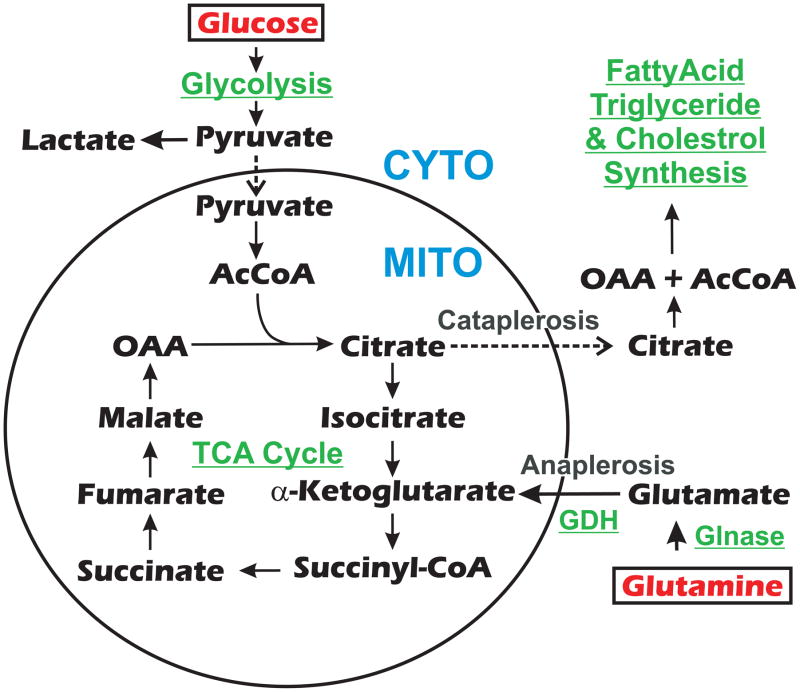
Intermediary metabolism has been resurrected in the minds of many of us who left metabolic studies in the 1970s to pursue the molecular biology revolution. This awakening has been spurred by the discoveries over the past five to eight years in the area of cancer biology, where metabolism has been shown to be significantly altered by oncogenesis 1–6. Additionally, modern technologies in metabolomics (see Glossary), combined with classical biochemistry, cell biology and molecular biology, provide a great opportunity for these studies, at this time. However, the realization that metabolism is dramatically altered in tumor cells derives from an old and classic observation, the Warburg effect. Otto Warburg and colleagues observed, in 1924, that cancer cells metabolize glucose very differently than normal cells 7. Cancer cells, like normal cells, use glycolysis to convert glucose to pyruvate (Figure 1); however, normal cells and cancer cells part dramatically in how they utilize pyruvate. Most normal cells allow pyruvate to enter the tricarboxylic acid (TCA) cycle, where it is catabolized to CO2 and promotes oxidative phosphorylation. Warburg observed that in cancer cells pyruvate is converted to lactate (Figure 1), rather than entering into the TCA cycle, even when there is sufficient oxygen to support mitochondrial oxidative phosphorylation 8. It is now known that some of the pyruvate is also converted to acetyl CoA (AcCoA) in the mitochondrion and begins the TCA cycle by enzymatically combining with oxaloacetic acid (OAA) to form citrate (Figure 1). However, the citrate is shuttled from the TCA cycle and the mitochondrion to be converted back to OAA and AcCoA in the cytoplasm where the AcCoA can be used biosynthetically, primarily for fatty acid synthesis. The development and progression of cancer is often associated with a remarkable increase in de novo fatty acid synthesis 9–11.
Figure 1.
Aspects of glucose and glutamine metabolism and fatty acid synthesis. Reactions within the circle occur in the mitochondrion (MITO), while those outside the circle occur in the cytoplasm (CYTO). Movement across the mitochontrial membrane is indicated by dashed arrows. Enzymes and general enzymatic pathways are shown in green. Abbreviations: TCA Cycle, tricarboxylic acid cycle; Glnase, glutaminase; GDH, glutamate dehydrogenase; AcCoA, acetyl coenzyme A; CoA, coenzyme A.
Such utilization of glucose has several important implications. First, the production of pyruvate from glucose via glycolysis produces just two ATP molecules per molecule of glucose. In contrast, if glucose-derived pyruvate proceeds through the TCA cycle, the NADH produced would have supported oxidative phosphorylation and an additional 36 ATP molecules would have been made. While this appears to be an inefficient utilization of glucose energetically, the shift from the complete breakdown of glucose by cancer cells (and other rapidly proliferating cells) promotes the use of glucose carbon for macromolecule biosynthesis, for example the synthesis of fatty acids needed to construct progeny cells 5, 8.
The second implication of this utilization of glucose is that it deprives the TCA cycle of glucose-derived carbon, which not only limits ATP production, but also limits the production of TCA cycle intermediates which are important biosynthetic precursors, e.g. for amino acids. Cancer cells, or any cells that choose to utilize glucose in this way, must overcome these limitations. It is now known that in many cancer cells exogenous glutamine is used as a carbon source for maintenance of the TCA cycle 1–3. This is accomplished via glutaminolysis, which requires the enzyme glutaminase (Glnase) to deaminate glutamine to glutamate, and glutamate dehydrogenase (GDH) to convert glutamate to -ketoglutarate (Figure 1). Thus glutamine anaplerotically maintains the TCA cycle intermediates (Figure 1) and, in the process, maintains NADH generation for oxidative phosphorylation and ATP production1, 2. In contrast, most normal, quiescent cells use only a small amount of consumed glutamine in this way. Thus both glucose and glutamine metabolism are dramatically altered in tumor cells 2, 3, 12.
Increasing evidence suggests that a variety of infectious agents, including bacteria, parasites and viruses, contribute to oncogenesis 13. Mechanistically these agents might contribute to oncogenesis directly or indirectly. For example, some viruses encode proteins which can transform cells by affecting major cellular growth control proteins such as p53 and the retinoblastoma proteins (RB)14, while others transform the cell by using modified host genes which have integrated into the viral genomes 15. Other viruses, as well as bacteria and parasites, could function more indirectly, through immunosuppression, chronic inflammation, suppression of apoptosis and induction of genetic instability 13. In truth, we have only begun to discover the extent to which infectious agents could contribute to oncogenesis, especially via indirect routes.
In this review we will examine the effects of human cytomegalovirus (HCMV) on cellular metabolism; HCMV is one of the first viruses on which modern metabolic analysis has been performed. The startling result is that HCMV’s modification of glucose and glutamine metabolism is very much like that seen in tumor cells.
HCMV, potentially a subtle co-factor in many maladies
The basics of HCMV and its known pathogenesis are provided in Box 1. In order for HCMV’s replicative cycle to be successful the virus needs to alter the host cell to establish an environment that can accommodate the increased demands for nutrients, energy and macromolecular synthesis that accompany viral infection. These alterations also result in cellular stress that induces stress responses, the consequences of which could be deleterious to the viral infection; for example, most stress responses inhibit translation. Thus during the long HCMV growth cycle the virus must not only induce means to increase energy and macromolecule synthesis, but also alter stress responses to trick the cell into ignoring stress signaling. HCMV is adept at taking control of key cellular processes, e.g. signaling pathways, stress response signaling and metabolism, that affect broad aspects of cellular synthesis, growth and survival 21–42. Thus it is not difficult to see how the pathologic effects of these alterations could implicate HCMV as a subtle cofactor in far more maladies then presently considered. For example, it has long been suggested that HCMV could contribute to atherosclerosis 43–47, and with the advent of more sensitive methods for detection, HCMV has been implicated in the development of several cancers, including glioblastoma 48, 49, colorectal cancer 50 and prostate cancer 51. While there are reports in the literature suggesting that HCMV could be able to transform cells by a hit and run mechanism 52, the virus is not considered to be able to frankly transform cells. However, the recent data proposing associations with cancer have led to the suggestion that HCMV might mediate more subtle means of manipulating cells towards oncogenesis (oncopotentiation), or tumor cells toward more serious malignancy (oncomodulation) 53, 54. The counter argument in any discussion of viral association with tumorogenesis is that tumor tissue could simple provide a favorable tissue niche in which a virus can replicate and that the presence of the virus has little to do with oncogenesis. At this point one cannot rule out this possibility for HCMV; however, during immune suppression HCMV is capable of infecting and replicating in a wide variety of tissues and organs, thus it does not seem to need a favorable tissue niche.
Box 1. HCMV and its pathogenesis: the basics.
HCMV is the prototype β-herpesvirus, characterized by slow growth and restricted species and tissue specificity. It is the largest human herpesvirus, with a genome of greater than 200,000 bp, capable of encoding an estimated 200 or more open reading frames 16. The expression of HCMV genes is temporally regulated where groups of genes are activated more or less in unison at different times post-infection. These include the immediate early (IE) or α genes which are expressed first; followed by early (E) or β gene expression; and finally the late (L) or γ genes are expressed 17. Expression of each temporal class affects the initiation of expression of the next class 17. It is well established that IE gene products function in preparing the cell to execute the viral infection; thus they would be expected to have significant effects on cellular processes. The early genes primarily function in viral DNA synthesis, and the late genes primarily encode virion structural proteins needed to encapsidate the DNA and form the final virion.
HCMV is a significant human pathogen which infects most of the human population by puberty, the primary infection can be unnoticed. Like all herpesviruses, HCMV establishes lifelong latency after the primary infection; the primary sites of HCMV latency are the mononuclear cells of myeloid lineage, from CD34+ haematopoietic stem cells through to peripheral blood monocytes 18–20. HCMVs subsequent reactivation from latency accounts for much of the morbidity associated with the virus which can have severe outcomes: infection in utero may result in central nervous system damage, severe birth defects, deafness or death; infection of immunocompromised individuals can result in severe disease, transplant rejection, organ damage and death.
In the following discussion we will examine the effects of HCMV infection on glucose and glutamine metabolism and the role these play in potentiating fatty acid synthesis needed by the viral infection. Although it remains to be established how HCMV might be involved in oncopotentiation and oncomodulation, the changes in metabolism induced by HCMV are very similar to those seen in tumor cells; thus HCMV-mediated alteration in metabolism could benefit a transforming cell.
Glucose transport in HCMV-infected cells
The first indication that HCMV might alter intermediary metabolism came from early studies showing that glucose uptake was significantly increased in infected cells 55. The transport of glucose requires specific carrier molecules, the glucose transporters (GLUTs). There are 13 different GLUTs in mammalian cells, GLUT 1–12, plus the proton (H+)-myoinositol cotransporter (HMIT) 56, 57. Structurally, the GLUTs can be divided into three classes: GLUT1-4 (class I); GLUT5, 7, 9 and 11 (class II) and GLUT6, 8, 10, 12 and HMIT (class III) 56, 57; all have a differing tissue specificities and affinities for glucose.
The best characterized are the class I GLUTS, GLUT1-4. GLUT1 is ubiquitously distributed and is abundant in fibroblasts, erythrocytes, and endothelial cells 58–60. It is the major GLUT found in human fibroblasts, used in many HCMV studies 36. GLUT2 is a low-affinity transporter for glucose, found primarily in the intestines, pancreatic β-cells, kidney and liver 61. GLUT3 protein distribution is restricted to brain and testis 62, and has the highest affinity for glucose. GLUT4 is the major glucose transporter in adipose tissue, as well as in skeletal and cardiac muscle. Under conditions where GLUT4 is not needed on the cell surface for transport, it resides in intracellular vesicles. Its translocation to the cell surface for glucose transport requires activation of Akt kinase, a consequence of insulin signaling 63, 64.
Examination of GLUTs in HCMV-infected human fibroblasts has shown that the predominant GLUT, GLUT1, is eliminated from the infected cells and replaced by GLUT4, which has a greater transport capacity but is normally not expressed in these cells 36. This change in GLUTs appears to be vital for the success of the HCMV infection in fibroblasts, since specific inhibition of GLUT4 function not only lowers glucose uptake but also dramatically inhibits formation of infectious progeny virions. The mechanism for the replacement of GLUT1 with GLUT4 utilizes HCMV protein IE72, the 72 kDa major immediate early protein, which appears to modulate the mRNA levels, eliminating GLUT1 mRNA and greatly increasing GLUT4 mRNA 36. Previous studies have suggested that overexpression of GLUT4 alone can increase GLUT4 levels on the cell surface65, thus circumventing the Akt-mediated translocation step described above. Thus the dramatic increase in GLUT4 in infected cells might drive GLUT4 onto the cell surface, assuring that glucose uptake increases regardless of the activity of Akt.
Glucose utilization in HCMV-infected cells
For many years it was assumed that the increased uptake of glucose resulted in increased glucose breakdown to promote oxidative phosphorylation, supplying ATP to support the viral infection. However, a number of studies beginning in 2006 dramatically altered the view of glucose metabolism in infected cells. Metabolic flux analysis 34, 35 using liquid chromatography–tandem mass spectrometry (LC-MSMS) analysis and 13C-labeled glucose showed that HCMV institutes its own metabolic program, altering the metabolism pattern observed under normal conditions and considerably stimulating glycolysis, the TCA cycle, and the pyrimidine biosynthetic pathway 34. Of particular note was the efflux of glucose carbon from the TCA cycle, in the form of citrate, to support fatty acid biosynthesis 35 (Figure 1). This sets up a situation which is much like that in tumor cells (Figure 1). Specifically, the increased amounts of glucose taken up by HCMV-infected cells proceed through glycolysis to pyruvate, some of which is converted to lactate in the cytoplasm and some to AcCoA in the mitochondrion. The AcCoA combines with OAA to form citrate, much of which shuttles out of the mitochondrion and is converted back to OAA and AcCoA in the cytoplasm, thus supplying cytoplasmic AcCoA for fatty acid and sterol synthesis. Microarray analysis of gene expression suggested that the metabolic and biosynthetic enzymes needed to increase glycolysis and biosynthesis are upregulated by HCMV 34. Additionally, it was shown that this diversion of glucose carbon for fatty acid synthesis was critical for HCMV infection. Specifically, pharmacological inhibition of fatty acid biosynthesis suppressed HCMV replication 35. Thus in HCMV-infected cells glucose metabolism is altered such that glucose carbon is used primarily for fatty acid biosynthesis and is not broken down in the TCA cycle to promote oxidative phosphorylation.
Glutamine utilization in HCMV-infected cells
The removal of citrate from the citric acid cycle in HCMV-infected cells has the same consequence as it does in tumor cells: there is a great decrease in glucose-derived carbon in the TCA cycle. This presents virally-infected cells with the same problem as tumor cells, the production of TCA cycle intermediates is limited. Since the biosynthesis of these intermediates is the source of NADH for oxidative phosphorylation, the production of ATP is also limited. However, it has been demonstrated that HCMV-infected cells, similar to tumor cells, activate a means to use glutamine anaplerotically to maintain the TCA cycle 29(Figure 1).
That an alternative carbon source must be used in HCMV-infected cells came from the observation that normal human fibroblasts infected with HCMV for at least 12 hours remained viable upon glucose deprivation 29, while uninfected cells rapidly lose viability without glucose 29, 66. Since tumor cells use glutamine as the alternative carbon source to maintain the TCA cycle, it was suspected that glutamine might be used for anaplerosis in HCMV-infected cells 34. Viral growth analyses supported this hypothesis, showing that glutamine deprivation stopped the formation of infectious virions; in addition, glutamine was found to be a major source of ATP in infected human fibroblasts, but not in uninfected cells 29. HCMV-induced glutamine utilization was also indicated by increased glutamine uptake and increased ammonia production, which results from glutaminolysis. The increase in glutamine utilization corresponded to increased levels and enzymatic activities of glutaminase and glutamate dehydrogenase, the two enzymes that must be activated to convert glutamine to -ketoglutarate for entry into the TCA cycle (Figure 1). Finally, confirmation that glutamine is used to maintain the TCA cycle was established by the finding that the TCA cycle intermediate -ketoglutarate, the product of glutaminolysis (Figure 1), could rescue ATP production and viral growth under conditions of glutamine deprivation 29. Thus in HCMV-infected cells, as in many tumor cells, a program is activated whereby glutamine uptake is increased for the purpose of maintaining the TCA cycle and ATP production, thereby allowing glucose to be used biosynthetically, e.g. for fatty acid synthesis.
Mechanisms by which HCMV controls glycolysis, glutaminolysis and citrate cataplerosis
At this point, the mechanisms by which HCMV alters cellular metabolism are largely speculative; however, we can propose models for experimental analysis.
Glycolysis
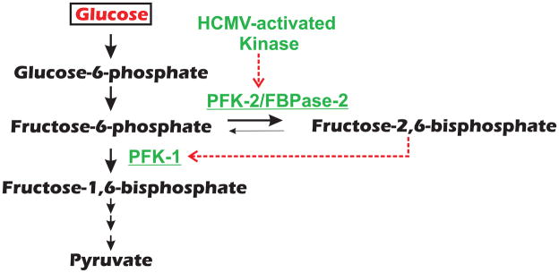
Microarray analysis suggests that HCMV infection increases the levels of mRNA encoding enzymes in the glycolytic pathway 34, providing one means of increasing glycolysis. Whether this is due to transcriptional activation or increased mRNA stability remains to be determined. The virus might also mediate mechanisms that allosterically activate glycolytic enzymes. In this regard a potential means for HCMV to activate glycolysis is to control the production of fructose 2,6-bisphosphate (F-2,6-P2), a potent positive glycolytic signaling molecule (reviewed in 67, 68). As shown in Figure 2, the first step of glycolysis is the conversion of glucose to fructose-6-phosphate (F-6-P). To proceed in glycolysis the enzyme 6-phosphofructo-1-kinase (PFK-1) converts F-6-P to fructose-1,6-bisphosphate (F-1,6-P2); this is the first committed step in glycolysis, thus it is tightly controlled. Interestingly, negative effectors of PFK-1 include citrate and ATP, two molecules that HCMV-infected cells are striving to produce; thus a mechanism to overcome negative effectors seems appropriate. The positive effector of PFK-1 is F-2,6-P2 (Figure 2); the activity of PFK-1 is very low unless there are physiological (micromolar) concentrations of F-2,6-P2 to activate PFK-1 and to relieve inhibition.
Figure 2.
The control of glycolysis by fructose-2,6-bisphosphate. The bifunctional enzyme 6-phosphofructo-2-kinase (PFK-2)/fructose-2,6-bisphosphatase (FBPase-2) can both phosphorylate and dephosphorylate fructose-6-phosphate at the 2 position. A number of kinases (some of which are activated during HCMV infection) can phosphorylate and activate the kinase activity of the enzyme. This increases the levels of fructose-2,6-bisphosphate, which allosterically activates 6-phosphofructo-1-kinase (PFK-1), a key enzyme in glycolytic control. Activation steps are indicated by dashed red arrows.
Conditions known to increase glycolysis in the heart are accompanied by concomitant increases in F-2,6-P2. The concentration of F-2,6-P2 is determined by the bifunctional enzyme 6-phosphofructo-2-kinase (PFK-2)/fructose-2,6-bisphosphatase (FBPase-2), which controls both phosphorylation and dephosphorylation at the 2 position. Mammalian tissues have distinct isozymes of PFK-2/FBPase-2 and in most cases phosphorylation determines whether the predominant activity of the enzyme is as a protein kinase or a phosphatase. Phosphorylation usually leads to increased activity of the PFK-2 kinase activity, thus increasing F-2,6-P2 levels, which would activate PFK-1 and advance glycolysis. PFK-2/FBPase-2 is the target of many protein kinases, including AMP-activated protein kinase (AMPK), mitogen activated protein kinase activated protein kinase 1 (MAPKAPK-1), p70 ribosomal S6 kinase (p70S6K) and Akt. In HCMV-infected cells many of the above kinases are known to be active, e.g. AMPK 23, Akt 24,69 and p70S6K 22. Thus one prediction is that HCMV-infected cells have increased levels of F-2,6-P2 due to activation of PFK-2, possibly due to HCMV-mediated activation of PFK-2-activating kinases (Figure 2).
In this regard a recent study established that inhibition of calmodulin-dependent kinase kinase (CaMKK) blocks HCMV-mediated activation of glycolysis and restricts HCMV replication 70. That a calcium-dependent kinase could be involved is supported by the observations that HCMV infection mobilizes calcium stores 71 and successful infection is very dependent on the calcium homeostasis set up during infection 27. The mechanism by which CaMKK is utilized by HCMV to activate glycolysis remains to be determined; however, one possibility is CaMKK-mediated activation of calmodulin-dependent kinase 1 (CaMK1), another kinase suggested to be able to phosphorylate PFK-270.
Glutaminolysis
Initially, glutamine transport is increased in infected cells 29; the mechanisms used by HCMV to increase glutamine uptake has not yet been studied. However, it is known that glutaminase (Glnase) and glutamate dehydrogenase (GDH) enzyme levels and enzymatic activities are greatly increased in infected cells to promote the conversion of glutamine to -ketoglutarate (Figure 1). The mechanism by which HCMV increases the levels of these enzymes has not yet been determined; however, the virus could utilize the same mechanism used by tumor cells. Recent studies have shown that oncogenic levels of c-Myc induce a transcriptional program that promotes glutamine uptake and glutamine conversion to -ketoglutarate, resulting in cellular addiction to glutamine as a bioenergetic substrate 3. In this regard, many tumor cells show increased c-Myc levels and glutamine dependence 3, 66. Several studies have suggested that c-Myc mRNA and protein levels are increased in HCMV-infected cells 72–74.
Citrate cataplerosis
An important step in the metabolic program set up by HCMV is the induction of citrate cataplerosis, specifically the activation of mechanisms promoting the efflux of citrate from the mitochondria to the cytosolic compartment, where it is converted by ATP-citrate lyase to OAA and AcCoA (Figure 1). Mitochondrion/cytoplasmic shuttling mechanisms have been characterized that shuttle citrate and pyruvate or citrate and malate 75, 76; these could be targets for HCMV manipulation. A number of HCMV proteins traffic through the mitochondria-associated membrane (MAM) compartment and localize in mitochondrial membranes (reviewed in 77). It has been proposed that viral proteins affecting the MAM might manipulate cellular processes including calcium signaling, lipid synthesis and transfer, bioenergetics, metabolic flow, and apoptosis 77. Thus citrate shuttling could also be affected.
Fatty acid synthesis
The means by which HCMV activate fatty acid synthesis remains undefined. However, the increased fatty acid synthesis detected in HCMV-infected cells 35 requires the activation of a number of lipogenic enzymes, similar to the events seen during adipocyte differentiation 78. Thus it is possible that the virus induces a modified form of adipocyte differentiation in infected cells. This is supported by the fact that HCMV-mediated upregulation of GLUT4 36, the glucose transporter found in abundance in adipocytes, is induced during adipocyte differentiation 78.
Why does the virus need such an increase in fatty acid synthesis?
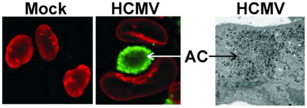
Since HCMV is an enveloped virus, there are clearly fatty acid requirements for membranes from which the envelopes can be derived. However, examination of HCMV-infected cells suggests that membrane formation is needed for many other aspects of viral infection. It is well known that infected cells are enlarged (cytomegalia), which would require additional plasma membrane. In addition, the virion egress and assembly process appears to require a significant amount of membrane. The immunofluorescence micrographs in Figure 3 show two striking features of HCMV-infected cells. First is the remodeled nucleus, which is much enlarged and often takes on a characteristic concave, kidney shape. The significantly enlarged nucleus in comparison to mock infected cells indicates another requirement for additional membranes for nuclear expansion. The second feature shown in Figure 3 is a perinuclear structure known as the cytoplasmic assembly compartment or assembly complex (AC) nestled against the concave surface of the kidney-shaped nucleus 79–81. Current models of HCMV egress and assembly suggest that nucleocapsids, which form in the remodeled nucleus, exit the nucleus and move through the AC where the virion tegument layer and envelope are acquired. The AC is a highly vesicular viral organelle composed of viral proteins as well as components from the endoplasmic reticulum, Golgi, trans-Golgi network and early endosomes 39, 81–83. Numerous viral structural proteins have been identified as part of the AC, e.g. tegument proteins (pp28, pp65) 79 and viral glycoproteins (gB, gH, gL, gO, gp65) 79, 82, 84. A rigorous study of AC structure 81 resulted in a three-dimensional model proposing that the AC is cylindrical and composed of organelle-specific vesicles which form nested cylinders. Each vesicular cylinder is proposed to contain a specific set of virion structural proteins (tegument proteins) which transfer to nucleocapsids as they move toward the center of the AC 81. In agreement with this model, ultrastructural studies suggest that full tegumentation requires the nucleocapsids to migrate through circularly arrayed vesicles 85–87. The electron micrograph in Figure 3 shows the vesicular nature of the AC. Thus another need for membranes in the infected cells is for the vesicles concentrated within the AC and found throughout the cytoplasm of infected cells.
Figure 3.
The enlargement and structural changes in the nucleus and the formation of the assembly compartment during HCMV infection. The two immunofluorescence micrographs show normal nuclei in mock infected cells (left) and the significantly enlarged and kidney shaped nuclei seen in HCMV-infected cells (middle, the nuclei are visualized by staining for lamin B, red). In infected cell nuclei, a perinuclear structure known as the cytoplasmic assembly compartment or assembly complex (AC; visualized by staining for the HCMV tegument protein pp28, green) is seen nestled against the concave surface of the kidney-shaped nucleus 79–81. The immunofluorescence micrographs are reproduced from Ref. 38 with permission from the Journal of Virology. As shown in the electron micrograph (right), the AC is made of many vesicular structures; the bar indicates 2 m.
Considering the enlargement of the infected cell and the infected cell nucleus, as well as the vesicular nature of the AC, it is not difficult to understand why the virus must alter glucose and glutamine utilization in order to allow glucose to be used biosynthetically for the production of fatty acids to fulfill the significant membrane requirement in infected cells.
Concluding remarks
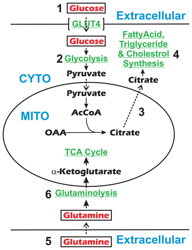
HCMV infection dramatically alters cellular metabolism, and in this review we have focused on how the virus alters glucose and glutamine metabolism and fatty acid synthesis (Figure 4). Changes to glucose metabolism include increased glucose uptake through the induction of the glucose transporter GLUT4, upregulation of glycolytic enzymes and likely allosteric activation of glycolysis. Glucose is not completely broken down by the TCA cycle but maintained intact for biosynthetic purposes. This is primarily accomplished by the transport of citrate from the mitochondrion to the cytoplasm. In order to counter this loss of glucose carbon from the TCA cycle, HCMV affects glutamine metabolism by increasing exogenous glutamine uptake and inducing glutaminolysis enzymes to convert the imported glutamine to -ketoglutarate to anapleroetically maintain the TCA cycle. In the cytoplasm, HCMV upregulates fatty acid and sterol synthesis so that AcCoA, derived from the cytoplasmic citrate, is used in fatty acid synthesis. The induction of fatty acid synthetic enzymes and the induction of GLUT4 could be related, as each would occur if HCMV infection induces a form of adipocyte differentiation.
Figure 4.
HCMV-mediated modifications in cellular processes that lead to alterations of glucose and glutamine metabolism and fatty acid synthesis. (1) Glucose uptake increases through the induction of the glucose transporter GLUT4 which replaces the transporter GLUT1. (2) Glycolytic enzymes are upregulated and it is likely that glycolysis is allosterically activated by infection. (3) Glucose carbons are diverted from the TCA cycle through transport of citrate from the mitochondrion to the cytoplasm. (4) Fatty acid and sterol synthesis are upregulated so that AcCoA derived from cytoplasmic citrate can be used for fatty acid synthesis. The induction of fatty acid synthesis enzymes and the induction of GLUT4 may be related, as each would occur if HCMV infection induces a form of adipocyte differentiation. (5) Exogenous gutamine uptake is increased in infected cells. (6) Glutaminolysis enzymes are induced to convert the imported glutamine to -ketoglutarate to anapleroetically maintain the TCA cycle. Abbreviations: CYTO, cytoplasm; MITO, mitochondrion.
While these changes are quite dramatic they are likely to represent only a fraction of the metabolic changes wrought by HCMV infection. Such changes are likely to contribute to forms of HCMV pathogenesis that could be very different from those that have been studied and documented to this point. For example, HCMV and other viruses which induce metabolic changes similar to HCMV might play a greater role in oncogenesis than previously recognized.
At this point our understanding of the mechanisms used by HCMV to alter metabolic processes is very limited and requires targeted study. It is essential that the metabolic alterations wrought by HCMV infection be worked out using a combination of modern mass spectrometric based metabolic flux analysis, classical biochemistry and enzymology, and cellular and molecular biological techniques. In a broader sense, similar studies must be initiated with all viral families; it is likely that other viruses will take very different approaches in their interactions with host metabolism. The complete understanding of viral interactions with host metabolism will provide new targets for antiviral therapy, for example drugs that might inhibit GLUT4, glutamine uptake or key metabolic enzymes with which the viral infection is dependent. As metabolism is experiencing a renaissance, it is unfortunate to find that many find it intimidating or see it as boring, we hope that this review shows it otherwise.
Acknowledgments
The authors would like to thank Sherrill Adams and Alan Diehl for helpful discussions and comments on the manuscript. JCA acknowledges funding through Public Health Service Grant R01 CA157679-01 awarded by the National Cancer Institute.
Glossary
- Anaplerosis and cataplerosis
anaplerosis is the process of replenishing TCA cycle intermediates that have been removed for cataplerosis, i.e. removed from the cycle for biosynthetic reactions. For example, in HCMV-infected cells citrate is cataplerotically removed from the TCA for fatty acid synthesis, while glutamine is anaplerotically used to fill the TCA cycle to compensate for the lost citrate
- Glutaminolysis
the set of metabolic reactions that converts glutamine to -ketogluterate, allowing glutamine to be used anaploretically to form the intermediates of the TCA cycle and support oxidative phosphorylation
- Glycolysis
the metabolic pathway that converts glucose to pyruvate
- Metabolomics
the systematic study of a cell’s or organism’s small-molecule metabolite profiles. Using isotopically-labeled compounds, for example 13C-labeled glucose or glutamine, the metabolites resulting from metabolism can be detected by techniques involving gas chromatography-mass spectrometry (GC-MS) or nuclear magnetic resonance (NMR) spectroscopy. The resulting data can define the metabolic flux and the metabolome, the collection of all metabolites in a biological sample
- Tricarboxylic acid (TCA) cycle
also known as the Kreb’s cycle and the citric acid cycle and was established by the work of Albert Szent-Györgyi and Hans Krebs. It is a succession of enzyme-catalyzed reactions that occur in the mitochondia which are part of a metabolic pathway involved in the chemical conversion of carbohydrates, fats and proteins into carbon dioxide and water to generate usable energy. The cycle begins with combination of the two-carbon acetyl group from acetyl-CoA (derived from pyruvate via glycolysis) to the four carbons of oxaloacetate to form the six-carbon citrate molecule. The citrate is biochemically altered as it moves through the TCA cycle, losing two carboxyl groups as CO2, returning to the four carbon oxaloacetate to allow the cycle to run again with more AcCoA derived from glucose by glycolysis. As a result of the oxidative reactions of the TCA cycle, electrons are transferred to NAD+ to form NADH. In turn, NADH transfers its electrons to the oxidative phosphorylation pathway which, along with oxygen, forms ATP
Footnotes
Publisher's Disclaimer: This is a PDF file of an unedited manuscript that has been accepted for publication. As a service to our customers we are providing this early version of the manuscript. The manuscript will undergo copyediting, typesetting, and review of the resulting proof before it is published in its final citable form. Please note that during the production process errors may be discovered which could affect the content, and all legal disclaimers that apply to the journal pertain.
Referances
- 1.DeBerardinis RJ, et al. Beyond aerobic glycolysis: Transformed cells can engage in glutamine metabolism that exceeds the requirement for protein and nucleotide synthesis. Proc Natl Acad Sci USA. 2007;104:19345–19350. doi: 10.1073/pnas.0709747104. [DOI] [PMC free article] [PubMed] [Google Scholar]
- 2.DeBerardinis RJ, et al. Brick by brick: metabolism and tumor cell growth. Current Opinion in Genetics & Development. 2008;18:54–61. doi: 10.1016/j.gde.2008.02.003. [DOI] [PMC free article] [PubMed] [Google Scholar]
- 3.Wise DR, et al. Myc regulates a transcriptional program that stimulates mitochondrial glutaminolysis and leads to glutamine addiction. Proc Natl Acad Sci USA. 2008;105:18782–18787. doi: 10.1073/pnas.0810199105. [DOI] [PMC free article] [PubMed] [Google Scholar]
- 4.Wellen KE, et al. ATP-citrate lyase links cellular metabolism to histone acetylation. Science. 2009;324:1076–1080. doi: 10.1126/science.1164097. [DOI] [PMC free article] [PubMed] [Google Scholar]
- 5.Vander Heiden MG, et al. Evidence for an alternative glycolytic pathway in rapidly proliferating cells. Science. 2010;329:1492–1499. doi: 10.1126/science.1188015. [DOI] [PMC free article] [PubMed] [Google Scholar]
- 6.Ward PS, et al. The common feature of leukemia-associated IDH1 and IDH2 mutations is a neomorphic enzyme activity converting alpha- ketoglutarate to 2-hydroxyglutarate. Cancer Cell. 2010;17:225–234. doi: 10.1016/j.ccr.2010.01.020. [DOI] [PMC free article] [PubMed] [Google Scholar]
- 7.Warburg O, et al. Über den Stoffwechsel der Carcinomzelle. Biochem Z. 1924;152:319–344. [Google Scholar]
- 8.Vander Heiden MG, et al. Understanding the Warburg effect: the metabolic requirements of cell proliferation. Science. 2009;324:1029–1033. doi: 10.1126/science.1160809. [DOI] [PMC free article] [PubMed] [Google Scholar]
- 9.Kuhajda FP. Fatty acid synthase and cancer: new application of an old pathway. Cancer Res. 2006;66:5977–5980. doi: 10.1158/0008-5472.CAN-05-4673. [DOI] [PubMed] [Google Scholar]
- 10.Menendez JA, Lupu R. Fatty acid synthase and the lipogenic phenotype in cancer pathogenesis. Nat Rev Cancer. 2007;7:763–777. doi: 10.1038/nrc2222. [DOI] [PubMed] [Google Scholar]
- 11.Swinnen JV, et al. Increased lipogenesis in cancer cells: new players, novel targets. Curr Opin Clin Nutr Metab Care. 2006;9:358–365. doi: 10.1097/01.mco.0000232894.28674.30. [DOI] [PubMed] [Google Scholar]
- 12.Newsholme EA, et al. The role of high rates of glycolysis and glutamine utilization in rapidly dividing cells. Biosci Rep. 1985;5:393–400. doi: 10.1007/BF01116556. [DOI] [PubMed] [Google Scholar]
- 13.zur Hausen H. The search for infectious causes of human cancers: Where and why. Virology. 2009;392:1–10. doi: 10.1016/j.virol.2009.06.001. [DOI] [PubMed] [Google Scholar]
- 14.O’Shea CC. DNA tumor viruses -- the spies who lyse us. Current Opinion in Genetics & Development. 2005;15:18–26. doi: 10.1016/j.gde.2004.12.010. [DOI] [PubMed] [Google Scholar]
- 15.Chen HR, Barker WC. Nucleic acid sequence database VI: Retroviral oncogenes and cellular proto-oncogenes. DNA. 1985;42:171–182. doi: 10.1089/dna.1985.4.171. [DOI] [PubMed] [Google Scholar]
- 16.Murphy E, et al. Coding potential of laboratory and clinical strains of human cytomegalovirus. Proc Natl Acad Sci USA. 2003;25:14976–14981. doi: 10.1073/pnas.2136652100. [DOI] [PMC free article] [PubMed] [Google Scholar]
- 17.Mocarski ES, Courcelle CT. Cytomegaloviruses and their replication. Fields Virology. 2001;2:2629–2673. [Google Scholar]
- 18.Goodrum F, et al. Human cytomegalovirus sequences expressed in latently infected individuals promote a latent infection in vitro. Blood. 2007;110:937–945. doi: 10.1182/blood-2007-01-070078. [DOI] [PMC free article] [PubMed] [Google Scholar]
- 19.Mendelson M, et al. Detection of endogenous human cytomegalovirus in CD34+ bone marrow progenitors. J Gen Virol. 1966;77:3099–3102. doi: 10.1099/0022-1317-77-12-3099. [DOI] [PubMed] [Google Scholar]
- 20.Reeves MB, et al. Latency, chromatin remodeling, and reactivation of human cytomegalovirus in the dendritic cells of healthy carriers. Proc Natl Acad Sci U S A. 2005;102:4140–4145. doi: 10.1073/pnas.0408994102. [DOI] [PMC free article] [PubMed] [Google Scholar]
- 21.Kudchodkar SB, et al. Human cytomegalovirus infection alters the substrate specificities and rapamycin sensitivities of raptor- and rictor-containing complexes. Proc Natl Acad Sci USA. 2006;103:14182–14187. doi: 10.1073/pnas.0605825103. [DOI] [PMC free article] [PubMed] [Google Scholar]
- 22.Kudchodkar S, et al. Human cytomegalovirus infection induces rapamycin insensitive phosphorylation of downstream effectors of mTOR kinase. J Virol. 2004;78:11030–11039. doi: 10.1128/JVI.78.20.11030-11039.2004. [DOI] [PMC free article] [PubMed] [Google Scholar]
- 23.Kudchodkar SB, et al. AMPK-mediated inhibition of mTOR kinase is circumvented during immediate-early times of human cytomegalovirus infection. J Virol. 2007;81:3649–3651. doi: 10.1128/JVI.02079-06. [DOI] [PMC free article] [PubMed] [Google Scholar]
- 24.Yu Y, Alwine JC. Human cytomegalovirus major immediate-early proteins and simian virus 40 large T antigen can inhibit apoptosis through activation of the phosphatidylinositide 3′-OH kinase pathway and cellular kinase Akt. J Virol. 2002;76:3731–3738. doi: 10.1128/JVI.76.8.3731-3738.2002. [DOI] [PMC free article] [PubMed] [Google Scholar]
- 25.Alwine JC. Modulation of host cell stress responses by human cytomegalovirus. In: Shenk TE, Stinski MF, editors. Current Topics in Microbiology and Immunology, Human Cytomegalovirus. Springer; 2008. pp. 263–279. [DOI] [PubMed] [Google Scholar]
- 26.Isler JA, et al. Human cytomegalovirus infection activates and regulates the unfolded protein response. J Virol. 2005;79:6890–6899. doi: 10.1128/JVI.79.11.6890-6899.2005. [DOI] [PMC free article] [PubMed] [Google Scholar]
- 27.Isler JA, et al. Production of infectious HCMV virions is inhibited by drugs that disrupt calcium homeostasis in the endoplasmic reticulum. J Virol. 2005;79:15338–15397. doi: 10.1128/JVI.79.24.15388-15397.2005. [DOI] [PMC free article] [PubMed] [Google Scholar]
- 28.Yu Y, et al. Effects of simian virus 40 large and small tumor antigens on mammalian target of rapamycin (mTOR) signaling: Small tumor antigen mediates hypophosphorylation of eIF4E-binding protein 1 late in infection. J Virol. 2005;79:6882–6889. doi: 10.1128/JVI.79.11.6882-6889.2005. [DOI] [PMC free article] [PubMed] [Google Scholar]
- 29.Chambers JW, et al. Glutamine metabolism is essential for human cytomegalovirus infection. J Virol. 2010;84:1867–1873. doi: 10.1128/JVI.02123-09. [DOI] [PMC free article] [PubMed] [Google Scholar]
- 30.Hakki M, et al. Binding and nuclear relocalization of protein kinase R by human cytomegalovirus TRS1. Journal of Virology. 2006;80:11817–11826. doi: 10.1128/JVI.00957-06. [DOI] [PMC free article] [PubMed] [Google Scholar]
- 31.Walsh D, et al. Regulation of the translation initiation factor eIF4F by multiple mechanisms in human cytomegalovirus-infected cells. J Virol. 2005;79:8057–8064. doi: 10.1128/JVI.79.13.8057-8064.2005. [DOI] [PMC free article] [PubMed] [Google Scholar]
- 32.Moorman NJ, et al. Human cytomegalovirus protein UL38 inhibits host cell stress responses by antagonizing the tuberous sclerosis protein complex. Cell Host & Microbe. 2008;3:1–10. doi: 10.1016/j.chom.2008.03.002. [DOI] [PMC free article] [PubMed] [Google Scholar]
- 33.Xuan B, et al. Human cytomegalovirus protein pUL38 induces ATF4 expression, inhibits persistent JNK phosphorylation, and suppresses endoplasmic reticulum stress-induced cell death. J Virol. 2009;83:3463–3474. doi: 10.1128/JVI.02307-08. [DOI] [PMC free article] [PubMed] [Google Scholar]
- 34.Munger J, et al. Dynamics of the cellular metabolome during human cytomegalovirus infection. PLoS Pathogens. 2006;2:1165–1175. doi: 10.1371/journal.ppat.0020132. [DOI] [PMC free article] [PubMed] [Google Scholar]
- 35.Munger J, et al. Systems-level metabolic flux profiling identifies fatty acid synthesis as a target for antiviral therapy. Nature Biotechnology. 2008;26:1179 – 1186. doi: 10.1038/nbt.1500. [DOI] [PMC free article] [PubMed] [Google Scholar]
- 36.Yu Y, et al. Human cytomegalovirus activates glucose transporter 4 expression to increase glucose uptake during infection. J Virol. 2011;85:1573–1580. doi: 10.1128/JVI.01967-10. [DOI] [PMC free article] [PubMed] [Google Scholar]
- 37.McArdle J, et al. Inhibition of calmodulin-dependent kinase kinase blocks human cytomegalovirus-induced glycolytic activation and severely attenuates production of viral progeny. Journal of Virology. 2011;85:705–714. doi: 10.1128/JVI.01557-10. [DOI] [PMC free article] [PubMed] [Google Scholar]
- 38.Buchkovich NJ, et al. Role of the endoplasmic reticulum chaperone BiP, SUN domain proteins, and dynein in altering nuclear morphology during human cytomegalovirus infection. J Virol. 2010;84:7005–7017. doi: 10.1128/JVI.00719-10. [DOI] [PMC free article] [PubMed] [Google Scholar]
- 39.Buchkovich NJ, et al. Human cytomegalovirus specifically controls the levels of the endoplasmic reticulum chaperone BiP/GRP78 which is required for virion assembly. J Virol. 2008;82:31–39. doi: 10.1128/JVI.01881-07. [DOI] [PMC free article] [PubMed] [Google Scholar]
- 40.Buchkovich NJ, et al. The endoplasmid reticulum chaperone BiP/GRP78 is important in the structure and function of the HCMV assembly compartment. J Virol. 2009;83:11421–11428. doi: 10.1128/JVI.00762-09. [DOI] [PMC free article] [PubMed] [Google Scholar]
- 41.Buchkovich NJ, et al. Human cytomegalovirus induces the endoplasmic reticulum chaperone BiP through increased transcription and activation of translation by using the BiP internal ribosome entry site. J Virol. 2010;84:11479–11486. doi: 10.1128/JVI.01330-10. [DOI] [PMC free article] [PubMed] [Google Scholar]
- 42.Buchkovich NJ, et al. The TORrid affairs of viruses: effects of mammalian DNA viruses on the PI3K-Akt-mTOR signaling pathway. Nature Reviews Microbiology. 2008;6:265–275. doi: 10.1038/nrmicro1855. [DOI] [PMC free article] [PubMed] [Google Scholar]
- 43.Melnick JL, et al. Cytomegalovirus and atherosclerosis. Arch Immunol Therapiae Exper. 1996;44:297–302. [PubMed] [Google Scholar]
- 44.Speir E. Cytomegalovirus gene regulation by reactive oxygen species. Agents of atherosclerosis. Ann NY Acad Sci. 2000;889:363–374. doi: 10.1111/j.1749-6632.2000.tb06200.x. [DOI] [PubMed] [Google Scholar]
- 45.Mattila KJ, et al. Role of infection as a rick factor for atherosclerosis, myocardiak infarction, and stroke. Clin Inf Diseases. 1998;26:719–734. doi: 10.1086/514570. [DOI] [PubMed] [Google Scholar]
- 46.Cinatl J, et al. Modulatory effects of human cytomegalovirus infection on malignant properties of cancer cells. Intervirology. 1997;39:259–269. doi: 10.1159/000150527. [DOI] [PubMed] [Google Scholar]
- 47.Streblow DN, et al. Mechanisms of cytomegalovirus-accelerated vascular disease: induction of paracrine factors that promote angiogenesis and wound healing. Current Topics in Microbiology & Immunology. 2008;325:397–415. doi: 10.1007/978-3-540-77349-8_22. [DOI] [PMC free article] [PubMed] [Google Scholar]
- 48.Cobbs CS, et al. Modulation of oncogenic phenotype in human glioma cells by cytomegalovirus IE1-mediated mitogenicity. Cancer Research. 2008;68:724–730. doi: 10.1158/0008-5472.CAN-07-2291. [DOI] [PubMed] [Google Scholar]
- 49.Mitchell DA, et al. Sensitive detection of human cytomegalovirus in tumors and peripheral blood of patients diagnosed with glioblastoma. Neuro-Oncology. 2008;10(1):10–18. doi: 10.1215/15228517-2007-035. [DOI] [PMC free article] [PubMed] [Google Scholar]
- 50.Harkins L, et al. Specific localisation of human cytomegalovirus nucleic acids and proteins in human colorectal cancer. Lancet. 2002;360:1557–1563. doi: 10.1016/S0140-6736(02)11524-8. [DOI] [PubMed] [Google Scholar]
- 51.Samanta M, et al. High prevalence of human cytomegalovirus in prostatic intraepithelial neoplasia and prostatic carcinoma. Journal of Urology. 2003;170:998–1002. doi: 10.1097/01.ju.0000080263.46164.97. [DOI] [PubMed] [Google Scholar]
- 52.Shen Y, et al. Human cytomegalovirus IE1 and IE2 proteins are mutagenic and mediate “hit-and-run” oncogenic transformation in cooperation with the adenovirus E1A proteins. Proc Natl Acad Sci USA. 1997;94:3341–3345. doi: 10.1073/pnas.94.7.3341. [DOI] [PMC free article] [PubMed] [Google Scholar]
- 53.Michaelis M, et al. The story of human cytomegalovirus and cancer: increasing evidence and open questions. Neoplasia. 2009;11:1–9. doi: 10.1593/neo.81178. [DOI] [PMC free article] [PubMed] [Google Scholar]
- 54.Michaelis M, et al. Oncomodulation by human cytomegalovirus: evidence becomes stronger. Med Microbiol Immunol. 2009;198:79–81. doi: 10.1007/s00430-009-0107-8. [DOI] [PubMed] [Google Scholar]
- 55.Landini MP. Early enhanced glucose uptake in human cytomegalovirus-infected cells. J Gen Virology. 1984;65:1229–1232. doi: 10.1099/0022-1317-65-7-1229. [DOI] [PubMed] [Google Scholar]
- 56.Joost HG, et al. Nomenclature of the GLUT/SLC2A family of sugar/polyol transport facilitators. Am J Physiol Endocrinol Metab. 2002;282:E974–976. doi: 10.1152/ajpendo.00407.2001. [DOI] [PubMed] [Google Scholar]
- 57.Uldry M, Thorens B. The SLC2 family of facilitated hexose and polyol transporters. Pflugers Arch. 2004;447:480–489. doi: 10.1007/s00424-003-1085-0. [DOI] [PubMed] [Google Scholar]
- 58.Birnbaum MJ, et al. Cloning and characterization of a cDNA encoding the rat brain glucose-transporter protein. Proc Natl Acad Sci U S A. 1986;83:5784–5788. doi: 10.1073/pnas.83.16.5784. [DOI] [PMC free article] [PubMed] [Google Scholar]
- 59.Fukumoto H, et al. Sequence, tissue distribution, and chromosomal localization of mRNA encoding a human glucose transporter-like protein. Proc Natl Acad Sci U S A. 1988;85:5434–5438. doi: 10.1073/pnas.85.15.5434. [DOI] [PMC free article] [PubMed] [Google Scholar]
- 60.Pardridge WM, et al. Brain-type glucose transporter (GLUT-1) is selectively localized to the blood-brain barrier. Studies with quantitative western blotting and in situ hybridization. J Biol Chem. 1990;265:18035–18040. [PubMed] [Google Scholar]
- 61.Thorens B, et al. Cloning and functional expression in bacteria of a novel glucose transporter present in liver, intestine, kidney, and beta-pancreatic islet cells. Cell. 1988;55:281–290. doi: 10.1016/0092-8674(88)90051-7. [DOI] [PubMed] [Google Scholar]
- 62.Haber RS, et al. Tissue distribution of the human GLUT3 glucose transporter. Endocrinology. 1993;132:2538–2543. doi: 10.1210/endo.132.6.8504756. [DOI] [PubMed] [Google Scholar]
- 63.Birnbaum MJ. Identification of a novel gene encoding an insulin-responsive glucose transporter protein. Cell. 1989;57:305–315. doi: 10.1016/0092-8674(89)90968-9. [DOI] [PubMed] [Google Scholar]
- 64.Langfort J, et al. Time course of GLUT4 and AMPK protein expression in human skeletal muscle during one month of physical training. Scand J Med Sci Sports. 2003;13:169–174. doi: 10.1034/j.1600-0838.2003.20120.x. [DOI] [PubMed] [Google Scholar]
- 65.Carvalho E, et al. GLUT4 overexpression or deficiency in adipocytes of transgenic mice alters the composition of GLUT4 vesicles and the subcellular localization of GLUT4 and insulin-responsive aminopeptidase. J Biol Chem. 2004;279:21598–21605. doi: 10.1074/jbc.M312269200. [DOI] [PubMed] [Google Scholar]
- 66.Yuneva M, et al. Deficiency in glutamine but not glucose induces MYC-dependent apoptosis in human cells. Journal of Cell Biology. 2007;178:93–105. doi: 10.1083/jcb.200703099. [DOI] [PMC free article] [PubMed] [Google Scholar]
- 67.Hue L, et al. Myocardial energy metabolism in health and disease insulin and ischemia stimulate glycolysis by acting on the same targets through different and opposing signaling pathways. J Mol Cell Cardiol. 2002;34:1091–1097. doi: 10.1006/jmcc.2002.2063. [DOI] [PubMed] [Google Scholar]
- 68.Rider MH, et al. 6-Phosphofructo-2-kinase/fructose-2,6-bisphosphatase: head-to-head with a bifunctional enzyme that controls glycolysis. Biochem J. 2004;381:561–579. doi: 10.1042/BJ20040752. [DOI] [PMC free article] [PubMed] [Google Scholar]
- 69.Johnson RA, et al. Human cytomegalovirus up-regulates the phosphatidylinositol 3-kinase (PI3-K) pathway: inhibition of PI3-K activity inhibits viral replication and virus-induced signaling. J Virol. 2001;75:6022–6032. doi: 10.1128/JVI.75.13.6022-6032.2001. [DOI] [PMC free article] [PubMed] [Google Scholar]
- 70.McArdle J, et al. Inhibition of calmodulin-dependent kinase kinase blocks human cytomegalovirus-induced glycolytic activation and severely attenuates production of viral progeny. J Virol. 2011;85:705–714. doi: 10.1128/JVI.01557-10. [DOI] [PMC free article] [PubMed] [Google Scholar]
- 71.Sharon-Friling R, et al. Human cytomegalovirus pUL37x1 induces the release of endoplasmic reticulum calcium stores. Proc Natl Acad Sci USA. 2006;130:19117–19122. doi: 10.1073/pnas.0609353103. [DOI] [PMC free article] [PubMed] [Google Scholar]
- 72.Monick MM, et al. The immediate early genes of human cytomegalovirus upregulate expression of the cellular genes myc and fos. American Journal of Respiratory Cell & Molecular Biology. 1992;7:251–256. doi: 10.1165/ajrcmb/7.3.251. [DOI] [PubMed] [Google Scholar]
- 73.Boldogh I, et al. Transcriptional activation of cellular oncogenes fos, jun, and myc by human cytomegalovirus. J Virol. 1991;65:1568–1571. doi: 10.1128/jvi.65.3.1568-1571.1991. [DOI] [PMC free article] [PubMed] [Google Scholar]
- 74.Blaheta RA, et al. Human cytomegalovirus infection alters PC3 prostate carcinoma cell adhesion to endothelial cells and extracellular matrix. Neoplasia. 2006;8:807–816. doi: 10.1593/neo.06379. [DOI] [PMC free article] [PubMed] [Google Scholar]
- 75.Eboli ML, Galeotti T. Evidence for the occurrence of the malate-citrate shuttle in intact Ehrlich ascites tumor cells. Biochimica et Biophysica Acta. 1981;638:75–79. doi: 10.1016/0005-2728(81)90187-0. [DOI] [PubMed] [Google Scholar]
- 76.Farfari S, et al. Glucose-regulated anaplerosis and cataplerosis in pancreatic beta-cells: possible implication of a pyruvate/citrate shuttle in insulin secretion. Diabetes. 2000;49:718–726. doi: 10.2337/diabetes.49.5.718. [DOI] [PubMed] [Google Scholar]
- 77.Williamson CD, Colberg-Poley AM. Access of viral proteins to mitochondria via mitochondria-associated membranes. Reviews in Medical Virology. 2009;19:147–164. doi: 10.1002/rmv.611. [DOI] [PMC free article] [PubMed] [Google Scholar]
- 78.Muruganandan S, et al. Adipocyte differentiation of bone marrow-derived mesenchymal stem cells: Cross talk with the osteoblastogenic program. Cell Mol Life Sci. 2009;66:236–253. doi: 10.1007/s00018-008-8429-z. [DOI] [PMC free article] [PubMed] [Google Scholar]
- 79.Sanchez V, et al. Accumulation of virion tegument and envelope proteins in a stable cytoplasmic compartment during human cytomegalovirus replication: characterization of a potential site of virus assembly. J Virol. 2000;74:975–986. doi: 10.1128/jvi.74.2.975-986.2000. [DOI] [PMC free article] [PubMed] [Google Scholar]
- 80.Sanchez V, et al. Human cytomegalovirus pp28 (UL99) localizes to a cytoplasmic compartment which overlaps the endoplasmic reticulum-golgi-intermediate compartment. J Virol. 2000;74:3842–3851. doi: 10.1128/jvi.74.8.3842-3851.2000. [DOI] [PMC free article] [PubMed] [Google Scholar]
- 81.Das S, et al. Three-dimensional structure of the human cytomegalovirus cytoplasmic virion assembly complex includes a reoriented secretory apparatus. J Virol. 2007;81:11861–11869. doi: 10.1128/JVI.01077-07. [DOI] [PMC free article] [PubMed] [Google Scholar]
- 82.Das S, Pellett PE. Members of the HCMV US12 family of predicted heptaspanning membrane proteins have unique intracellular distributions, including association with the cytoplasmic virion assembly complex. Virology. 2007;361:263–273. doi: 10.1016/j.virol.2006.11.019. [DOI] [PubMed] [Google Scholar]
- 83.Homman-Loudiyi M, et al. Envelopment of human cytomegalovirus occurs by budding into Golgi-derived vacuole compartments positive for gB, Rab 3, trans Golgi network 46, and mannosidase II. J Virol. 2003;77:3191–3203. doi: 10.1128/JVI.77.5.3191-3203.2003. [DOI] [PMC free article] [PubMed] [Google Scholar]
- 84.Theiler RN, Compton T. Distinct glycoprotein O complexes arise in a post-Golgi compartment of cytomegalovirus-infected cells. J Virol. 2002;76:2890–2898. doi: 10.1128/JVI.76.6.2890-2898.2002. [DOI] [PMC free article] [PubMed] [Google Scholar]
- 85.Pignatelli S, et al. Cytomegalovirus primary envelopment at large nuclear membrane infoldings: what’s new? J Virol. 2007;81:7320–7321. doi: 10.1128/JVI.00503-07. [DOI] [PMC free article] [PubMed] [Google Scholar]
- 86.Severi B, et al. Human cytomegalovirus morphogenesis: an ultrastructural study of the late cytoplasmic phases. Arch Virol. 1988;98:51–64. doi: 10.1007/BF01321005. [DOI] [PubMed] [Google Scholar]
- 87.Severi B, et al. A study of the passage of human cytomegalovirus from the nucleus to the cytoplasm. Microbiologica. 1979;2:265–273. [Google Scholar]


