

Abstract
Proteolytic cleavage activation of influenza virus hemagglutinin (HA0) is required for cell entry via receptor-mediated endocytosis. Despite numerous studies describing bacterial protease-mediated influenza A viral activation in mammals, very little is known about the role of intestinal bacterial flora of birds in hemagglutinin cleavage/activation. Therefore, the cloaca of wild waterfowl was examined for (i) representative bacterial types and (ii) their ability to cleave in a “trypsin-like” manner the precursor viral hemagglutinin molecule (HA0). Using radiolabeled HA0, bacterial secretion-mediated trypsin-like conversion of HA0 to HA1 and HA2 peptide products was observed to various degrees in 42 of 44 bacterial isolates suggestive of influenza virus activation in the cloaca of wild waterfowl. However, treatment of uncleaved virus with all bacterial isolates gave rise to substantially reduced emergent virus progeny compared with what was expected. Examination of two isolates exhibiting pronounced trypsin-like conversion of HA0 to HA1 and HA2 peptide products and low infectivity revealed lipase activity to be present. Because influenza virus possesses a complex lipid envelope, the presence of lipid hydrolase activity could in part account for the observed less-than-expected level of viable progeny. A thorough characterization of respective isolate protease HA0 hydrolysis products as well as other resident activities (i.e., lipase) is ongoing such that the role of these respective contributors in virus activation/inactivation can be firmly established.
INTRODUCTION
Avian influenza viruses preferentially replicate in cells lining the intestinal tract, giving rise to little or no sign of disease and high concentrations of virus in the feces (9, 10, 14, 26–28, 33). “Trypsin-like” proteolytic cleavage of hemagglutinin (HA0), a viral glycoprotein located on the surface of the surrounding viral membrane to HA1 and HA2 peptides is required for entry of the virus into the cell via receptor-mediated endocytosis (29). Although viral infection in host cells of the small intestine, colon, and cecum has been demonstrated (13, 14, 27, 33), the proteases responsible for viral activation remain unknown (11). Thus, the fundamental question arises—could microbes present in the lower digestive tract provide proteases capable of cleaving hemagglutinin much like those found in the avian, swine, and human respiratory tracts (3, 4, 16, 17, 19, 23, 25, 30, 31)? Several studies have been carried out describing indigenous avian intestinal microflora; however, these studies focused primarily on diseases affecting commercial poultry and the potential of free-ranging birds to transport and disseminate pathogenic microorganisms to humans (5, 12, 32). Therefore, the primary focus of work described in this report assesses proteolytic cleavage of HA0 by secreted bacterial proteases in the lower digestive tract of wild ducks. Secondarily, we observed lipase activity in two representative bacterial secretions that could account for the inability of activated (i.e., proteolytically cleaved) virus to give rise to progeny virus.
MATERIALS AND METHODS
Isolation of protease-secreting bacteria from cloacal samples.
Cloacal samples were collected from 112 hunter-harvested ducks: mallard (Anas platyrhynchos; n = 64), blue-winged teal (Anas discors; n = 32), northern pintail (Anas acuta; n = 9), and green-winged teal (Anas carolinensis; n = 7). Samples were collected with sterile cotton fiber swabs, suspended in 1 ml Gram-negative (GN) broth, and transported to the laboratory. Using a 10-μl calibrated loop, samples were four-quadrant streaked onto a set of agar media selected to allow growth of a range of bacteria. MacConkey's agar (Fisher Scientific, Pittsburgh, PA) and Columbia CNA agar (Fisher Scientific, Pittsburgh, PA) supplemented with 5% (vol/vol) sheep blood were used to differentiate Gram-negative and Gram-positive bacteria, respectively. Detection of Gram-negative proteolytic bacteria was determined by using standard methods caseinate agar (SMCA; Fisher Scientific, Pittsburgh, PA) (18), with modification of the published recipe by addition of 1.5 g bile salt 3 and 1.0 mg crystal violet. Gram-positive proteolytic organisms were identified with phenylethyl alcohol (PEA; Fisher Scientific, Pittsburgh, PA) agar supplemented with 10 g sodium caseinate. Culture plates were incubated aerobically for 24 to 72 h at 37°C and observed every 24 h. Colonies exhibiting different morphologies were placed on SMCA and evaluated for proteolytic activity (18). Proteolytic isolates were streaked for purity on tryptic soy agar (Fisher Scientific, Pittsburgh, PA) supplemented with 5% (vol/vol) sheep blood.
Identification of protease-secreting bacteria from cloacal samples.
Following Gram staining, isolates were identified using a Vitek 2 Compact automated identification system (bioMérieux, Inc., Durham, NC). For bacterial isolates identified with confidence levels of <85% or isolates not identified by using the Vitek 2 Compact system, sequence analysis of 16S rRNA was utilized for identification (2). Bacterial nucleic acids were isolated with a High Pure PCR template preparation kit (Roche Applied Science, Indianapolis, IN). A ∼1,500-bp region coding for 16S rRNA was PCR amplified with the following conserved primers (Integrated DNA Technologies, Skokie, IL): 8F (5′-AGAGTTTGATCCTGGCTCAG) and 1492R (5′-ACGGTTACCTTGTTACGACTT). Each amplification mixture contained 24.3 μl double-distilled H2O (ddH2O), 5.0 μl 10× PCR buffer, 5.0 μl primer mix (5 μM), 4 μl MgCl2 (25 mM), 4 μl premixed deoxynucleoside triphosphates (25 mM each), 2.5 μl dimethyl sulfoxide (DMSO) (100% [vol/vol]), 0.2 μl Taq polymerase (5.0 U/μl; Fisher Scientific, Pittsburgh, PA), and 5 μl DNA template for a total reaction volume of 50 μl. The PCR cycling conditions consisted of initial denaturation at 95°C for 5 min followed by 30 cycles of 94°C for 1 min, 63°C for 1 min, and 72°C for 1.15 min, with final extension at 72°C for 10 min. Amplified products were purified by using a High Pure PCR product purification kit (Roche Applied Science, Indianapolis, IN) according to the manufacturer's instructions and sequenced by using an Applied Biosystems 3730XL DNA analyzer (University of Arizona). 16S rRNA sequences were investigated with ChromasLite, and contigs were constructed with ChromasPro (Technelysium Pty., Ltd.). Sequences were compared with available GenBank sequences by using the gapped BLASTN 2.2.21 program through the National Center for Biotechnology Information Server. Identified isolates were placed in Cryocare Bacterial Preservers (Key Scientific Products, Stamford, TX) according to the manufacturer's instructions and stored at −80°C.
Preparation of bacterial supernatants containing secreted proteases.
Bacterial isolates were incubated in 15 ml brain heart infusion broth (Fisher Scientific, Pittsburgh, PA) for 36 to 72 h at 37°C with shaking (250 rpm). Samples were clarified by centrifugation (9,000 × g for 10 min), and supernatant material was filtered through sterile 0.2-μm-pore cellulose acetate membrane syringe filters. Samples were concentrated to approximately 1 ml by ultrafiltration using Centriprep 10-kDa molecular mass-cutoff concentrators (Millipore, Tullagreen, Ireland). Concentrated culture supernatant material was aliquoted (100 μl) and stored at −80°C.
Detection of protease activity in bacterial supernatants.
Concentrated bacterial culture supernatants were evaluated for proteolytic activity by agar gel diffusion. Agar gels contained 25 mM Tris (pH 7.2), 150 mM NaCl, 0.6% (wt/vol) casein sodium salt, and 1% (wt/vol) Bacto agar poured to a depth of 4 mm (approximately 23 ml) in 100- by 15-mm petri dishes. Aliquots (10 μl) of concentrated bacterial culture supernatant material were placed in 3-mm-diameter wells and incubated for 18 h at 37°C. Plates were overlaid with 3% (vol/vol) acetic acid, and proteolytic activity was noted as a clear zone or a zone of precipitated casein products (para-κ-, αs1-, and β-caseins) around the sample well. Proteolytic activity was determined by measuring the diameter of the proteolytic zone around the respective sample wells. Tosyl-l-phenylalanyl chloromethyl ketone (TPCK; Sigma-Aldrich, St. Louis, MO) trypsin (10 μg/ml) served as a positive control.
Virus.
A low-pathogenicity laboratory-derived reassortant virus construct (combination of A/Indonesia/5/2005 H5N1 and A/PR8/34 H1N1 viruses) was kindly provided by Ruben Donis from the Centers for Disease Control and Prevention and was used for in vitro HA0 cleavage assays and in vivo influenza virus activation experiments. This virus contains the low-pathogenicity HA0 cleavage site (single basic amino acid) of of A/Indonesia/5/2005 and grows well in MDCK cell lines.
Uncleaved virus stock preparation.
MDCK cells were infected with allantoic fluid-activated virus at a multiplicity of infection of 1 in virus production–serum-free medium (VP-SFM; Gibco, NY). After 1 h of incubation, the inoculum was removed and the cells were washed five times with warm phosphate-buffered saline (PBS; pH 7.4). Fresh VP-SFM0 was added before incubating the cells for 24 to 48 h. Cell supernatants containing uncleaved virions were initially clarified by centrifugation at 8,000 rpm (Beckman Alerga 25R, A-10.250 rotor) for 20 min at 5°C to remove cell debris. The resulting supernatant was concentrated by centrifugation at 48,000 × g for 4 h at 5°C on a sucrose cushion using a precooled Beckman type 19 rotor and centrifuge. Concentrated virus was collected and stored at −80°C until needed.
Preparation of radiolabeled HA0.
Confluent MDCK cells (American Type Culture Collection, Manassas, VA) were infected with allantoic fluid-activated virus at a multiplicity of infection of 1. After allowing viral attachment and penetration for 1 h, the inoculum was removed and the cells were washed once with warmed phosphate-buffered saline (PBS). The monolayers were incubated in a mixture of [35S]methionine and -cysteine (0.1 mCi/ml specific activity; Perkin Elmer, Waltham, MA) at 37°C with 5% (vol/vol) CO2 overnight. The resulting cells and supernatant were pelleted by centrifugation at 850 × g for 2 min, and HA0 was extracted with a membrane protein extraction kit (Pierce Protein Research Products, Rockford, IL) supplemented with 100 μl of 10× protease inhibitor cocktail (Sigma-Aldrich, St. Louis, MO) and 1 mM phenylmethylsulfonyl fluoride (PMSF; Sigma-Aldrich, St. Louis, MO) to prevent inadvertent cleavage of HA0 by liberated cytosolic proteases. Radiolabeled HA0 was immunoprecipitated with anti-H5 monoclonal antibodies (Rockland Immunochemicals, Inc., Gilbertsville, PA) linked to Dynabeads-protein A (Invitrogen, Oslo, Norway) per the manufacturer's instructions, ensuring all washes were free of protease inhibitors.
HA0 cleavage assay.
[35S]HA0 (4.8 nCi, ∼10,600 dpm) was treated with 10 μl concentrated bacterial supernatant and incubated for 60 min at 37°C. Trypsin (10 μg/ml) served as a positive control. Following incubation, respective samples were mixed with Laemmli sample buffer containing 5% (vol/vol) 2-β-mercaptoethanol, boiled for 5 min, separated by SDS-PAGE, and autoradiographed using a Typhoon 9400 variable mode imager (GE Healthcare) and phosphor storage screens per the manufacturer's instructions.
In vivo influenza virus activation.
A/Indonesia/5/2005 H5N1 reassortant virus was treated with bacterial supernatants to determine the effect on influenza virus infectivity. Uncleaved virus was diluted in PBS to a titer of 3.0 × 10⁴ PFU/ml, and 5-μl aliquots were incubated with 10 μl of the respective concentrated bacterial supernatants at 37°C for 60 min, followed by layering on MDCK cells grown in 6-well plates for double-layer overlay analysis (34). Trypsin (2.5 μg) was used as a positive control. After incubation at 37°C for 60 min allowing viral attachment, the inoculum was removed and the cells were rinsed with PBS. A 1.5-ml trypsin-free VP-SFM 1% (wt/vol) agarose overlay was added to the monolayer. After 24 h of incubation, a second 1.5-ml agarose overlay (1% [wt/vol]) containing trypsin (5 μg) was added to the wells. After incubation for 48 to 72 h at 37°C in a 5% (vol/vol) CO2 atmosphere, cells were fixed with 10% (vol/vol) buffered formalin, the agarose layer was removed, and the fixed cells were stained with 2% (wt/vol) crystal violet in 70% (vol/vol) ethanol prior to counting. PFU values were determined in triplicate by the method of Gray (8).
Assay for lipase activity.
Lipase activity was determined using API ZYM substrate assay strips (bioMérieux, Inc., Durham, NC). Briefly, 65 μl of the respective supernatant preparations was added to substrate wells, and enzymatic activity was determined per the manufacturer's instructions. Phospholipase C (100 μg/ml) from Bacillus cereus (Sigma-Aldrich, St. Louis, MO) was used as a positive control.
Nucleotide sequence accession numbers.
Representative 16S rRNA sequences have been submitted to GenBank under accession no. GQ478402 to GQ478426.
RESULTS
Identification of protease-secreting bacteria from cloacal samples.
Summarized in Table 1 are the proteolytic activities of the 44 bacterial isolates found in the cloaca of 67 ducks out of a total of 112 birds examined. Concentrated bacterial supernatant from Pseudomonas aeruginosa exhibited the most intense caseinolytic activity, with a clearing zone of 29 mm, while Staphylococcus aureus exhibited the least intense caseinolytic activity, with a clearing zone of 7 mm (Table 1). Citrobacter freundii was the only bacterial isolate that exhibited a localized clouding of the gel (no clear zone of proteolysis). Of the 67 ducks possessing protease-secreting bacteria, 40 exhibited 2 or more protease-secreting bacteria. Forty-five ducks were observed to exhibit no protease-secreting bacteria (data not shown). Of the 44 bacteria identified, 11 were Gram-positive bacilli, 16 were Gram-positive cocci, and 17 were Gram-negative bacilli (Table 2). Twenty-two of the 44 isolates were identified by 16S rRNA sequencing (Table 2). Aeromonas sobria was the most frequently observed protease-secreting Gram-negative bacterium isolated from 24 of 67 samples (36%), while Bacillus pumilus was the most frequently encountered Gram-positive protease-secreting bacterium (13 samples constituting 19%) (Table 2).
Table 1.
Proteolytic activity of bacterial supernatantsa
| Bacterial species | Diameter (mm) of clearing zoneb |
|---|---|
| Acinetobacter haemolyticus | 20 |
| Aerococcus viridans | 14 |
| Aeromonas caviae | 10 |
| Aeromonas hydrophila | 13 |
| Aeromonas sobria | 16 |
| Aeromonas veronii | 13 |
| Bacillus spp. | 14 |
| Bacillus amyloliquefaciens | 15 |
| Bacillus cereus | 14 |
| Bacillus coagulans | 13 |
| Bacillus licheniformis | 14 |
| Bacillus megaterium | 15 |
| Bacillus pumilus | 18 |
| Bacillus subtilis | 25 |
| Cellulomonas spp. | 14 |
| Cellulosimicrobium spp. | 16 |
| Citrobacter freundiic | 16 |
| Enterobacter cloacae | 20 |
| Enterococcus faecalis | 20 |
| Gemella morbillorum | 16 |
| Hafnia alvei | 16 |
| Klebsiella pneumoniae subsp. pneumoniae | 17 |
| Kocuria kristinae | 19 |
| Kocuria rosea | 15 |
| Lactococcus lactis subsp. lactis | 23 |
| Lysinibacillus sphaericus | 11 |
| Microbacterium oxydans | 20 |
| Microbacterium spp. | 17 |
| Pantoea agglomerans | 22 |
| Pseudomonas aeruginosa | 29 |
| Pseudomonas alcaligenes | 22 |
| Pseudomonas fluorescens | 23 |
| Raoutella ornithinolytica | 20 |
| Rhizobium radiobacter | 19 |
| Staphylococcus aureus | 7 |
| Staphylococcus cohnii subsp. cohnii | 14 |
| Staphylococcus sciuri | 14 |
| Staphylococcus warneri | 14 |
| Stenotrophomonas maltophilia | 16 |
| Streptococcus gallolyticus subsp. gallolyticus | 14 |
| Streptococcus gallolyticus subsp. pasteurianus | 15 |
| Streptococcus hyointestinalis | 16 |
| Streptococcus pneumoniae | 18 |
| Vibrio vulnificus | 19 |
Protease activity in bacterial supernatants was determined as described in Materials and Methods.
Values represent the diameter of the clearing zone on SMCA produced by 10 μl concentrated bacterial supernatant material. The trypsin (10 μg/ml) control gave rise to a clearing zone of 19 mm.
Citrobacter freundii produced a cloudy zone 16 mm in diameter.
Table 2.
Summary of protease-secreting bacteria identified from wild ducksa
| Bacterial species | No. of isolates fromb: |
||||
|---|---|---|---|---|---|
| All ducks (n = 112) | Mallards (n = 64) | BWT (n = 32) | Pintails (n = 9) | GWT (n = 7) | |
| Gram negative | |||||
| Acinetobacter haemolyticus | 2 | 0 | 1 | 0 | 1 |
| Aeromonas caviae | 5 | 0 | 4 | 0 | 1 |
| Aeromonas hydrophila | 13 | 0 | 13 | 0 | 0 |
| Aeromonas sobria | 24 | 1 | 18 | 3 | 2 |
| Aeromonas veronii | 8 | 0 | 7 | 1 | 0 |
| Citrobacter freundii | 2 | 0 | 2 | 0 | 0 |
| Enterobacter cloacae | 15 | 9 | 4 | 2 | 0 |
| Hafnia alvei | 6 | 1 | 5 | 0 | 0 |
| Klebsiella pneumoniae subsp. pneumoniae | 1 | 1 | 0 | 0 | 0 |
| Pantoea agglomerans | 4 | 1 | 1 | 0 | 2 |
| Pseudomonas aeruginosa | 4 | 1 | 0 | 1 | 2 |
| Pseudomonas alcaligenes | 2 | 0 | 1 | 0 | 1 |
| Pseudomonas fluorescens | 4 | 1 | 0 | 1 | 2 |
| Raoultella ornithinolytica | 1 | 0 | 1 | 0 | 0 |
| Rhizobium radiobacter | 2 | 1 | 0 | 1 | 0 |
| Stenotrophomonas maltophilia | 1 | 1 | 0 | 0 | 0 |
| Vibrio vulnificus | 2 | 0 | 2 | 0 | 0 |
| Gram positive | |||||
| Aerococcus viridans | 1 | 0 | 1 | 0 | 0 |
| Bacillus spp. | 4 | 2 (2) | 1 (1) | 0 | 1 (1) |
| Bacillus amyloliquefaciens | 3 | 0 | 3 | 0 | 0 |
| Bacillus cereus | 3 | 3 | 0 | 0 | 0 |
| Bacillus coagulans | 2 | 0 | 0 | 0 | 2 |
| Bacillus licheniformis | 2 | 0 | 2 (2) | 0 | 0 |
| Bacillus megaterium | 9 | 1 | 7 | 0 | 1 |
| Bacillus pumilus | 13 | 3 | 9 | 1 | 0 |
| Bacillus subtilis | 1 | 1 | 0 | 0 | 0 |
| Cellulomonas spp. | 1 | 0 | 1 (1) | 0 | 0 |
| Cellulosimicrobium spp. | 10 | 2 (2) | 7 (7) | 0 | 1 (1) |
| Enterococcus faecalis | 1 | 1 | 0 | 0 | 0 |
| Gemella morbillorum | 6 | 5 | 1 | 0 | 0 |
| Kocuria kristinae | 11 | 6 | 2 | 0 | 3 |
| Kocuria rosea | 3 | 1 | 0 | 1 | 1 |
| Lactococcus lactis subsp. lactis | 1 | 0 | 1 | 0 | 0 |
| Lysinibacillus sphaericus | 1 | 0 | 1 (1) | 0 | 0 |
| Microbacterium oxydans | 1 | 0 | 1 | 0 | 0 |
| Microbacterium spp. | 4 | 0 | 1 (1) | 1 (1) | 2 (2) |
| Staphylococcus aureus | 1 | 0 | 0 | 1 | 0 |
| Staphylococcus cohnii subsp. cohnii | 1 | 0 | 1 | 0 | 0 |
| Staphylococcus sciuri | 1 | 0 | 1 | 0 | 0 |
| Staphylococcus warneri | 1 | 0 | 1 | 0 | 0 |
| Streptococcus gallolyticus subsp. gallolyticus | 3 | 3 | 0 | 0 | 0 |
| Streptococcus gallolyticus subsp. pasteurianus | 1 | 1 | 0 | 0 | 0 |
| Streptococcus hyointestinalis | 2 | 2 | 0 | 0 | 0 |
| Streptococcus pneumoniae | 2 | 1 | 0 | 0 | 0 |
Protease-secreting bacteria from free-range ducks were identified as described in Materials and Methods.
BWT, blue-winged teal; GWT, green-winged teal. Numbers in parentheses indicate isolates identified by 16S RNA sequence analysis.
HA0 cleavage by protease-secreting bacteria isolated from ducks.
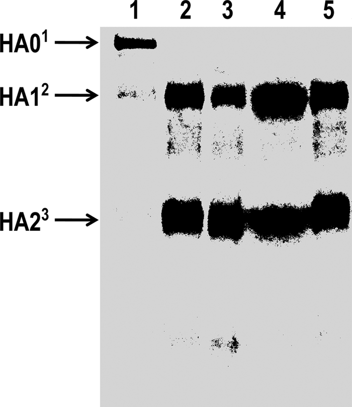
Trypsin-like cleavage of HA0 (i.e., the disappearance of HA0 with the appearance of labeled HA1 and HA2 peptides) is characteristic of viral activation. Shown in Fig. 1 is representative proteolytic conversion of HA0 to HA1 and HA2 peptides (approximately 58 and 26 kDa, respectively [cf. reference 24]) observed for 42 of 44 isolates. Trypsin-like conversion of HA0 to HA1 and HA2 peptides varied in band intensity, suggesting different degrees of hydrolysis by the respective isolates. Two of the 44 isolates appear to promote extensive proteolysis, as evidenced by complete disappearance of radiolabeled HA0, with little to no appearance of labeled HA1 and HA2 peptide bands (Fig. 1, lanes 8 and 10, respectively). Although not the focus of this work, labeled hydrolysate bands migrating primarily between HA1 and HA2 peptides also differing in band intensity were also observed.
Fig. 1.

SDS-PAGE analysis of polypeptide fragment patterns obtained following incubation of HA0 with supernatant material from protease-secreting bacterial isolates. [35S]HA0 was incubated with bacterial supernatant material for 60 min, subjected to SDS-PAGE analysis, and autoradiographed as described in Materials and Methods. Lanes: 1, PBS negative control; 2, trypsin, 10 μg/ml; 3, Streptococcus hyointestinalis (isolate 95-11); 4, Aerococcus viridans (isolate 135-8); 5, Lysinibacillus sphaericus (isolate 135-12); 6, Bacillus amyloliquefaciens (isolate 135-4); 7, Kocuria kristinae (isolate 107-14); 8, Bacillus pumilus (isolate 136-9); 9, Enterobacter cloacae (isolate 99-3); 10, Cellulosimicrobium sp. (isolate 111-15); 11, Aeromonas sobria (isolate 124-1); 12, Aeromonas hydrophila (isolate 119-3). The numbered arrows indicate the established molecular masses for HA0 and trypsin hydrolysis products HA1 and HA2 (80, 58, and 26 kDa, respectively) (24).
In vivo assay.
In order to assess the effects of HA0 cleavage by concentrated bacterial supernatants on infectivity of influenza virus, uncleaved virus was treated with all 44 bacterial supernatants, and MDCK cell monolayers were subsequently inoculated and double-layer plaque assay analysis carried out as described by Zhirnov et al. (34). All isolates were evaluated for toxic effects on MDCK cells. Only one isolate (Pseudomonas aeruginosa) was observed to exhibit deleterious effects on the monolayer (data not shown). Shown in Fig. 2 are double-layer in vivo plaque assay analyses corresponding to the 10 HA0 in vitro degradation gel profiles shown in Fig. 1. In vivo data are listed from highest PFU value (left, isolate 2) to the lowest PFU value (right, isolate 11). The PBS control (indicated by the dotted line) corresponded to 3.4 × 10³ PFU/ml, indicating the presence of previously activated virions. Comparing the PBS and trypsin controls, the uncleaved viral stock contained approximately 12% active (i.e., proteolytically cleaved) HA0. The trypsin control (lane 1, 28,000 PFU) was in excellent agreement with the viral titer of the beginning stock (30,000 PFU/ml). Surprisingly, the 10 respective bacterial supernatants shown in Fig. 2 as well as the 33 profiles not shown all gave rise to progeny PFU values less than that of the PBS control (Pseudomonas aeruginosa-treated MDCK cells exhibited cytopathic effects and were not included). In light of the less-than-expected background progeny PFU values for endogenous activated virus following exposure to supernatants from bacterial isolates, the involvement of some additional component was suggested.
Fig. 2.

Infectivity of influenza A/Indonesia/5/2005(H5N1) virus following incubation with concentrated supernatants from duck cloacal bacterial isolates. Uncleaved influenza A/Indonesia/5/2005 (H5N1) virus was incubated with respective bacterial supernatants for 60 min followed by layering onto MDCK monolayers for double-layer plaque assay analysis as described in Materials and Methods. Lanes: 1, trypsin, 10 μg/ml; 2, Lysinibacillus sphaericus (isolate 135-12); 3, Streptococcus hyointestinalis (isolate 95-11); 4, Aerococcus viridans (isolate 135-8); 5, Bacillus amyloliquefaciens (isolate 135-4); 6, Kocuria kristinae (isolate 107-14); 7, Enterobacter cloacae (isolate 99-3); 8, Aeromonas hydrophila (isolate 119-3); 9, Bacillus pumilus (isolate 136-9); 10, Cellulosimicrobium sp. (isolate 111-15); 11, Aeromonas sobria (isolate 124-1). The dotted line represents the PBS control (3.4 × 10³ PFU/ml).
Lipase assessment.
Due to the membrane-enveloped nature of the influenza virus, we were desirous of examining representative isolates which exhibited low PFU values but pronounced trypsin-like cleavage of HA0 for the presence of lipase activity. Aeromonas sobria and Aeromonas hydrophila isolates were assayed for lipase activity and observed to exhibit high levels (4+) of esterase (C8) and lipase (C14) activity (data not shown). Shown in Fig. 3 (lanes 2 and 4) is the effect of these two bacterial isolate supernatants on trypsin-activated virus. Post-supernatant incubation plaque counts indicated substantially reduced infectivity compared to the trypsin-only control (lane 1). Likewise, treatment of trypsin-activated virus with phospholipase C (lane 3) indicated decreased infectivity (∼ 80%). As shown in Fig. 4, trypsin-treated, radiolabeled HA0 treated with phospholipase C and bacterial supernatants yielded a similar cleavage pattern to that shown in Fig. 1 (lanes 11 and 12), suggesting not only competent HA0 cleavage but no additional digestion of HA1 and HA2 peptide fragments. Although endogenous lipase activity could account in part for lower-than-expected emergent virus following proteolytic activation, phospholipase C treatment and subsequent reduction of infectivity are only suggestive and not proof of lipase involvement.
Fig. 3.

Infectivity of trypsin-activated influenza A/Indonesia/5/2005(H5N1) virus following incubation with supernatant material from two lipase-secreting cloacal bacteria isolates. Trypsin-treated influenza A/Indonesia/5/2005 (H5N1) virus was incubated with bacterial supernatants for 60 min, followed by layering onto MDCK monolayers for double-layer plaque assay analysis as described in Materials and Methods. Lanes: 1, trypsin-only control (10 μg/ml); 2, supernatant material from Aeromonas hydrophila (isolate 119-3); 3, phospholipase C-only control (100 μg/ml); 4, supernatant material from Aeromonas sobria (isolate 124-1).
Fig. 4.
SDS-PAGE analysis of polypeptide fragment patterns obtained following incubation of trypsin-activated [35S]HA0 with supernatant material from two lipase-secreting cloacal bacterial isolates. Preparation of radiolabeled HA0, HA0 cleavage with trypsin, SDS-PAGE analysis, and autoradiography were carried out as previously described in Materials and Methods. Lanes: 1, [35S]HA0, untreated; 2, trypsin-only control (10 μg/ml); 3, phospholipase C-only control (100 μg/ml); 4, supernatant material from Aeromonas sobria (isolate 124-1); 5, supernatant material from Aeromonas hydrophila (isolate 119-3). The superscripted numbered arrows indicate the established molecular masses for HA0 and trypsin hydrolysis products HA1 and HA2 (80, 58, and 26 kDa, respectively) (24).
DISCUSSION
We report here that bacterial enzyme secretion mediates trypsin-like conversion of HA0 to HA1 and HA2 products, suggestive of influenza virus activation in the cloaca of wild waterfowl. In general, bacterial supernatants produced zones of hydrolysis comparable to that of trypsin (19 mm) (Table 1). Additionally, all bacterial supernatants were evaluated using PepTag (Promega Corp, Madison, WI) artificial peptide substrates to rule out false positives due to clouding of the agar medium arising from changes in pH (data not shown). The most frequently observed protease-secreting bacterium was Aeromonas sobria (Table 2). In addition to A. sobria, three other aeromonad species identified in this study (Aeromonas caviae, Aeromonas hydrophila, and Aeromonas veronii) have previously been isolated from wild waterfowl (1). Several species belonging to the genus Bacillus were isolated, with B. pumilus observed in all ducks, except green-winged teal (Table 2). Numerous proteolytic isolates of Enterobacter cloacae were encountered, as were isolates of Kocuria kristinae (formerly Micrococcus kristinae) and Cellulosimicrobium (formerly assigned to the genera Oerskovia and Nocardia).
Radiolabeled HA0 was cleaved in a trypsin-like manner to various degrees by supernatants from 42 of 44 duck cloacal isolates. Although, trypsin-like cleavage was observed, additional analysis of HA1 and HA2 peptides is required in order to rule out the possibility that small alterations arising from incorrect cleavage initially and/or subsequent removal of residues have not occurred resulting in loss of function and thus lower-than-expected in vivo infectivity data. Bacillus pumilus and Cellulosimicrobium spp. secreted proteases that extensively degraded the HA0 glycoprotein and HA1 and HA2 peptide hydrolysis products.
Utilization of MDCK cell monolayers and the double-overlay plaque assay as described by Zhirnov and coworkers (34) simulated conditions similar to that of the lower gastrointestinal tract of birds, eliminating (i) trypsin activation of viral particles as is the case for standard plaque assays and (ii) proteases found in the allantoic fluid of embryonated chicken eggs (7), enzymes not found in distal portions of the avian intestinal tract (21). Thus, activation of virus arose solely from proteolytic cleavage by the bacterial supernatant. Interestingly, virus stock used for in vivo experiments contained cleaved HA0 (∼12%), which proved advantageous in that the negative effect of bacterial supernatants on these cleaved (i.e., activated) virions was observed and assessed (Fig. 2). Despite producing a trypsin-like cleavage pattern, plaque counts less than that of the control, which contained cleaved (i.e., activated) virions, were observed for all isolates tested.
Because influenza virus is surrounded by a membrane envelope, we examined the possible presence of lipolytic activity in two isolates that exhibited a pronounced trypsin-like hydrolysis pattern but reduced infectivity of endogenous activated virus. Esterase (C8) and lipase (C14) activities were observed in both isolates. As shown in Fig. 3, trypsin-activated virions incubated with supernatants from these two bacterial isolates (lanes 2 and 4) or phospholipase C (lane 3) gave rise to significantly reduced plaque formation compared to that of the trypsin control (lane 1), albeit higher than that observed following treatment of influenza virus with supernatants from these two isolates as shown in Fig. 2, lanes 8 and 11. Trypsin-treated radiolabeled HA0 incubated with these bacterial isolate supernatants or phospholipase C (Fig. 4, lanes 3, 4, and 5) exhibited trypsin-like cleavage patterns similar to those previously observed (Fig. 1, lanes 11 and 12), suggesting that activation (i.e., cleavage of HA0 to HA1 and HA2 peptides) occurred. However, due to the complex nature of these bacterial supernatants, the lack of predicted infectivity could arise from contributors other than proteases either individually or in combination. For example, removal of membrane components as well as specific sugars from the HA0 glycoprotein by glycosidases present in the bacterial supernatants could also occur, resulting in decreased infectivity. β-Galactosidase, α-mannosidase, and N-acetyl-β-glucosaminidase activities were observed in these isolates (data not shown). Thus, disruption of the viral membrane or incorrect proteolytic cleavage, as well as possible removal of sugars required for viral binding to the cell surface receptor, could account for the observed disparate surveillance numbers between virus isolation and real-time PCR (6, 20, 22). Higher rates of detection are associated with molecular screening methods than with cultured samples because PCR detects viral RNA from both viable as well as nonviable viruses (15).
Previous studies of influenza virus and coinfecting proteolytic bacteria in the respiratory tract demonstrated Aerococcus viridans, Staphylococcus aureus, and Stenotrophomonas maltophilia to activate influenza virus in vivo (16, 23, 30, 31). We also observed these three organisms in the avian lower digestive tract. As indicated in Fig. 1 and 2 (lane 4), Aerococcus viridans exhibited the expected HA1 and HA2 hydrolysis products but with infectivity (PFU/ml) values less than that of the PBS control, like that of Staphylococcus aureus and Stenotrophomonas maltophilia (data not shown).
In the present study, we describe identification of protease-secreting bacteria from the gastrointestinal tracts of different waterfowl and their capability to cleave HA0 both in vitro and in vivo. Despite producing trypsin-like cleavage patterns consistent with that of viral activation, none of these isolates gave rise to expected progeny virus. Thus, the contribution of microbial proteases to influenza virus activation and other bacterially derived activities (e.g., lipase) to virus inactivation warrants further research.
ACKNOWLEDGMENTS
Lieutenant Colonel King was the recipient of a predoctoral fellowship from the United States Air Force Institute of Technology.
We thank John Gaines of the Air Force Institute for Occupational Health, Brooks City-Base, TX, for the use of the Vitek automated identification system. Special thanks goes to Cory Mason and Matthew Symmank, Texas Parks and Wildlife Department, for assistance with migratory bird collections.
Footnotes
Published ahead of print on 29 April 2011.
REFERENCES
- 1. Aguirre A. A., Quan T. J., Cook R. S., McLean R. G. 1992. Cloacal flora isolated from wild black-bellied whistling ducks (Dendrocygna autumnalis) in Laguna La Nacha, Mexico. Avian Dis. 36:459–462 [PubMed] [Google Scholar]
- 2. Bodour A. A., Drees K. P., Maier R. M. 2003. Distribution of biosurfactant-producing bacteria in undisturbed and contaminated arid Southwestern soils. Appl. Environ. Microbiol. 69:3280–3287 [DOI] [PMC free article] [PubMed] [Google Scholar]
- 3. Byrum B. R., Slemons R. D. 1995. Detection of proteolytic bacteria in the upper respiratory tract flora of poultry. Avian Dis. 39:622–626 [PubMed] [Google Scholar]
- 4. Callan R. J., Hartmann F. A., West S. E., Hinshaw V. S. 1997. Cleavage of influenza A virus H1 hemagglutinin by swine respiratory bacterial proteases. J. Virol. 71:7579–7585 [DOI] [PMC free article] [PubMed] [Google Scholar]
- 5. Fallacara D. M., Monahan C. M., Morishita T. Y., Wack R. F. 2001. Fecal shedding and antimicrobial susceptibility of selected bacterial pathogens and a survey of intestinal parasites in free-living waterfowl. Avian Dis. 45:128–135 [PubMed] [Google Scholar]
- 6. Ferro P. J., et al. 2008. Avian influenza surveillance in hunter-harvested waterfowl from the Gulf Coast of Texas (November 2005-January 2006). J. Wildl. Dis. 44:434–439 [DOI] [PubMed] [Google Scholar]
- 7. Gotoh B., et al. 1990. An endoprotease homologous to the blood clotting factor X as a determinant of viral tropism in chick embryo. EMBO J. 9:4189–4195 [DOI] [PMC free article] [PubMed] [Google Scholar]
- 8. Gray J. 1999. Assays for virus infection, p. 81–109 In Cann A. J. (ed.), Virus culture: a practical approach. Oxford University Press, Oxford, United Kingdom [Google Scholar]
- 9. Hinshaw V. S., Webster R. G., Turner B. 1980. The perpetuation of orthomyxoviruses and paramyxoviruses in Canadian waterfowl. Can. J. Microbiol. 26:622–629 [DOI] [PubMed] [Google Scholar]
- 10. Hinshaw V. S., Webster R. G., Turner B. 1979. Water-bone transmission of influenza A viruses? Intervirology 11:66–68 [DOI] [PubMed] [Google Scholar]
- 11. Horimoto T., Kawaoka Y. 2001. Pandemic threat posed by avian influenza A viruses. Clin. Microbiol. Rev. 14:129–149 [DOI] [PMC free article] [PubMed] [Google Scholar]
- 12. Hubalek Z. 2004. An annotated checklist of pathogenic microorganisms associated with migratory birds. J. Wildl. Dis. 40:639–659 [DOI] [PubMed] [Google Scholar]
- 13. Keawcharoen J., et al. 2008. Wild ducks as long-distance vectors of highly pathogenic avian influenza virus (H5N1). Emerg. Infect. Dis. 14:600–607 [DOI] [PMC free article] [PubMed] [Google Scholar]
- 14. Kida H., Yanagawa R., Matsuoka Y. 1980. Duck influenza lacking evidence of disease signs and immune response. Infect. Immun. 30:547–553 [DOI] [PMC free article] [PubMed] [Google Scholar]
- 15. Krafft A. E., et al. 2005. Evaluation of PCR testing of ethanol-fixed nasal swab specimens as an augmented surveillance strategy for influenza virus and adenovirus identification. J. Clin. Microbiol. 43:1768–1775 [DOI] [PMC free article] [PubMed] [Google Scholar]
- 16. Mancini D. A., et al. 2008. Influenza virus and proteolytic bacteria co-infection in respiratory tract from individuals presenting respiratory manifestations. Rev. Inst. Med. Trop. Sao Paulo 50:41–46 [DOI] [PubMed] [Google Scholar]
- 17. Mancini D. A., Mendonca R. M., Dias A. L., Mendonca R. Z., Pinto J. R. 2005. Co-infection between influenza virus and flagellated bacteria. Rev. Inst. Med. Trop. Sao Paulo 47:275–280 [DOI] [PubMed] [Google Scholar]
- 18. Martley F. G., Jayashankar S. R., Lawrence R. C. 1970. An improved agar medium for the detection of proteolytic organisms in total bacterial counts. J. Appl. Bacteriol. 33:363–370 [DOI] [PubMed] [Google Scholar]
- 19. McCullers J. A. 2006. Insights into the interaction between influenza virus and pneumococcus. Clin. Microbiol. Rev. 19:571–582 [DOI] [PMC free article] [PubMed] [Google Scholar]
- 20. Munster V. J., et al. 2009. Practical considerations for high-throughput influenza A virus surveillance studies of wild birds by use of molecular diagnostic tests. J. Clin. Microbiol. 47:666–673 [DOI] [PMC free article] [PubMed] [Google Scholar]
- 21. Philips S. M., Fuller R. 1983. The activities of amylase and a trypsin-like protease in the gut contents of germ-free and conventional chickens. Br. Poult. Sci. 24:115–121 [DOI] [PubMed] [Google Scholar]
- 22. Runstadler J. A., et al. 2007. Using RRT-PCR analysis and virus isolation to determine the prevalence of avian influenza virus infections in ducks at Minto Flats State Game Refuge, Alaska, during August 2005. Arch. Virol. 152:1901–1910 [DOI] [PMC free article] [PubMed] [Google Scholar]
- 23. Scheiblauer H., Reinacher M., Tashiro M., Rott R. 1992. Interactions between bacteria and influenza A virus in the development of influenza pneumonia. J. Infect. Dis. 166:783–791 [DOI] [PubMed] [Google Scholar]
- 24. Skehel J. J., Waterfield M. D. 1975. Studies on the primary structure of the influenza virus hemagglutinin. Proc. Natl. Acad. Sci. U. S. A. 72:93–97 [DOI] [PMC free article] [PubMed] [Google Scholar]
- 25. Slemons R., Byrum B., Swayne D. 2003. Bacterial proteases and co-infections as enhancers of virulence, p. 203–208 In Swayne D. E., Slemons R. D. (ed.), Proceedings of the Fourth International Symposium on Avian Influenza. U.S. Animal Health Association, St. Joseph, MO [Google Scholar]
- 26. Slemons R. D., Easterday B. C. 1977. Type-A influenza viruses in the feces of migratory waterfowl. J. Am. Vet. Med. Assoc. 171:947–948 [PubMed] [Google Scholar]
- 27. Slemons R. D., Easterday B. C. 1978. Virus replication in the digestive tract of ducks exposed by aerosol to type-A influenza. Avian Dis. 22:367–377 [PubMed] [Google Scholar]
- 28. Slemons R. D., Johnson D. C., Osborn J. S., Hayes F. 1974. Type-A influenza viruses isolated from wild free-flying ducks in California. Avian Dis. 18:119–124 [PubMed] [Google Scholar]
- 29. Steinhauer D. A. 1999. Role of hemagglutinin cleavage for the pathogenicity of influenza virus. Virology 258:1–20 [DOI] [PubMed] [Google Scholar]
- 30. Tashiro M., Ciborowski P., Klenk H. D., Pulverer G., Rott R. 1987. Role of Staphylococcus protease in the development of influenza pneumonia. Nature 325:536–537 [DOI] [PubMed] [Google Scholar]
- 31. Tashiro M., et al. 1987. Synergistic role of staphylococcal proteases in the induction of influenza virus pathogenicity. Virology 157:421–430 [DOI] [PubMed] [Google Scholar]
- 32. Tsiodras S., Kelesidis T., Kelesidis I., Bauchinger U., Falagas M. E. 2008. Human infections associated with wild birds. J. Infect. 56:83–98 [DOI] [PMC free article] [PubMed] [Google Scholar]
- 33. Webster R. G., Yakhno M., Hinshaw V. S., Bean W. J., Murti K. G. 1978. Intestinal influenza: replication and characterization of influenza viruses in ducks. Virology 84:268–278 [DOI] [PMC free article] [PubMed] [Google Scholar]
- 34. Zhirnov O. P., Ovcharenko A. V., Bukrinskaya A. G. 1982. A modified plaque assay method for accurate analysis of infectivity of influenza viruses with uncleaved hemagglutinin. Arch. Virol. 71:177–183 [DOI] [PubMed] [Google Scholar]