Abstract
The group II azoreductase BTI1 utilizes NADPH to directly cleave azo bonds in water-soluble azo dyes, including quenchers of fluorescence. Unexpectedly, optimal reduction was dye specific, ranging from a pH of <5.5 for Janus green B, to pH 6.0 for methyl red, methyl orange, and BHQ-10, to pH >8.3 for flame orange.
TEXT
Azo dyes are vivid colorants that consist of aromatic rings connected by one or more azo bonds. Thanks to the lack of native fluorescence, the DAB(C/S)YL and Black Hole Quencher (BHQ) azo dyes can be used as true dark quenchers of fluorescence in genetic assays, such as real-time quantitative PCR (5). Three groups of azoreductases (EC 1.7.1.6) cleave azo dyes into their colorless aromatic amines. Group II flavodoxin-related enzymes have been described from Bacillus sp. strain OY1-2, Bacillus subtilis, and others. They exist in a dimer-tetramer equilibrium, utilize flavin mononucleotide (FMN) as a noncovalently bound cofactor, and spend NADPH for direct reduction (1, 2, 4, 10, 11, 13). To date, azoreductase substrate specificities have been assessed mostly around physiological pH, which possibly underestimates optimal reactivity. Data on the pH-dependent reduction by the group II BTI1 azoreductase (Biosearch Technologies Inc.) of water-soluble azo dye quenchers are here presented.
Enzyme.
The cloning of BTI1, based on the Bacillus OY1-2 azoreductase (11), as well as expression and verification of the functionalized BTI10 variant used throughout this study as a tetrameric FMN-containing azoreductase is described in the supplemental material.
Azoreductase activity measurements.
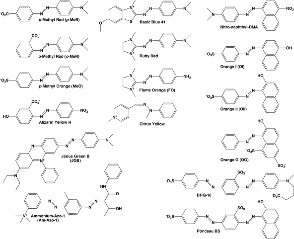
Potential substrates (Fig. 1) in fresh double-distilled water (ddH2O) at 1.0 or 0.10 mM were kept at +4°C until used at 20 to 40 μM for substrate susceptibility testing and at 1.0 to 20 μM for kinetics measurements. Stock solutions of β-NADH and β-NADPH (5 mM) in ddH2O were prepared fresh weekly, stored at −20°C (12), and used at 200 μM. Reduction was carried out in 200-μl reaction mixtures in 96-well plates, with 0.5 to 2 μg of enzyme for substrate susceptibility testing and 10.0 ng to 1.0 μg of enzyme for kinetics measurements. Reaction rates were calculated by fitting the initial part of the absorbance decay by linear regression to find the slope. Extinction coefficients and absorbance maxima (λmax) are in Table 1. All experiments were repeated at least three times, with similar results. The data were processed and plotted and kinetic parameters calculated by using Origin 6.0 (OriginLab, Corp., Northampton, MA).
Fig. 1.
Structures of dyes tested for reduction. The full names and details on the synthesis of BHQ-10 are given in the supplemental material.
Table 1.
Absorption and kinetic data of dyes reduced by BTI10
| Substrate | λmax (nm) | ε (M−1·cm−1) | Vmax (μM·s−1·mg−1)b | Km (μM) | Vmax/Km |
|---|---|---|---|---|---|
| BHQ-10 | 515 | 35,600 | 290 | 59 | 4.9 |
| Janus green B | 594a | 38,000 | ND | ND | ND |
| Flame orange | 483 | 28,359 | 150 | 510 | 0.29 |
| Methyl orange | 466 | 24,846 | 510 | 300 | 1.7 |
| o-Methyl red | 435 | 23,400 | 130 | 9.7 | 13.5 |
Loss of absorbance was measured at 660 nm.
ND, not determined.
Reaction conditions.
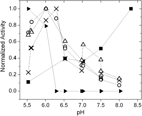
The initial reduction by BTI10 of the test dye methyl orange (MeO) in 20 mM sodium phosphate (pH 7.0) buffer, 250 μM β-NADPH at room temperature, and a 20 μM concentration of the substrate (11) was slow (0.80 μmol·min−1·mg−1 protein) and failed to reach completion. Addition of more NADPH, but not NADH, extended the reaction but did not affect the rate, verifying hydride donor depletion (4, 7). Upon cessation of reduction, the absorbance increased, suggesting azo bond reformation from a short-lived hydrazo intermediate (13). The effect of pH on the reaction was explored in buffers at a 20 mM final concentration (see the supplemental material). BTI10 optimally reduced MeO at pH 6.0 (Fig. 2).
Fig. 2.
Reduction by BTI10 as a function of pH of JGB (filled triangles), o-MeR (open triangles), MeO (open circles), BHQ-10 (×), and FO (filled squares). The data were normalized for each dye to its maximal activity (μmol dye reduced min−1·mg−1 protein) at optimal pH (for BHQ-10, 1.35 at pH 6.29; for MeO, 0.95 at pH 6.01; for o-MeR, 0.95 at pH 6.01; and for FO, 0.168 at pH 8.32). The maximal activity for JGB was empirically defined as a 25.1% loss of absorbance at 660 nm and at pH 5.5 after 20 min.
Substrate specificity is influenced by pH.
Known and potential azo fluorescence quenchers (Fig. 1) expected to be water soluble were tested. The disappearance of absorbance at the dyes' λmax was monitored at pHs 5.5 to 8.3 over time. Several dyes could not be tested due to insufficient solubility or sensitivity to NADPH (see the supplemental material). Ammonium-azo-1 (Am-Azo-1), citrus yellow, orange G, Ponceau BS, and ruby red were inert to reduction with BTI10 (pHs 5.5 to 8.3). A lack of reduction of the last two is surprising because they have been reduced by other azoreductases (2, 3, 8, 9). At pH 6, BTI10 efficiently reduced MeO, o-methyl red (o-MeR), and BHQ-10 but flame orange (FO) and Janus green B (JGB) only poorly. The reduction rate was found to be both dye and pH specific (Fig. 2); BHQ-10 and o-MeR reduction peaked at pHs 6.0 to 6.5, similarly to that of MeO, whereas the FO reduction rate increased up to at least pH 8.3, and reduction of JGB in a 20-min endpoint assay could be observed only at pHs 5.5 and 6.0.
Kinetic analysis.
Michaelis-Menten and Lineweaver-Burke kinetic analyses were applied to determine the Vmax and the Km values for MeO, o-MeR, and BHQ-10 at pH 6.0 and for FO at pH 8.3 (Table 1). Kinetic parameters for the reduction of JGB could not be obtained due to slow reduction. The Vmax of reduction (μM concentration of dye reduced s−1·mg−1 enzyme) ranged from 131 for o-MeR to 508 for MeO. The o-MeR reduction rate is comparable to that published for Rhodobacter sphaeroides azoreductase at pH 7.0 (408 μM s−1·mg−1 enzyme) (6) but considerably faster than those reported at pH 7.5 for B. subtilis YhdA, Geobacillus stearothermophilus, and Bacillus OY1-2 azoreductases (12.0, 13.1, and 10.4, respectively) (10). The measured Km values (μM) showed a wide (53-fold) range, from 9.7 for o-MeR to 511 for FO. The reported Km for o-MeR at pH 7.0 is 270 μM for R. sphaeroides azoreductase (6) and at pH 7.5 is 91.7 μM for B. subtilis YhdA, 134 μM for G. stearothermophilus azoreductase, and 395 μM for Bacillus OY1-2 azoreductase (10, 11). Efficient o-MeR reduction by BTI10 may thus be due to tight substrate binding.
Reaction at suboptimal pH.
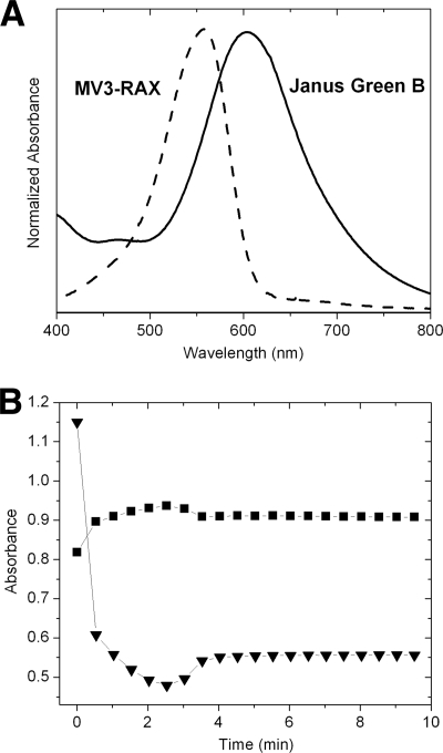
Reduction of the JGB (λmax, 594 nm) azo bond releases 4-(dimethyl-amino)-aniline and methylene violet 3RAX (MV-3RAX) (λmax, 557 nm) (Fig. 3 A), enabling simultaneous observation of substrate reduction and product formation at wavelengths distinct from those of the cofactors. Upon addition of azoreductase at the suboptimal pH of 6.0 and an amount of NADPH insufficient for complete reduction, the A594 rapidly decreased for less than a minute and then slowly decreased for up to 2.5 min, after which it increased slightly and then leveled off at 4 min (Fig. 3B). Reciprocal changes were seen at 557 nm. While no distinct absorption spectrum for the semireduced product intermediate was observed, its spectrum is expected to be very similar to that of MV-3RAX, also lacking azo bond conjugation. The coupled absorbance changes for JGB and MV-3RAX upon NADPH depletion is consistent with the reformation of JGB from a semireduced hydrazo intermediate.
Fig. 3.
Reduction at a suboptimal pH. (A) Absorbance spectra of JGB and MV-3RAX recorded at pH 6.0. (B) The effect of NADPH depletion on the reduction of JGB (30 μM) by BTI10 (73 ng·liter−1) in MES (morpholineethanesulfonic acid; pH 6.0) with 200 μM NADPH. Change in absorbance at the JGB λmax (594 nm) (triangles) and at the MV-3RAX λmax (557 nm) (squares) was monitored over 10 min. Shown are the results from a representative experiment (out of three).
The results obtained in the present study with BTI10 revealed a profound pH-dependent substrate specificity. Although the active substrates differ both in structure and charge, their absorption spectra were refractory to a change in pH from 5.5 to 8.3, consistent with their known and calculated pKas. Conversely, a lack of color change is consistent with maintained charge and tautomer equilibria and can thus not explain the pH-dependent substrate specificity, leaving pH-dependent dye-enzyme interactions as the most likely explanation.
Supplementary Material
Acknowledgments
Alex Herrault is thanked for assistance with mass spectrometry, and Jerry Ruth is thanked for support.
The project described was supported by award number R43/R44GM076843 from the National Institute of General Medical Sciences.
The content is solely the responsibility of the authors and does not necessarily represent the official views of the National Institute of General Medical Sciences or the National Institutes of Health.
Footnotes
Supplemental material for this article may be found at http://aem.asm.org/.
Published ahead of print on 29 April 2011.
REFERENCES
- 1. Binter A., et al. 2009. A single intersubunit salt bridge affects oligomerization and catalytic activity in a bacterial quinone reductase. FEBS J. 276:5263–5274 [DOI] [PubMed] [Google Scholar]
- 2. Chen H., Hopper S. L., Cerniglia C. E. 2005. Biochemical and molecular characterization of an azoreductase from Staphylococcus aureus, a tetrameric NADPH-dependent flavoprotein. Microbiology 151:1433–1441 [DOI] [PMC free article] [PubMed] [Google Scholar]
- 3. Chen H., Wang R. F., Cerniglia C. E. 2004. Molecular cloning, overexpression, purification, and characterization of an aerobic FMN-dependent azoreductase from Enterococcus faecalis. Protein Expr. Purif. 34:302–310 [DOI] [PMC free article] [PubMed] [Google Scholar]
- 4. Deller S., et al. 2006. Characterization of a thermostable NADPH:FMN oxidoreductase from the mesophilic bacterium Bacillus subtilis. Biochemistry 45:7083–7091 [DOI] [PubMed] [Google Scholar]
- 5. Johansson M. K. 2006. Choosing reporter-quencher pairs for efficient quenching through formation of intramolecular dimers. Methods Mol. Biol. 335:17–29 [DOI] [PubMed] [Google Scholar]
- 6. Liu G., et al. 2007. Azoreductase from Rhodobacter sphaeroides AS1.1737 is a flavodoxin that also functions as nitroreductase and flavin mononucleotide reductase. Appl. Microbiol. Biotechnol. 76:1271–1279 [DOI] [PubMed] [Google Scholar]
- 7. Liu G., et al. 2008. Site-directed mutagenesis of substrate binding sites of azoreductase from Rhodobacter sphaeroides. Biotechnol. Lett. 30:869–875 [DOI] [PubMed] [Google Scholar]
- 8. Pricelius S., et al. 2007. Enzymatic reduction of azo and indigoid compounds. Appl. Microbiol. Biotechnol. 77:321–327 [DOI] [PubMed] [Google Scholar]
- 9. Pricelius S., et al. 2007. Enzymatic reduction and oxidation of fibre-bound azo-dyes. Enzyme Microbial Technol. 40:1732–1738 [Google Scholar]
- 10. Sugiura W., Yoda T., Matsuba T., Tanaka Y., Suzuki Y. 2006. Expression and characterization of the genes encoding azoreductases from Bacillus subtilis and Geobacillus stearothermophilus. Biosci. Biotechnol. Biochem. 70:1655–1665 [DOI] [PubMed] [Google Scholar]
- 11. Suzuki Y., Yoda T., Ruhul A., Sugiura W. 2001. Molecular cloning and characterization of the gene coding for azoreductase from Bacillus sp. OY1-2 isolated from soil. J. Biol. Chem. 276:9059–9065 [DOI] [PubMed] [Google Scholar]
- 12. Wu J. T., Wu L. H., Knight J. A. 1986. Stability of NADPH: effect of various factors on the kinetics of degradation. Clin. Chem. 32:314–319 [PubMed] [Google Scholar]
- 13. Yan B., et al. 2004. Expression and characteristics of the gene encoding azoreductase from Rhodobacter sphaeroides AS1.1737. FEMS Microbiol. Lett. 236:129–136 [DOI] [PubMed] [Google Scholar]
Associated Data
This section collects any data citations, data availability statements, or supplementary materials included in this article.


