Abstract
A Clostridium botulinum type A strain (A661222) in our culture collection was found to produce the botulinum neurotoxin subtype A5 (BoNT/A5). Its neurotoxin gene was sequenced to determine its degree of similarity to available sequences of BoNT/A5 and the well-studied BoNT/A1. Thirty-six amino acid differences were observed between BoNT/A5 and BoNT/A1, with the predominant number being located in the heavy chain. The amino acid chain of the BoNT/A from the A661222 strain was superimposed over the crystal structure of the known structure of BoNT/A1 to assess the potential significance of these differences—specifically how they would affect antibody neutralization. The BoNT/A5 neurotoxin was purified to homogeneity and evaluated for certain properties, including specific toxicity and antibody neutralization. This study reports the first purification of BoNTA5 and describes distinct differences in properties between BoNT/A5 and BoNT/A1.
INTRODUCTION
Clostridium botulinum produces botulinum neurotoxin (BoNT), which is the most potent neurotoxin known. BoNTs are characterized as category A select agents because of their potential as a bioterrorism threat as listed by the Centers for Disease Control and Prevention (1). BoNTs can be immunologically distinguished by homologous antitoxins into seven primary serotypes, designated A to G. Among these serotype distinctions, there is considerable genetic variation, as demonstrated by the recognition of at least 24 subtypes (3, 8, 11, 17). These subtypes have been distinguished based on their degree of genetic variation, with subtypes having a minimum of 2.6% divergence at the amino acid level (3), but except for BoNT subtypes A1 (BoNT/A1) and -A2, they have not been purified and analyzed at the protein level, which is important to delineate functional differences between the subtypes (15). The purification and characterization of the biochemical, toxicological, and molecular mechanisms of the subtype toxins of various serotypes will provide valuable information as to their biochemical, immunological, and cell biology properties.
Recently, a new subtype of BoNT/A was identified and named “BoNT/A5”; there are five strains known to possess the gene encoding BoNT/A5 (3, 8). Among these five strains, four of them have neurotoxin sequences that are identical, and the fifth strain has a neurotoxin sequence that is 99.8% identical to the others at the amino acid level. The subtype features both a high degree of similarity to BoNT/A1 and a hemagglutinin (HA)-type gene cluster which is present in only BoNT/A1 clusters and none of the other BoNT/A subtypes. The Eric A. Johnson (E.A.J.) laboratory identified an additional A5 strain of C. botulinum, A661222, which possessed a gene for BoNT/A5 identical to that of strain IBCA94-0216 (8).
To investigate the BoNT/A5 of this strain, the neurotoxin gene and its associated genes were completely sequenced and analyzed at both the nucleotide and amino acid level. Thirty-six amino acid differences were observed between BoNT/A1 and BoNT/A5, with most of them in the heavy chain (HC) (8). Three-dimensional (3D) molecular modeling was performed comparing BoNT/A5 with the established BoNT/A1 subtype. These modeling studies on BoNT/A focused on determining if amino acid differences observed in BoNT/A5 would have an effect on known antibody epitope sites. The BoNT/A5 protein was then purified from culture, and its toxicity was determined. The ability of BoNT/A1-specific antibodies to neutralize BoNT/A5 was also tested by mouse bioassay.
MATERIALS AND METHODS
Bacterial strains and growth conditions.
The Clostridium botulinum strains A661222 and ATCC 3502 included in this study were from the E.A.J. strain collection. The A661222 strain was grown from a lyophilized culture which was received by H. Sugiyama from the Lanzhou Institute, China in 1981. No information is available regarding the environmental source and other properties of the isolated strain. The original source of the strain is unknown. Cultures were grown in 10 ml of sterile TPGY media (which contains [per liter] 50 g Trypticase peptone, 5 g Bacto peptone, 4 g d-glucose, 20 g yeast extract, and 1 g cysteine-HCl [pH 7.4]) for 2 days at 37°C under anaerobic conditions.
Total genomic DNA isolation.
Total genomic DNA was isolated from C. botulinum by lysozyme and proteinase K treatment as described previously (6). DNA was then diluted to a concentration of 50 ng/μl and used for PCR amplification.
PCR amplification and DNA sequencing.
PCR amplifications were performed using the GeneAmp high-fidelity PCR system (Applied BioSystems). The PCR cycles were as follows: 95°C for 2 min, followed by 25 cycles of 95°C for 1 min, an annealing step for 45 s at 48°C, and 72°C for extension, followed by 1 cycle of 72°C extension for 10 min. The extension time depended on the length of the fragment being amplified. Following amplification, PCR products were isolated with the PureLink PCR purification kit (Invitrogen). Sequencing was performed using conditions advised by the University of Wisconsin Biotechnology Center with the ABI PRISM BigDye cycle sequencing kit (Applied BioSystems). The primers used for PCR and sequencing for the HA cluster, ntnh and the bont/A gene, are the same as those used previously (12). PCRs were performed in a staggered manner such that the amplicons produced overlapping products for each of the genes in the neurotoxin cluster. Appropriate primers were then used for sequencing each PCR product. Correct assembly of the contigs was verified by using overlapping sequence data, with each region of the sequence being analyzed at least four times. Sequencing analysis was performed at the University of Wisconsin Biotechnology Center, and final sequencing results were analyzed with the Vector NTI Suite program (Invitrogen).
Sequence alignment.
The amino acid sequences of BoNT/A subtypes A1 to A5 were aligned with ClustalW and MEGA software to produce an unweighted pair group method with arithmetic mean (UPGMA) phylogeny tree of the subtypes as a whole and for their heavy chains.
Molecular modeling.
The first model comparing the BoNT/A proteins was generated with the program Coot (9), using the crystal structures of BoNT/A1 (Protein Data Bank [PDB] code 1BTA) (14). Pymol was used to generate illustrations (5).
Purification of BoNT/A5 and determination of toxicity.
BoNT/A5 was purified by using the previously described protocol used for the purification of BoNT/A1 (4). The purified BoNT/A5 was visualized by 4 to 12% NuPage SDS-PAGE (Invitrogen) under reducing and nonreducing conditions to assess protein purity.
The specific toxicity of the purified BoNT/A5 was determined by intraperitoneal (i.p.) injection using four toxin concentrations of 15, 10, 6.67, and 4.45 pg per mouse. The toxin was diluted in 0.5 ml gelatin phosphate buffer, and four mice were injected with each concentration and observed for 4 days for symptoms. The 50% lethal dose (LD50)/mg of toxin was calculated by the method described by Reed and Muench (16).
Neutralization of BoNT/A5 using anti-BoNT/A1 antibody.
Serum from rabbits immunized with BoNT/A1 toxoid was fractionated by protein A chromatography to obtain polyclonal anti-BoNT/A1 IgG. The titer of the resulting IgG preparation was determined so that 1 μl of this antibody can neutralize 5,000 LD50 of BoNT/A1. In this study, 2 μl of antibody was used to neutralize 16,000, 12,000, 10,000, 5,000, 2,500, and 1,250 LD50, respectively, either with BoNTA1 or BoNT/A5 to compare antibody neutralization between BoNT/A1 and BoNT/A5. Toxin was diluted with gelatin phosphate to achieve the appropriate LD50 concentrations. The different mixtures of toxin and antibody were incubated at 37°C for 90 min prior to intraperitoneal (i.p.) injection. Two mice were injected with 0.5 ml of the toxin-antibody mixture, respectively, and were observed for 4 days for botulinal symptoms and death.
Nucleotide sequence accession number.
Sequences for the neurotoxin cluster genes and bont/A from A661222 were determined and have been deposited in the GenBank database under accession no. HM153705.1.
RESULTS
Identification and sequencing of the neurotoxin gene and its associated cluster genes.
PCR and sequencing reactions were performed on the neurotoxin and the associated neurotoxin cluster genes of C. botulinum A661222, and the results were compiled using the Vector NTI Suite program. These studies involved a process of amplifying overlapping pieces of the neurotoxin and its associated cluster. Based on this work, it was determined that the A661222 strain contained only one neurotoxin gene cluster consisting of a complete HA cluster with ha70, ha17, ha33, botR, ntnh, and bont/A. This arrangement is consistent with the cluster arrangement identified in other BoNT/A5-producing strains (3, 8).
Comparison of the neurotoxin and associated HA cluster protein sequences between strain A661222 and the A5 strains IBCA94-0216 and H04402 065.
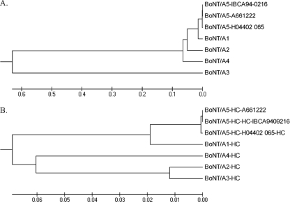
When the neurotoxin gene cluster of strain A661222 was compared to that of the A5 strains IBCA94-0216 and H04402 065, it was observed that the neurotoxin cluster genes and neurotoxin gene from strain A661222 were identical to those from strain IBCA94-0216 (8). A 1% amino acid sequence difference between strain A661222 and strain H04402 065 (3) was observed for all of the genes except ha17 and botR, which were identical at the nucleotide level. At the amino acid level, the neurotoxins of the two strains were 99.8% similar and identical. HA70 was 99.2% similar and identical and HA33 was 99.7% similar and identical between the two strains. The NTNH amino acid sequences of the two strains were 99.9% similar and identical. Sequence comparisons of the BoNT/A proteins demonstrated the high degree of homology among the BoNT/A5 strains as they grouped together and were clearly separated from the other type A subtypes for both the entire length of the protein and the heavy chain portion (Fig. 1 A and B). The origins of A661222 are unknown, aside from it being received from the Lanzhou Institute in western China. Additional studies confirmed that A661222 had both a deletion of the botR promoter and the presence of BoNT/B3 downstream of the BoNT/A5 cluster.
Fig. 1.
Graphical representation of degree of relatedness among the amino acid sequences of the BoNT/A1 to -A5 subtypes. The evolutionary history was inferred by UPGMA with a phylogeny tree drawn to scale. Branch lengths are in the same units as the evolutionary distances that were computed by the Poisson correction method, and these units represent the number of amino acid substitutions per site. (A) Comparison of the entire BoNTs among all five C. botulinum type A subtypes. (B) Comparison of the heavy chains of toxins of all five C. botulinum type A subtypes. A1 is from the ATCC 3502 strain, A2 is from the Kyoto F strain, A3 is from the CDC/A3 strain, A4 is from the 657Ba strain, and the BoNT/A5 sequence was derived from the strain listed in the figure.
Comparison of the neurotoxin and associated HA cluster proteins between strain A661222 and the C. botulinum A1 strain ATCC 3502.
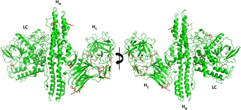
Analysis of the bont/A gene sequences of C. botulinum strain A661222 (BoNT/A5) and strain ATCC 3502 (BoNT/A1) demonstrated significant homology with the amino acid sequences, having 97.1% and 97.9% identity and similarity, respectively, similar to previous results (3). There were only 36 amino acid differences from ∼1,500 amino acids between BoNT/A1 and BoNT/A5 produced by the two strains, and the differences were mainly located in the heavy chain of the BoNTs, which spanned between the translocation domain and the binding domain (Fig. 2). The differences in the heavy chain are as follows. For the heavy chain N-terminal sequence (comprising amino acids 438 to 872), the differences were A567V, R581S, K592R, D707G, D767G, and E775D. For the heavy chain C-terminal sequence (comprising amino acids 873 to 1296), the differences were K897E, V926I, R948K, N954S, S955K, S957N, M968I, T990N, Q991K, E992Q, I993N, K994I, I1005V, N1006A, V1017I, T1063P, H1064Q, D1103N, V1143I, M1144V, R1156M, A1208V, T1232R, A1259D, L1278F, and R1294S.Only four differences were located in the light chains (LC) (comprising amino acids 1 to 437): D102E, E171D, G268E, and K381E. This high degree of homology made it possible to generate a model for the BoNT/A5 subtype based on already known BoNT/A structures (14, 18).
Fig. 2.
3D model of the BoNT/A5 subtype from A661222. The varied residues are shown as sticks in red. Overall structure is displayed as ribbon diagram with a green Cα. HC, heavy chain C terminal; HN, heavy chain N terminal.
The HA cluster genes demonstrated high homology between ATCC 3502 and A661222. The ha70 genes were 98% identical at the nucleotide level and 97.4% similar and 97.3% identical at the amino acid level. The ha17 genes were 97.7% identical at the nucleotide level and 97.3% similar and identical at the amino acid level. Also, the botR genes were 98.3% identical at the nucleotide level between the two strains and 97.2% similar and 96.6% identical at the amino acid level. The ntnh genes were 98.4% identical between the two strains and 98.2% and 97.8% similar and identical at the amino acid level, respectively. The ha33 genes demonstrated 95.0% nucleotide identity between the two strains, but only 91.5% similarity and 90.5% identity at the amino acid level.
Antibody recognition.
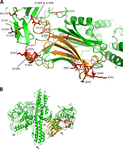
An epitope comparison was done utilizing certain specific peptides of the A2 HC domain based on previous work (10). Although the percentages of identity between the BoNT/A5 and BoNT/A2 sequences for the overall HC domain were low, the specific identity for those peptide regions was around 98% between the A5 and A2 subtypes, compared to only 83% between the A5 and A1 subtypes. Four different regions in the HC of the protein were selected for analysis (Table 1). These peptides are known to be important for antibody recognition since they were previously characterized as highly sensitive epitopes (2, 7, 10, 13, 17, 19). The antibodies generated against A1 could be significantly affected by the differences in these regions, even though the identity of the entire amino acid chains of A1 and A5 is close to 97%. Previous work has shown that the differences observed in A2 in this region are sufficient to disturb the binding of the antibodies (10, 17). Forty percent of the differences observed in BoNT/A5 compared to BoNT/A1 were found in these important regions (Fig. 3).
Table 1.
Comparison of specific peptides of BoNT/A1, -A2, and -A5 that are known to be targets for antibody neutralization
| Amino acid positions | % identity for comparison ofa: |
||
|---|---|---|---|
| A1 vs A2 | A5 vs A1 | A5 vs A2 | |
| 925–957 | 88 | 85 | 97 |
| 967–1013 | 85 | 83 | 98 |
| 1051–1069 | 79 | 90 | 85 |
| 1275–1296 | 86 | 91 | 96 |
All numbers are the percentages of identity between the specific subtypes analyzed. Strain sources of subtypes: A1, ATCC 3502; A2, Kyoto F; A5, A661222.
Fig. 3.
(A) Ribbon diagram of the area containing the important epitopes. The epitopes already identified are displayed in orange, and the varied residues are shown as sticks in red. The identities of key residues in the A5 model from strain A661222 are indicated. (B) This view localizes the area in the overall structure of the protein. HC, heavy chain C terminal; HN, heavy chain N terminal.
Garcia-Rodriguez et al. (10) have shown that several amino acids are important to optimize the interactions between certain antibodies and BoNT/A1. Some of these were different in BoNT/A5, making it similar to BoNT/A2, but several residues were also conserved (Table 2). The seven most energetically important residues are indicated in Table 2. The key residue 1064 differs between A1 (His), A2 (Arg), and A5 (Gln). The energetically important threonine 1063 is replaced with a proline in A5. The different equilibrium dissociation constant (KD) values were measured and proposed to affect the importance of these residues for antibody affinity. The most significant amino acid was H1064, which is deeply buried in the interface between the Fab and the toxin (Fig. 4 A). It has previously been reported that the truncation of the side chain of H1064 to alanine lowers the affinity of AR2 and CR1 for BoNT/A1 by more than 200,000-fold (10). The introduction of a mutated H1064R into the BoNT/A1 HC has been reported to reduce the affinity for AR2 and CR1 by only 41- and 188-fold. This decrease is less profound probably due to the fact that the arginine can interact with the Fab amino acids as the modeling shows (Fig. 4B) (10). In BoNT/A5, the difference is predicted to be more pronounced because the histidine (H1064) in A1 is substituted for a glutamine in A5, which is not able to pi stack with F36 on the CR1 antibody light chain. Furthermore, Q1064 has just one positive charge to interact with D102 on the CR1 antibody heavy chain, and thus the stability of that loop decreases at the complex interface (Fig. 4C). The affinity of the antibody to A5 is predicted to be lower than to the H1064R A1. That indicates that the antibodies developed against that BoNT/A1 area could have significant differences in their interactions with A5.
Table 2.
Amino acids known to be located in the interface between the CR1 antibody and BoNT/A1a
| BoNT/A1 amino acid position | Amino acid in: |
|
|---|---|---|
| BoNT/A2 | BoNT/A5 | |
| Ser 902 | Asp | Ser |
| Phe 917 | Ile | Phe |
| Asn 918 | Asn | Asn |
| Leu 919 | Leu | Leu |
| Glu 920 | Glu | Glu |
| Phe 953 | Phe | Phe |
| Asn 954 | Ser | Ser |
| Ser 955 | Lys | Lys |
| Ile 956 | Ile | Ile |
| Lys 1056 | Lys | Lys |
| Asp 1058 | Asp | Asp |
| Arg 1061 | Arg | Arg |
| Asp 1062 | Asp | Asp |
| Thr 1063 | Pro | Pro |
| His 1064 | Arg | Gln |
| Arg 1065 | Arg | Arg |
| Gly 1292 | Gly | Gly |
| Arg 1294 | Ser | Ser |
The five BoNT/A1 amino acids that differ in BoNT/A5 from A661222 and have direct contact with CR1 are in boldface. The seven residues with the most energetically important contributions to the binding between CR1 and BoNT/A1 are shaded gray.
Fig. 4.
Close-up view of sequence variability between H1064 of BoNT/A1 (green) (A), modeled R1064 of BoNT/A2 (green) (B), and Q1064 of BoNT/A5 from strain A661222 (green) (C) in complex with CR1 (light chain variable region [VL] in magenta and heavy chain variable region [VH] in yellow).
Purification of BoNT/A5 and determination of toxicity.
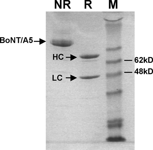
BoNT/A5 was purified by using the purification method previously used to isolate BoNT/A1. This was expected because BoNT/A5 is most closely related to BoNT/A1 among the type A subtypes, and they are the only subtypes to have an HA cluster arrangement associated with bont/A. Purified BoNT/A5 was confirmed by SDS-PAGE under reducing conditions (Fig. 5) and by mouse bioassay. SDS-PAGE data showed that ≥95% pure BoNT/A5 was obtained after the final chromatography step. The specific toxicity of the 150-kDa protein was determined to be ∼1.25 × 10⁸ LD50/mg.
Fig. 5.

Coomassie blue-stained SDS-PAGE gel of purified BoNT/A5 under reducing (R) and nonreducing (NR) conditions. M, marker; HC, heavy chain; LC, light chain.
Neutralization of BoNT/A1 and BoNT/A5 using an anti-BoNT/A1 polyclonal antibody.
The neutralization results showed that 2 μl anti-BoNT/A1 antibody was able to neutralize 10,000 LD50 of either BoNT/A1 or BoNT/A5 but was not able to completely neutralize 12,000 LD50 of either toxin subtype. The data indicate that both BoNT/A1 and BoNT/A5 have very similar binding reactions with anti-BoNT/A1 antibody. However, there were some differences in the times to death of mice between the mouse groups injected with the BoNT/A1-antibody mixture and the group injected with the BoNT/A5-antibody mixture. The mice injected with the BoNT/A5-antibody mixture died 1 day faster than those injected with a BoNT/A1-antibody mixture at an LD50 of 12,000. At the higher LD50 of 16,000, mice injected with the BoNT/A1-antibody mixture required ca. 2 days to die, while mice injected with the BoNT/A5-antibody mixture died within 4 h. Additionally, mice injected with 10,000 LD50 BoNT/A5-antibody mixture exhibited more severe symptoms than those injected with 10,000 LD50 BoNT/A1-antibody, even though all of the mice survived for 4 days.
DISCUSSION
Subtype BoNTs have been identified in numerous studies by nucleotide sequencing and the corresponding amino acid sequences. The criterion for a subtype has been stated to be a ≥2.6% difference in amino acid sequence. Initial modeling studies indicated that variable regions were present on BoNT/A subtypes A1 to A4, prior to the discovery of BoNT/A5. Although differences in nucleotide and amino acid sequences suggest that there may be differences in biochemical properties, structures, toxicities, and mechanisms, this cannot be definitively shown without the purification and analysis of the protein neurotoxins. A recent comparison of purified BoNT/1 and BoNT/A2 showed that there were differences in the kinetics of uptake into neuronal cells (15). This study showed that the purification and analysis of properties of BoNT subtypes will have high significance in mechanisms of action and immunogenicity, properties important for biodefense as well as the use of BoNTs as pharmaceuticals.
Prior studies identified a novel BoNT/A subtype, BoNT/A5. This designation was based on the level of divergence compared to known BoNT/A subtypes, but the importance of these differences was not identified at an amino acid level. We subsequently identified an A5 strain in E.A.J.'s laboratory that possessed the genes for an HA cluster containing the bont/A5 gene. In the present study, modeling was utilized to assess the effect on structure of amino acid differences in BoNT/A5 compared to BoNT/A1. There were 36 amino acid differences between the neurotoxins of strains A661222 and ATCC 3502 (BoNT/A1), with 32 of them present in the C terminus of the HC, the region responsible for binding to neural cells and a target for antibody neutralization.
BoNT/A5 is unique in certain properties compared to the other BoNT/A subtypes. It is highly similar to BoNT/A1 and also exhibits similarities to BoNT/A2 in regions that have been previously shown to affect antibody binding. The most important of these changes was at residue 1064. Previous studies have shown that mutating this residue from its original histidine to an alanine has the effect of decreasing binding by 200,000-fold in terms of the pi stacking between the histidine and F36 from the antibodies. In BoNT/A5, this residue is substituted for a glutamine, and the pi stacking is also not present based on protein modeling experiments. This led to the hypothesis that BoNT/A5 might have a different antibody neutralization profile from BoNT/A1.
BoNT/A5 was purified by the method used previously to purify BoNT/A1. Antibody neutralization experiments were also performed to compare the comparative neutralization by an antibody preparation raised against BoNT/A1. Both BoNT/A1 and BoNT/A5 were neutralized by 2 μl of anti-BoNT/A1 antibody at 10,000 LD50 but could not be completely neutralized at 12,000 LD50; however, differences in the times to death of mice were observed between the two toxins. The mice injected with a BoNT/A5-antibody mixture died 1 day faster than those injected with a BoNT/A1-antibody mixture at an LD50 of 12,000. At the higher LD50 of 16,000, it took mice injected with the BoNT/A1-antibody mixture 2 days to die, while mice injected with the BoNT/A5-antibody mixture died within 4 h. Additionally, mice injected with the 10,000-LD50 BoNT/A5-antibody mixture exhibited more severe symptoms than those injected with the 10,000-LD50 BoNT/A1-antibody mixture, even though all of the mice survived for 4 days. This indicates that the binding between BoNT/A1 and BoNT/A5 to anti-BoNT/A1 antibody might have slight differences, which were consistent with comparative structure predictions. These data also support that BoNT/A1 and BoNT/A5 may have different mechanisms of action, as was recently observed for BoNT/A1 and BoNT/A2 (15). Different immunological properties and mechanisms of action could have a high impact in the areas of biodefense and the increasing use of BoNT as a pharmaceutical. Further studies are necessary to purify and characterize the properties of BoNT subtypes to more fully appreciate the biology and structure of the subtype BoNTs.
ACKNOWLEDGMENTS
This work was sponsored by the NIH/NIAID Regional Center of Excellence for Bio-Defense and Emerging Infectious Diseases Research (RCE) Program. We acknowledge membership within and support from the Pacific Southwest Regional Center of Excellence (grant U54 AI065359). This work was also partially supported by grants from the Swedish Research Council (grant 2010-5200), the Wenner-Gren Foundations, and the Swedish Foundation for Strategic Research to P.S.
Footnotes
Published ahead of print on 22 April 2011.
REFERENCES
- 1. Arnon S. S., et al. 2001. Botulinum toxin as a biological weapon: medical and public health management. JAMA 285:1059–1070 [DOI] [PubMed] [Google Scholar]
- 2. Atassi M. Z., Dolimbek B. Z. 2004. Mapping of the antibody-binding regions on the HN-domain (residues 449–859) of botulinum neurotoxin A with antitoxin antibodies from four host species. Full profile of the continuous antigenic regions of the H-chain of botulinum neurotoxin A. Protein J. 23:39–52 [DOI] [PubMed] [Google Scholar]
- 3. Carter A. T., et al. 2009. Independent evolution of neurotoxin and flagellar genetic loci in proteolytic Clostridium botulinum. BMC Genomics 10:115. [DOI] [PMC free article] [PubMed] [Google Scholar]
- 4. DasGupta B. R., Sathyamoorthy V. 1984. Purification and amino acid composition of type A botulinum neurotoxin. Toxicon 22:415–424 [DOI] [PubMed] [Google Scholar]
- 5. Delano W. L. The PyMOL molecular graphics system. DeLano Scientific LLC, San Carlos, CA [Google Scholar]
- 6. Dineen S. S., Bradshaw M., Johnson E. A. 2003. Neurotoxin gene clusters in Clostridium botulinum type A strains: sequence comparison and evolutionary implications. Curr. Microbiol. 46:345–352 [DOI] [PubMed] [Google Scholar]
- 7. Dolimbek B. Z., Aoki K. R., Steward L. E., Jankovic J., Atassi M. Z. 2007. Mapping of the regions on the heavy chain of botulinum neurotoxin A (BoNT/A) recognized by antibodies of cervical dystonia patients with immunoresistance to BoNT/A. Mol. Immunol. 44:1029–1041 [DOI] [PubMed] [Google Scholar]
- 8. Dover N., Barash J. R., Arnon S. S. 2009. Novel Clostridium botulinum toxin gene arrangement with subtype A5 and partial subtype B3 botulinum neurotoxin genes. J. Clin. Microbiol. 47:2349–2350 [DOI] [PMC free article] [PubMed] [Google Scholar]
- 9. Emsley P., Cowtan K. 2004. Coot: model-building tools for molecular graphics. Acta Crystallogr. D. Biol. Crystallogr. 60:2126–2132 [DOI] [PubMed] [Google Scholar]
- 10. Garcia-Rodriguez C., et al. 2007. Molecular evolution of antibody cross-reactivity for two subtypes of type A botulinum neurotoxin. Nat. Biotechnol. 25:107–116 [DOI] [PubMed] [Google Scholar]
- 11. Hill K. K., et al. 2007. Genetic diversity among botulinum neurotoxin-producing clostridial strains. J. Bacteriol. 189:818–832 [DOI] [PMC free article] [PubMed] [Google Scholar]
- 12. Jacobson M. J., Lin G., Raphael B., Andreadis J., Johnson E. A. 2008. Analysis of neurotoxin cluster genes in Clostridium botulinum strains producing botulinum neurotoxin serotype A subtypes. Appl. Environ. Microbiol. 74:2778–2786 [DOI] [PMC free article] [PubMed] [Google Scholar]
- 13. Lacy D. B., Stevens R. C. 1999. Sequence homology and structural analysis of the clostridial neurotoxins. J. Mol. Biol. 291:1091–1104 [DOI] [PubMed] [Google Scholar]
- 14. Lacy D. B., Tepp W., Cohen A. C., DasGupta B. R., Stevens R. C. 1998. Crystal structure of botulinum neurotoxin type A and implications for toxicity. Nat. Struct. Biol. 5:898–902 [DOI] [PubMed] [Google Scholar]
- 15. Pier C. L., et al. 2011. Botulinum neurotoxin subtype A2 enters neuronal cells faster than subtype A1. FEBS Lett. 585:199–206 [DOI] [PMC free article] [PubMed] [Google Scholar]
- 16. Reed L. J., Muench H. 1938. A simple method of estimating fifty percent endpoints. Am. J. Hyg. (Lond.) 27:493–497 [Google Scholar]
- 17. Smith T. J., et al. 2005. Sequence variation within botulinum neurotoxin serotypes impacts antibody binding and neutralization. Infect. Immun. 73:5450–5457 [DOI] [PMC free article] [PubMed] [Google Scholar]
- 18. Stenmark P., Dupuy J. J., Imamura A., Kiso M., Stevens R. C. 2008. Crystal structure of botulinum neurotoxin type A in complex with the cell surface coreceptor GT1b—insight into the toxin-neuron interaction. PLoS Pathog. 4:e1000129. [DOI] [PMC free article] [PubMed] [Google Scholar]
- 19. Zarebski L. M., et al. 2008. Analysis of epitope information related to Bacillus anthracis and Clostridium botulinum. Expert Rev. Vaccines 7:55–74 [DOI] [PMC free article] [PubMed] [Google Scholar]


