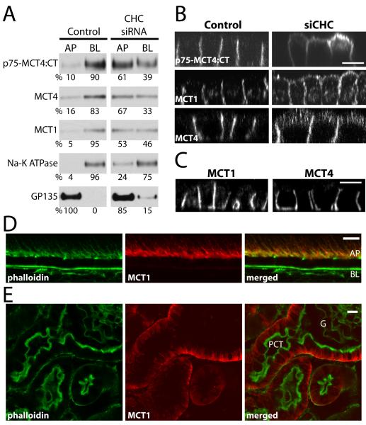
Figure 8. The steady state basolateral localization of MCTs is clathrin-dependent, but μ1B-independent.
MDCK cells stably expressing p75-MCT4:CT were transfected with siRNA against CHC or luciferase (Control) and the steady-state distribution of p75-MCT4:CT and endogenously expressed proteins was determined by domain-selective cell-surface biotinylation of polarized MDCK cells cultured on Transwell inserts and western blot (A) and LSCM (B). (C) Additionally, the polarity of endogenous MCT1 and MCT4 were determined by LSCM in cells where AP1B complex component μ1B was stably knocked down (20). Confocal images are shown as X-Z sections from the Z-stack. Mouse tissues sections were stained for MCT1 (red) and phalloidin (green) to show that even though neither the RPE (D) nor proximal convoluted tubule (E) express AP1B, the polarity of MCT1 is different between these tissues. AP=apical, BL=basolateral, PCT=proximal convoluted tubule; bar=10μm.

