Abstract
Pupil asymmetry or anisocoria can have benign or malignant causes, and be categorized as acute or chronic. It can also be a normal finding in about 20% of cases. Benign episodic unilateral mydriasis is an isolated benign cause of intermittent pupil asymmetry. The exact pathophysiology is not always understood. According to one hypothesis, it is due to discordance between the sympathetic and parasympathetic systems. It is occasionally seen in patients with migraine. Some authors consider it a limited form of ophthalmoplegic migraine. We report a case of benign episodic unilateral mydriasis diagnosed in a 30-year-old lady with a history of migraine who had extensive negative neurological evaluation.
Keywords: anisocoria, migraine, unilateral episodic mydriasis
Introduction
Benign episodic unilateral mydriasis (BEUM) is an isolated cause of intermittent pupil asymmetry that might be caused by hyperactivity of the sympathetic nervous system or hypoactivity of the parasympathetic system. An association between BEUM and migraine has been suggested in the literature. Our case report of BEUM diagnosed in a young lady with migraine adds to the literature suggesting such an association.
Case report
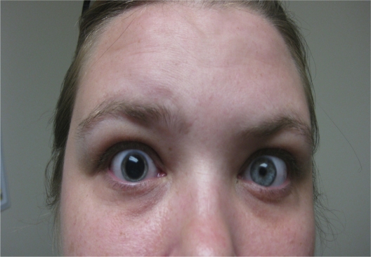
A 30-year-old lady with a history of migraine headache presented with a one-year history of chronic intermittent, bilateral, throbbing, and at times very severe, headache that often lasted for days and was associated with blurry vision, nausea, vomiting, photophobia, and phonophobia. Severe episodes were sometimes associated with transient right eye mydriasis (Figure 1), significant lower extremity weakness and at times confusion that all resolved with the resolution of her migraine episode. She denied any recent head trauma, fever, chills, or other complaints. Her past medical history was important for obesity, tobacco use, fibromyalgia, and depression. Her medications at presentation were duloxetine HCl 60 mg daily, trazodone HCl 100 mg at night, gabapentin 900 mg three times a day, and clonazepam 0.5 mg twice a day as needed. She denied using illicit drugs, herbal, or other medications, including ones over the counter. There was no family history of similar eye disturbance. Vital signs were normal. Physical examination revealed a fixed dilated right pupil (Figure 1), decreased proximal and distal muscular strength (3/5) in the lower extremities with normal reflexes. The rest of the neurological and physical examination was unremarkable. Her complete blood count, metabolic profile, and thyroid function test were unremarkable. She had normal cerebrospinal fluid analyses, head and neck magnetic resonance imaging and angiography, cervical, thoracic, and lumbar magnetic resonance imaging, electroencephalography, and electromyography of the lower extremities.
Figure 1.
Intermittent right pupil dilation.
Based on her presentation, laboratory and test results, she was diagnosed with an unusual case of migraine with benign episodic unilateral mydriasis. She was started on divalproex sodium, initially 500 mg then increased to 1000 mg daily, which led to significant improvement of her migraine headache as well as pupil disturbance.
Discussion
Causes of pupil asymmetry (anisocoria) range in seriousness from a normal, physiological condition (seen in 20% of normal people) to one that is immediately life-threatening, as seen with major stroke or intracranial bleed. Other causes include medications, infection, aneurysm, IIIrd nerve palsy, closed-angle glaucoma, and trauma.1,2
Benign episodic unilateral mydriasis is an isolated benign cause of pupil asymmetry.3 The episodes may be accompanied by blurred vision, orbital pain, headache, or photosensitivity.4 The underlying physiopathology is not always clear and may involve either parasympathetic deficiency or sympathetic hyperactivity affecting the iris.3,5 It can occasionally accompany migraine and some authors classify it as a limited form of ophthalmoplegic migraine. However, some cases have been described with no accompanying headache.3 The occurrence of mydriasis in ophthalmoplegic migraine can be due to functional exhaustion of parasympathetic fibers running within the IIIrd cranial nerve.6 Other possible mechanisms include ischemia or oculomotor nerve demyelination caused by neuropeptides secreted at the level of circle of Willis upon the activation of the trigeminovascular system causing edema and inflammation.7
According to one report of seven patients with BEUM associated with migraine, four had classic, one had common, and one had post-traumatic migraine. BEUM was also reported in migraine without aura or ophthalmoplegia, suggesting that it is a concomitant symptom.8,9 Furthermore, a family history of migraine or headache was reported in patients with BEUM.4,8
A detailed medical history, including active medications, full physical examination, including a careful ophthalmic and neurological system evaluation, and if indicated, imaging studies, should be performed to rule out other possible underlying conditions that sometimes can be life-threatening. Management and prognosis are determined by the underlying etiology. According to one case series report, patients with isolated benign episodic mydriasis appear to have a benign neurological prognosis, and do not require further neurodiagnostic studies.5
Here we report a rare case of BEUM diagnosed in a young lady with a history of migraine, supporting the association between the two conditions. We call for more research to understand fully the underlying pathophysiology which would help in managing this condition.
Footnotes
Disclosure
The authors report no conflicts of interest in this work.
References
- 1.Biousse V, Newman NJ. Neuro-Ophthalmology Illustrated. Stuttgart, Germany: Thieme Verlag; 2009. [Google Scholar]
- 2.Kawasaki A. Disorders of pupillary function, accommodation and lacrimation. In: Miller NR, Newman NJ, Biousse V, Kerrison JB, editors. Walsh and Hoyt’s Clinical Neuro-Ophthalmology. 6th ed. Baltimore, MD: Williams & Wilkins; 2005. [Google Scholar]
- 3.Balaguer-Santamaria JA, Escofet-Soteras C, Chumbe-Soto G, Escribano-Subias J. Episodic benign unilateral mydriasis. Clinical case in a girl. Rev Neurol. 2000;31:743–745. Spanish. [PubMed] [Google Scholar]
- 4.Jacobson DM. Benign episodic unilateral mydriasis. Clinical characteristics. Ophthalmology. 1995;102:1623–1627. doi: 10.1016/s0161-6420(95)30818-4. [DOI] [PubMed] [Google Scholar]
- 5.Johnston JA, Parkinson D. Intracranial sympathetic pathways associated with the sixth cranial nerve. J Neurosurg. 1974;40:236. doi: 10.3171/jns.1974.40.2.0236. [DOI] [PubMed] [Google Scholar]
- 6.Edelson RN, Levy DE. Transient benign unilateral pupillary dilation in young adults. Arch Neurol. 1974;31:12–14. doi: 10.1001/archneur.1974.00490370038003. [DOI] [PubMed] [Google Scholar]
- 7.Bek S, Genc G, Demirkaya S, Eroglu E, Odabasi Z. Ophthalmoplegic migraine. Neurologist. 2009;15:147–149. doi: 10.1097/NRL.0b013e3181870408. [DOI] [PubMed] [Google Scholar]
- 8.Woods D, O’Connor PS, Fleming R. Episodic unilateral mydriasis and migraine. Am J Ophthalmol. 1984;98:229–234. doi: 10.1016/0002-9394(87)90359-x. [DOI] [PubMed] [Google Scholar]
- 9.Evans RW, Jacobson DM. Transient anisocoria in migraineur. Headache. 2003;43:416–418. doi: 10.1046/j.1526-4610.2003.03081.x. [DOI] [PubMed] [Google Scholar]


