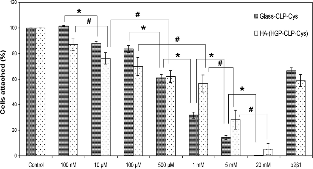
Figure 7. Inhibition of cell attachment.
Inhibition of hMSCs attachment with soluble CLP-Cys peptide and anti-α2β1 integrin antibody on Glass-CLP-Cys and HA-(HGP-CLP-Cys). Suspended cells were preincubated with different concentrations of the CLP-Cys peptide or anti-integrin α2β1 antibody (50 µg/mL) for 30 minutes at 37 °C and then allowed to attach to the Glass-CLP-Cys or HA-(HGP-CLP-Cys). The number of hMSCs attached to these surfaces, when the hMSCs were not preincubated with either the soluble CLP-Cys or anti-α2β1 integrin antibody was taken as 100 percent attachment. The fraction of cells attached was calculated relative to the fraction of cells that attached to the control surface. * and # are statistically different from each other (Student’s t test with p < 0.05). Each histogram represents the mean ± standard deviation of three repeats.

