Abstract
In this article we review the ‘state of the art’ with regards to biomarkers for prediction, diagnosis and prognosis in acute lung injury (ALI). We begin by defining biomarkers and the goals of biomarker research in ALI including their ability to define more homogenous populations for recruitment into trials of novel therapies as well as to identify important biological pathways in the pathogenesis of ALI. Progress along four general routes is then examined. First the results of wide-ranging existing protein biomarkers are reported. Secondly, we describe newer biomarkers awaiting or with strong potential for validation. Thirdly, we report progress in the fields of genomics and proteomics. Finally given the complexity and number of potential biomarkers, we examine the results of combining clinical predictors with protein and other biomarkers to produce better prognostic and diagnostic indices.
Keywords: biomarkers, clinical predictors, ALI, ARDS
INTRODUCTION
An invited commentary in the Lancet in 1997 noted that “despite two decades of intense effort, there is still no means of predicting reliably whether an individual patient will develop the acute respiratory distress syndrome (ARDS)”1. Shortly thereafter the NIH NHLBI Clinical Trials Network (ARDSnet) trial of lower tidal volume ventilation2 with an unprecedented 21% relative risk reduction in mortality led to renewed optimism in the field of Acute Lung Injury (ALI). In addition to guiding clinical management, this and subsequent ARDSnet studies have served as valuable sources of biological samples for large-scale validation of multiple biomarkers.3
In this article we will review the ‘state of the art’ with regards to biomarkers for prediction, diagnosis, prognosis and surrogate endpoints in ALI drawing on data from ARDSnet studies as well as other well-characterized patient populations. In addition to candidate biomarker studies, contributions from the ‘omics’ revolution with its many sub-genres – genomics, proteomics, metabolomics and others – will be discussed. Given the significant progress in the last decade, we are optimistic that the next decade will be marked by continued advancements in our ability to apply biomarkers to the diagnosis, treatment and prognostication in the clinical syndrome of ALI.
BIOMARKER RESEARCH IN ALI: DEFINITIONS AND GOALS
A widely cited definition for a biomarker came from the 1998 National Institutes of Health Biomarker Definitions Working Group: “a characteristic that is objectively measured and evaluated as a indicator of normal biological processes, pathogenic processes, or pharmacologic responses to a therapeutic intervention”4. This definition makes no supposition about the material nature of the characteristic in question. Reflecting this, the World Health Organization (WHO) suggests that a biomarker is “any substance, structure or process that can be measured in the body or its products and influence or predict the incidence of outcome or disease”.5 More broadly, the WHO proposed that a biomarker is “almost any measurement reflecting an interaction between a biological system and a potential hazard, which may be chemical, physical or biological. The response may be functional and physiological, biochemical at the cellular level, or a molecular interaction”.6 This definition provides a mechanistic framework for conceptualizing biomarkers in ALI and serves as a reminder that clinical signs such as pulse and blood pressure can also be biomarkers. The fundamental goal of biomarker research is to determine the relationship between a given biomarker and relevant clinical end-points.7
Several relevant clinical endpoints have been the focus of biomarker research in ALI. The most clinically important outcome is mortality8 and prediction of hospital or short-term mortality has been the predominant focus of biomarker research in the past decade.9 Another clinical endpoint of import is that of diagnosis – can a biomarker that is specific to lung injury facilitate the diagnosis in high-risk patients or distinguish between the high permeability pulmonary edema of ALI and cardiogenic edema? Related to diagnosis is the prediction of ALI in the ‘at risk’ patient. More accurate identification of patients likely to develop ALI would facilitate trials of novel agents or quality improvement initiatives for prevention of ALI. Similarly, identification of subgroups of patients either ‘at risk’ or with established ALI who may have a differential response to treatment could facilitate enrollment of more homogenous populations into clinical trials and represents an additional clinical end-point of interest.
A further potential role for biomarkers of ALI is as surrogate end-points in clinical trials.7 A biomarker response to treatment might substitute for a hierarchically more important clinical end-point such as mortality in early phase clinical trials that are not powered for mortality.10 However the use of surrogate end-points can be problematic in critical care. Improvements in surrogate end-points such as oxygenation and organ failures have not consistently been associated with mortality reductions in sepsis or ALI studies.2 Conversely, an absence of signal in a surrogate end-point does not necessarily imply a failure to improve mortality outcomes.11 In summary there are many potential roles for biomarkers in clinical ALI and these roles coalesce around predicting progression from the ‘at risk’ state, diagnosis, response to treatment, risk stratification and prognosis.
Biomarkers –illuminating biologic pathways
Another important goal of biomarker research is to shed light on the relative contribution of biologic pathways to ALI pathogenesis. Assays of candidate biomarkers that reflect various aspects of ALI pathogenesis derived from experimental models can provide confirmation that these pathways are important in the pathophysiology of human disease. Furthermore, modeling candidate biomarkers in a ‘head-to-head’ comparison has emerged as a powerful tool to determine the best performing biomarkers, an approach that can also provide important glimpses into pathogenesis.12
However, the apparent association of cytokines, biological pathways and clinical outcomes in ALI must be tempered by the knowledge that biomarkers such as cytokines are members of complex cascades and networks. Assessing levels of an individual cytokine dissociated from the levels of its antagonists or natural inhibitors may lead to the erroneous impression that an altered cytokine level reflects derangements in a biological pathway.13 In addition, immune-reactive assays that measure the presence of a protein may provide qualitatively different information than bioactivity assays that measure the functional, downstream signaling activity of the protein.14
Biomarker performance & validity
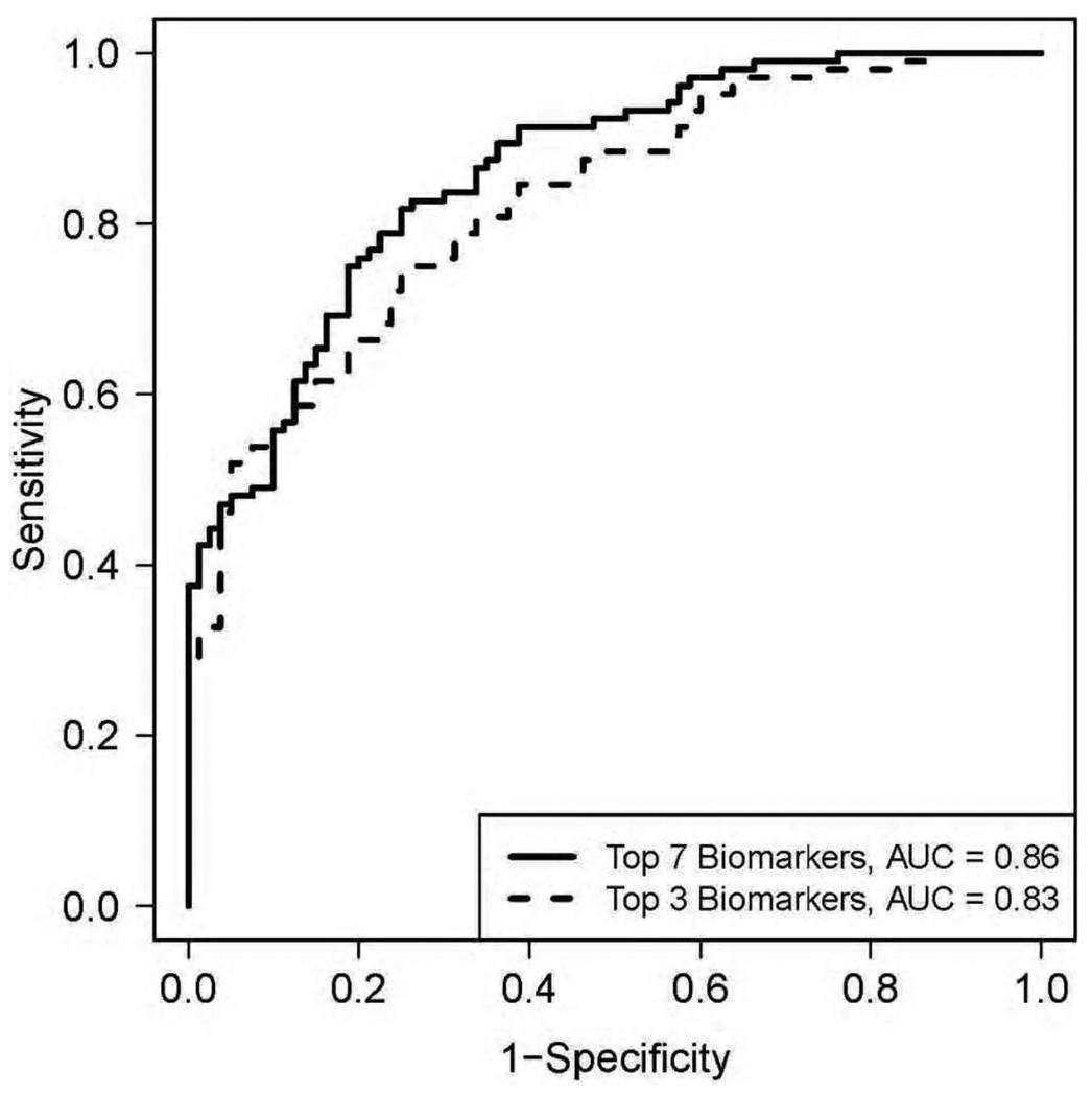
Assessment of biomarker performance is a function of sensitivity (the probability of a positive test given the presence of disease) and specificity (the probability of a negative test given the absence of disease). The ratio of sensitivity to 1-specificity (the false positive rate) yields a likelihood ratio. When the likelihood ratio exceeds 1 then the odds of the disease based on the test under examination is increased and the test has greater discriminatory value.10 The likelihood ratio is particularly useful as it can be examined at incremental values of the diagnostic test. When the values for the sensitivity and 1-specificifity are depicted graphically this results in a receiver operating characteristics (ROC) curve – Figure 1. The area under the curve (AUC) is a measure of performance. ROC curve analysis is particularly indicated to assess the diagnostic accuracy of a biomarker but newer statistical models suggest a role in disease prediction.15 A related term often used in the literature is accuracy. Accuracy is an aggregate (rather than a multiplicative) of sensitivity and specificity modified by the underlying prevalence of the disease.16
Figure 1.
Receiver Operator Characteristic curve analysis for the seven best performing biomarkers from a 21 biomarker panel for the diagnosis of trauma-induced acute lung injury. The three most discriminatory biomarkers were RAGE, BNP and PCP III with an AUC of 0.83. Figure reproduced from Fremont RD, Koyama T, Calfee CS, et al., J Trauma. May 2010; 68(5):1124, with permission.
Validity is an overarching term that incorporates aspects of precision, performance and reproducibility. Validity can be assessed at multiple levels. First is measurement validity: Is the biomarker measurable with precision and reproducibility? Second is internal validity: for a given study and clinical outcome how well does the biomarker under scrutiny perform. Third is external validity, what is the predictive power of the biomarker beyond its initial evaluation and its capacity for surrogacy – can the biomarker be used to stratify patient groups based on risk for ALI or responsiveness to therapy.7 A valid biomarker may be considered to have high effectiveness if it meets all aspects of validity.
Characteristics of the Ideal Biomarker - the “SMART” biomarker
The SMART criteria and mnemonic, a transplant from the business world, has been suggested for assessment of the disparate elements of quality control (performance, accuracy) and quality assurance (process measures such as reproducibility, accessibility, ease of use and internal validity). Shehabi and colleagues suggested that a SMART biomarker is Sensitive (and Specific), Measurable (with a high degree of precision), Available (Affordable and safely Attainable), Responsive (and Reproducible) in a Timely fashion (so as to expedite clinical decision making).17 Two additional words making the comparative SMARTER18 - Evaluate (validate) and Re-evaluate (revalidate) emphasize that biomarker research needs to be in a continuous cycle of appraisal and re-appraisal.
THE BIOMARKER & ALI INTERFACE
ALI and ARDS are complex, inflammatory syndromes of non-cardiogenic pulmonary edema. Non-pulmonary sepsis and pneumonia are the most common causes followed by major trauma, shock and aspiration of gastric contents.19 Injury to and permeabilization of endothelial and alveolar epithelial membranes, by a variety of injurious stimuli, leads to flooding of the alveolar compartment with protein rich edema fluid, neutrophils, cellular debris and inflammatory mediators. Multiple overlapping biologic pathways, cell and tissue types are deranged or injured reflecting both the local (site-specific) and systemic (site-independent) perturbations that arise in ALI. This complex pathophysiology and heterogeneity of cause yields a large number of potential biomarkers20
BIOMARKERS OF ACUTE LUNG INJURY – A RATIONAL CLASSIFICATION
Biomarkers of ALI can be classified according to clinical, molecular biological or pathophysiological dimensions. A clinical classification should take into consideration the underlying cause of lung injury, the phase of disease (early exudative or late fibroproliferative) and the site of sampling. Biomarkers of ALI may be measured in exhaled breath condensate, undiluted pulmonary edema fluid, saline-diluted broncho-alveolar lavage (BAL), plasma, serum, whole blood for gene expression analysis and urine. Additional considerations include the direction of change in the biomarker, the clinical outcome under investigation, the grade of evidence and the performance characteristics for the outcome of interest.
A molecular biological classification categorizes biomarkers by their place in the central dogma of molecular biology, namely the genome, transcriptome, proteome and metabolome. Most currently described biomarkers of ALI belong to the proteome and include proteins such as enzymes, receptors polypeptides lipoproteins and glycoproteins.21–24 It is likely that as our understanding of the genetics of ALI deepens, the focus may shift to genetic markers for identification of individuals or populations at risk.25
Mechanistically, biomarkers can be classified by their role in the pathophysiology of ALI which involves alveolar-capillary membrane injury, inflammation, activation of coagulation and increased permeability pulmonary edema.26,27 For example, biomarkers reflecting local injury to the alveolar-capillary membrane can be organized by compartment of origin (alveolar vs. vascular) and further organized according to the cell or tissue of origin (epithelial, endothelial, extracellular matrix) from which they are released. Surfactant proteins, for example, are released from alveolar epithelial cells and are considered markers of alveolar epithelial cell injury.3 Biomarkers mediating inflammation can be linked to their cell of origin (neutrophil, alveolar macrophage, platelets) or to their mode of action: cytokine, chemokine, protease, anti-protease and lipid signaling molecule. Biomarkers of coagulation can reflect activation of coagulation, endogenous anticoagulant systems or impaired fibrinolysis. Finally, increased pulmonary permeability results in a high ratio of protein in the pulmonary edema compared to plasma. This ratio in and of itself can be a useful biomarker for diagnosis of ALI.28 Alternatively biomarkers may track the repair and resolution pathways that mitigate or prolong the high permeability edema state.
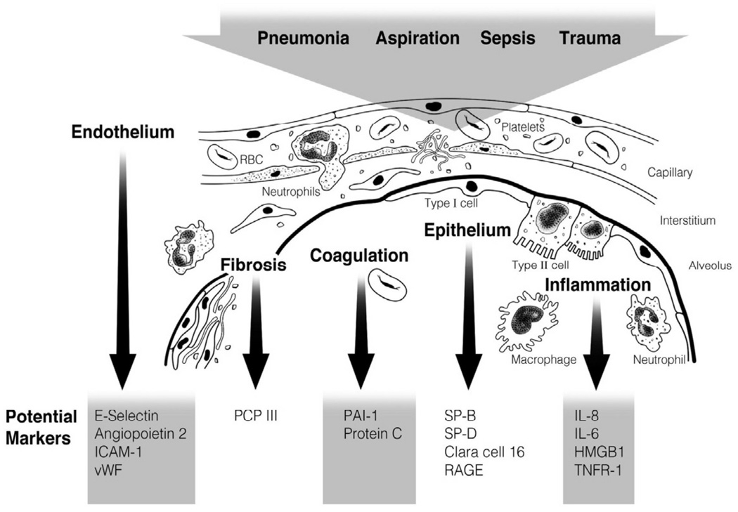
Although these classification systems create an orderly framework for consideration of ALI biomarkers, they may create artificial distinctions between overlapping pathways. The more integrated systems biology approach considers all these elements and classifications as interlinked, weaving together structural (molecular and cellular) and dynamic (pathophysiological) features into a unified whole.29 For example, epithelial repair mechanisms are instances of an anatomic or structural defect in the epithelium with clear pathogenic (dynamic) consequences. Potential biomarkers of ALI are summarized in Figure 2 according to pathophysiology and/or tissue of origin.
Figure 2.
Schematic representation of an alveolus and the alveolar-capillary interface demonstrating causes, pathophysiology and important potential biomarkers for prediction, diagnosis and prognosis in acute lung injury. Abbreviations, RBC = red blood cell. T1 = type 1 epithelial cell. T2 = type 2 epithelial cell. ICAM-1=Intercellular adhesion molecule 1. vWF = von Willebrand factor. PAI-1 = Plasminogen activator inhibitor 1. SP = surfactant protein. RAGE = Receptor for Advanced Glycation Products. HMGB1= Human Mobility Group Box 1 protein. TNFR-1 = Tumor Necrosis Factor Receptor 1.
PROTEIN BIOMARKERS FOR PREDICTION OF ALI IN THE ‘AT RISK’ PATIENT
Inflammation and cytokines
ALI is characterized by intra-alveolar inflammation mediated in part by pro-inflammatory cytokines. Differing cytokine profiles are characteristic of the early stages of ALI (termed the early response cytokines) compared to the later fibro-proliferative phase. Rising levels of inflammatory cytokines might be expected to precede the development of ALI in the ‘at risk’ patient population. Cytokines that have been identified in ALI include the interleukins IL-2, IL-6, IL-8, IL-10, IL-1β and its receptor antagonist IL-1ra, TNF-α and the soluble TNF 1 and 2 receptors – not strictly cytokines but an integral part of the downstream cytokine cascade.30,31 Remarkably for such central mediators, neither pro-inflammatory nor anti-inflammatory plasma cytokines have proved particularly useful in predicting ALI development.
Parsons and colleagues32 measured plasma levels of the anti-inflammatory cytokines IL-10 and IL-1 ra, both anti-inflammatory cytokines and found no association with disease prediction. Similarly, Bouros and colleagues33 failed to show a strong positive predictive value of either plasma or BAL IL-6 or IL-8 for development of ARDS in patients at risk. TNF-α, a pleiotropic and early phase cytokine, has also repeatedly failed to predict ALI development34,35 though issues with both the sensitivity and the internal validity of TNF-α assays (immuno-assay vs. bioassay) are well documented.34 In contrast, Takala and colleagues36 found elevated serum IL-8, IL-6 and soluble IL-2 receptor concentrations in ‘at risk’ patients who went on to develop ALI though none of the markers was able to discriminate between ARDS and non-ARDS patients. Donnelly and collegues37 measured plasma and BAL IL-8 in 29 consecutively enrolled ‘at risk’ patients. They showed significantly elevated BAL (but not plasma) IL-8 in the ‘at risk’ cohort who went on to develop ALI.
High Mobility Box-1 protein (HMGB1) is a DNA binding protein and inflammatory cytokine. HMGB1 is a ligand for and mediates part of its inflammatory effects through the Receptor for Advanced Glycation End Products (RAGE), a marker of epithelial injury. Cohen and colleagues38 showed that HMBG1 was released early (within 30 minutes) into the circulation of patients admitted to the emergency room after trauma and correlated with the development of acute organ dysfunction – including ALI.
Overall, the evidence to date indicates that cytokine levels are characteristic of but only weakly predictive for ALI. Causal heterogeneity, lack of statistical power and the observational nature of the studies undertaken suggest that there could still be some scope in determining the role of selected cytokine biomarkers (e.g. HMGB1) in ALI prediction.
Markers of Endothelial injury
Lung endothelial injury and activation leads to the increase in vascular permeability and influx of protein-rich edema fluid in ALI. Endothelial activation can be viewed as a three-fold process. Neutrophils are mobilized from the circulation to the alveolar space by bridging molecules that support neutrophil margination and adhesion (selectins and intercellular adhesion molecules (ICAMs)).39 This process is abetted by the secretion of angiopoietin-2 from endothelial cells which further destabilizes and permeabilizes the endothelial membrane – a process that is believed to be essential to allow endothelial cell migration and new vessel formation.40 Simultaneously the injured endothelium releases proteins such as von Willebrand factor that encourage vascular hemostasis.41 Endothelial activation and injury is thus an essential mechanism of non-cardiogenic pulmonary edema and biomarkers that reflect this process are promising in identifying the ‘at risk’ critically ill patient who will progress to ALI.
Angiopoietin-2
The angiopoietins are potent regulators of vascular permeability in critical illness, pulmonary diseases and beyond.40 Four ligands (Ang 1–4) have been identified but Ang-1 and Ang-2 are the best characterized.42 Both Ang-1 and Ang-2 bind to a common receptor termed Tie 2, a tyrosine kinase receptor present on the endothelial cell surface. Ang-1 is constitutively expressed in all vessels under quiescent conditions to maintain vessel wall stability and homeostasis. Injury to the vessel wall tilts the balance towards Ang-2 expression, which counteracts Ang-1 via Tie 2 inhibition.41 Ang-2 activates Rho kinase causing disaggregation of cell-cell junctions and potentiation of the inflammatory NfΚB pathway while silencing protective P13K/Akt (phosphoinositide-3 kinase) signaling vital to cell survival. The net result is capillary leak, neutrophil transmigration and angiogenesis.40
Clinical measurements of circulating Ang-2 have provided interesting results. Gallagher and colleagues43 in 63 ‘at risk’ surgical ICU patients showed higher median levels of Ang-2 in the patients who developed ALI compared to those who did not (10.1 vs. 3.7 ng/ml) and significantly higher median levels (19.8 ng/ml vs. 5.3, P = 0.004) in ALI patients who did not survive in samples obtained on the day of meeting ALI criteria.
Of particular interest are data linking polymorphisms in the Ang-2 gene (ANGPT2) to susceptibility to trauma-induced ALI.44,45 Using a large-scale candidate gene platform, the two single nucleotide polymorphisms most strongly associated with ALI were present in the Ang-2 gene. These findings were validated in two separate populations across two ethnicities. One of the ANGPT2 polymorphisms was associated with higher levels of a variant Ang-2 isoform in plasma.46
VEGF (Vascular endothelial growth factor)
VEGF is another novel mediator of vascular permeability and angiogenesis functionally related to the angiopoeitins with an adjuvant role in repair after lung injury.47 The inter-relation of VEGF and Ang-2 is complex. VEGF upregulates Ang-248 but also appears to induce shedding of Tie 2 to its soluble form thereby mitigating its effect on downstream signal transduction.49 However, in contrast to Ang-2, recent studies of VEGF have not corroborated earlier positive data and either shown no differences in plasma or edema fluid levels50 or a weak but non-significant signal for ALI or prediction of ALI.51
Von Willebrand factor antigen
Von Willebrand factor antigen (VWF) is a large multimeric glycoprotein involved in hemostasis. VWF performs the dual functions of coupling platelets to the endothelium via its platelet-binding domain and as a transport protein for factor VIII. It is synthesized principally in endothelial cells (where it resides within Weibel-Palade bodies) and to a lesser extent in platelets. While, it is constitutively expressed, release is greatly augmented by a wide variety of injurious stimuli.52. In a small cohort of 45 patients with non-pulmonary sepsis ‘at risk’ for ALI, a level above 450% of control had a positive predictive value of 80% for ALI.53 Bajaj and Tricomi54 and Moss and colleagues55 in broader study populations comprising both septic and non-septic patients could not replicate those findings, although the study populations may have differed in relation to chest x-ray findings. In the study by Rubin,53 the patients all had normal chest x-rays at enrollment while some of the patients in the other two studies already had chest x-ray abnormalities and thus may have had a sub-clinical stage of ALI.
Selectins
Selectins are cell-surface adhesion molecules involved in the early phase of neutrophil rolling and homing to a site of inflammation. There are three types E (endothelial), L (leucocyte) and P (platelets).56 E-selectin is selectively synthesized under conditions of cellular stress such as hypotension or organ hypoperfusion.57 Okajima and colleagues24 measured E-selectin by a rapid laboratory assay in 50 unselected patients with SIRS admitted to the emergency room. Higher levels of E-selectin had a positive predictive value of 68% and negative predictive value of 86% for the development of ALI. The assay was also predictive for other organ failures. Donnelly and colleagues58 measured all three selectins in plasma in 82 ‘at risk’ patients with trauma, pancreatitis or perforated bowel. Cleaved, soluble L-selectin (sL-selectin) was significantly lower in those patients who progressed to ARDS than those who did not. Unlike Okajima and colleagues they found no differences in plasma E- or P-selectin. The biologic rationale for lower sL-selectin is unknown since cleaved L-selectin is required for normal leukocyte migration but the authors speculated that sL-selectin may become bound to a ligand present on the endothelium, reducing circulating levels.
Markers of Epithelial injury
Epithelial injury is a pivotal step that contributes to inflammation and the influx of pulmonary edema fluid in ALI. Injury to the epithelium also compromises alveolar fluid clearance and surfactant production (from type II cells), facilitates bacterial transmigration into the systemic circulation and impairs alveolar repair mechanisms.59 Evidence of epithelial injury through release of an intracellular or cell-surface biomarker into the alveolar space and circulation represents a powerful potential tool for prediction of the ‘at risk’ patient.60,61
Surfactant proteins
Surfactant is a matrix of amphipathic lipoproteins and phospholipids whose major property is to lower surface tension at end-expiration preventing alveolar collapse. Four surfactant proteins have been identified lettered SP-A through SP-D. SP-A and SP-D are high-molecular-weight hydrophilic molecules with marked roles in innate immunity62 whereas SP-B and SP-C are low-molecular weight hydrophobic species essential for alveolar epithelial membrane integrity.63
BAL SP-A levels were low in one study of patients at risk for ALI with a negative predictive value of 100% for ALI64 when the levels remained above a cut-off of 1.2 mcg/ml. The same investigators found elevated plasma SP-A levels in at ‘at risk’ cohort who developed ARDS secondary to sepsis and aspiration but not trauma.65 These findings suggest that alveolar-capillary membrane permeability leading to leak of SP-A from the airspace into the plasma is a marker of lung epithelial injury.
In a single-centre study of 54 patients ‘at risk’ for ALI, Bersten and colleagues generated ROC curves for plasma SP-B with an AUC of 0.77 for all-cause prediction of ALI increasing to 0.87 for ALI from direct lung injury. However in contrast to the above study65 plasma SP-A was not predictive for ALI with an AUC 0.61.66
Interestingly, no further research on SP-B has ensued as a biomarker for detection of the at-risk patient, perhaps due to the difficulty of measuring this hydrophobic protein in the circulation. In addition, plasma SP-D has emerged in subsequent well-powered publications as a diagnostic and prognostic (but not predictive) biomarker in ALI (see later section on prognostic biomarkers).3
Clara cell protein
Clara cell protein (CC 16) is a small 16kDa anti-inflammatory protein secreted almost exclusively by Clara cells of the terminal bronchial epithelium.67 It is postulated to exert its actions through blockade of the phospholipase A2 second messenger system. Results from studies of CC16 as a biomarker for prediction of ALI have been somewhat discordant. In a small study of 22 patients with ventilator-associated pneumonia at risk for ALI/ARDS, an acute elevation in plasma CC 16 occurred prior to the diagnosis of the clinical syndrome of ALI.68 Sustained elevation of 30% or more yielded a diagnostic AUC of 0.91. However Kropski and colleagues found that plasma and pulmonary edema fluid CC16 levels were lower in patients with ALI/ARDS than control patients with cardiogenic pulmonary edema (CPE).69 Clearly prospective validation in larger scale trials of this interesting biomarker is still required.
PROTEIN BIOMARKERS FOR DIAGNOSIS OF ALI
Distinct from prediction of ALI in the ‘at risk’ patient is the use of biomarkers to confirm the diagnosis of ALI similar to the use of cardiac troponins for myocardial infarction.70 Ideally a biomarker would function and perform under all clinical conditions (i.e. be ‘universal’). However for biomarker characterization it is useful to specify the control group (cardiogenic pulmonary edema, patients with clear chest radiographs on the ICU, normal controls, patients ‘at risk’) against which cases are compared as well the phenotypic subtype (sepsis-induced, trauma-induced ALI, ventilator-induced lung injury) that is being evaluated. In addition it is important to note that the North American European Consensus Definitions of ALI and ARDS that are applied as the gold standard for assessment of diagnostic biomarkers have limitations in the area of specificity.71,72
Many individual biomarkers from diverse biologic ontologies have been tested that reflect the heterogeneity of ALI. Here we will mention some of the most promising markers with the strongest associations across important clinical end-points. First we will discuss the most straightforward method - measurement of total protein ratios.
Endothelial and epithelial injury – Edema fluid-to-plasma protein ratios and plasma protein levels
Protein-rich pulmonary edema due to increased permeability of the alveolar-capillary membrane is a pathophysiologic feature of ALI. Measurement of the pulmonary edema fluid to plasma protein (EF/PL) ratio is an intuitive, easy-to-perform and rapid way to distinguish between high and low permeability pulmonary edema. Ware and colleagues28 used a pre-defined EF/PL ratio of ≥ 0.65 and compared its performance by ROC analysis with expert clinical diagnosis as the gold standard in a large cohort of 390 patients. The AUC for discriminating ALI from cardiogenic edema was 0.81 increasing to 0.85 for measurements taken within three hours of endotracheal intubation. This technique is limited by the need to perform measurements early after intubation since fluid resorption mechanisms (if intact) tend to concentrate protein levels in the alveolar space over time, potentially confounding results.
Using a related approach, Aman and colleagues60,73 showed that low plasma levels of albumin and/or transferrin were predictive of a pulmonary leak index > 30 × 10−3/min with an AUC of 0.85 for albumin. However the pulmonary leak index is only a surrogate marker of extravascular lung water and pulmonary edema and is not currently part of the consensus criteria for ARDS.
Epithelial markers
Receptor for advanced glycation end-products (RAGE)
RAGE is a transmembrane protein of the immunoglobulin superfamily and multiligand receptor that binds modified glycoproteins including high-mobility group box-1 protein (HMGB-1) transmitting a pro-inflammatory downstream intracellular signal via NF-κB (60).74 Although it is ubiquitously expressed, expression levels are highest in the lung, RAGE is a specific marker of lung epithelial damage since it is anchored on the basolateral membrane of the alveolar type I cell. RAGE levels are increased in the plasma and pulmonary edema fluid of patients with ALI compared to patients with hydrostatic pulmonary edema.75 In a study of patients with severe trauma at high risk for ALI, RAGE was the best performing biomarker out of a panel of 21 biomarkers for distinguishing patients with ALI from those without ALI.76 In a much larger study of 676 patients from the ARDS Network trial of low tidal volume ventilation Calfee and colleagues60 showed that after adjusting for potential confounders, RAGE was also a marker of worse clinical outcomes (including mortality) only in patients in the higher tidal volume arm of the study. The overall impression is that RAGE is associated with alveolar epithelial injury and has diagnostic abilities both for ALI and worsening of ALI by ventilator-induced lung injury.
Laminin-5
Laminin-5, a polymorphic, polyfunctional epithelial cell adhesion molecule has recently been identified as a potential marker of early ALI. Laminins play an important role in cell adhesion, growth and differentiation.77 Katayama and colleagues78 showed that a degradation product of laminin-5, G2F, the terminal active portion of its γ2-chain, was significantly increased in the plasma of ALI patients as compared to patients with cardiogenic pulmonary edema. High levels were maintained in non-surviving patients.
Endothelial markers
Intercellular adhesion molecule-1 (ICAM-1)
As the name suggests this molecule mediates intercellular adhesion of leukocytes to the endothelium and epithelium where it co-locates to the cell membrane of all three cell types. ICAM-1 is upregulated in inflammatory states and facilitates movement of neutrophils across endothelial barriers to sites of inflammation.79 In a small pilot study ICAM-1 was elevated in both the plasma and edema fluid of patients with ALI as compared to patients with cardiogenic pulmonary edema.80 Earlier studies had found similar results but the elevation was confined to edema fluid only prompting the suggestion that this was a dual endothelial-epithelial membrane marker.81
Markers of coagulation and fibrinolysis
Plasminogen activator inhibitor-1 (PAI-1)
Plasminogen activator inhibitor-1 (PAI-1) is an antiprotease inhibitor of fibrinolysis that promotes fibrin deposition, one of the pathologic hallmarks of the acute respiratory distress syndrome.19 Prakhakaran and colleagues82 reported increases in plasma and pulmonary edema fluid of patients with early ALI as compared to patients with severe hydrostatic pulmonary edema.
Extracellular matrix
Procollagen peptide (PCP) III
PCP III is a marker of collagen synthesis. Two small studies83,84 have suggested an association of higher edema fluid levels of PCP III with ALI. In the above-mentioned trauma study,76 among a panel of 21 plasma biomarkers PCP III was the second best performing biomarker for distinguishing patients with ALI from severely injured controls without ALI. In another larger study, plasma and BAL PCP III levels decreased with steroid treatment85 suggesting that PCP III levels potentially mirror disease activity.
Inflammation
Lipopolysaccharide binding protein (LBP)
LPS binding protein (LBP) produced by alveolar epithelial type II cells is an acute phase reactant that mediates transduction of a pro-inflammatory response to LPS by binding of LPS from gram negative bacteria or lipotechoic acid from gram positive organisms.86 Abnormalities and activation of this protein in human sepsis syndrome have been reported for over a decade, though its role in the pathogenesis and as a biomarker of ARDS has recently attracted renewed attention. Sustained levels of LBP at 48 hrs post admission to the ICU were associated with ARDS (not ALI) but no ROC curves were generated to specify a particular cut-off in LBP levels to predict ARDS.87 In addition, admission LBP had no discriminatory value between survivors or non-survivors.
COMBINING BIOMARKERS FOR DIAGNOSIS OF ALI
Given the failure of a single biomarker to discriminate ALI with high accuracy, the question arises whether a composite of biomarkers that represent the most commonly identified and clinically validated biologic ontologies (inflammation, endothelial activation, lung epithelial injury and coagulation/altered fibrinolysis) might have better performance than any individual biomarker for diagnosis for ALI. As discussed above, Fremont and colleagues76 posed this question with regards to the diagnosis of ALI secondary to trauma. Using a backward elimination model, 21 biomarkers were reduced to the top-performing seven: RAGE, PCP III, brain natriuretic peptide (BNP), Ang-2, TNF-α, IL-10 and IL-8. A model that utilized these seven biomarkers generated an AUC of 0.86 for differentiating ALI/ARDS from a group of critically ill trauma patients without ALI who had normal chest radiographs or hydrostatic pulmonary edema. The top three performing markers (RAGE, PCP III and IL-8) had an AUC of 0.83 and excellent discriminatory power – Figure 1.
PROTEIN BIOMARKERS FOR PREDICTING OUTCOME IN ALI
Much of the strongest evidence in the field of ALI biomarkers comes from outcome prediction. A number of biomarkers belonging to the biologic ontologies discussed above have been validated in large multi-center clinical trials principally from the National Heart Lung and Blood Institute’s ARDSNet. The outcomes most commonly predicted are hospital, 30-, 60-day or 180-day mortality, ventilator and organ-failure free-days and assessment of response to low-tidal volume ventilation. The majority of this work has been performed in the last 10 years and is the strongest measure of progress in the field.
Markers of endothelial injury (vWF), epithelial injury (SP-D), leukocyte-endothelial interaction (ICAM-1), inflammation (IL-6, IL-8, TNFR1) and alterations in coagulation/fibrinolysis (Protein C, PAI-1) have the most robust associations with clinical outcomes such as mortality.
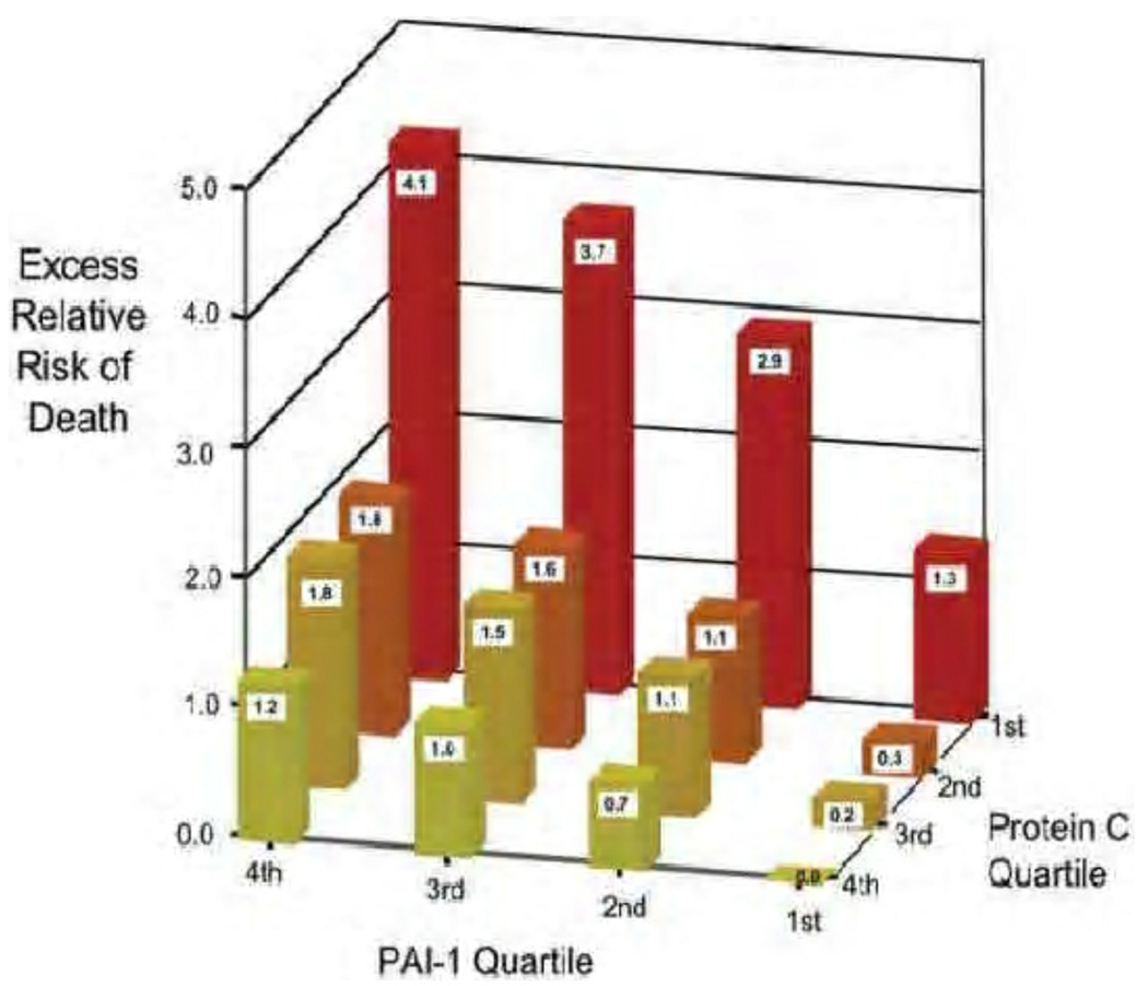
Elevations in plasma levels vWF were independently associated with hospital mortality to day 180 in acute lung injury in 559 patients even after controlling for illness severity, sepsis and ventilator strategy.88 However vWF levels were not responsive to a lower tidal volume strategy. Similarly, higher plasma SP-D levels in 565 patients were also independently associated with 180-day mortality and reduced ventilator and organ-failure free days.3 ICAM-1 in the larger multi-center arm of the study by Calfee and colleagues80 involving 778 patients again showed an independent association with the outcomes listed above. The same independent association of lower levels with survival and more ventilator and organ failure free days were replicated in 593 patients with ALI for IL-6, IL-8 and sTNFR-1.89 Finally in 779 patients, Ware and colleagues showed that lower enrollment levels of protein C and higher levels of PAI-1 were independently and synergistically associated with mortality and organ failure free days9 – Figure 3.
Figure 3.
Multi-dimensional representation charting plasma protein C and PAI-1 levels by quartile against excessive relative risk of death calculated as the difference between the highest PAI-1 and lowest protein C quartiles in 779 patients with acute lung injury. Reproduced from Ware LB, Matthay MA, Parsons PE, Thompson BT, Januzzi JL, Eisner MD. Pathogenetic and prognostic significance of altered coagulation and fibrinolysis in acute lung injury/acute respiratory distress syndrome. Crit Care Med. Aug 2007;35(8):1826, with permission.
Decoy receptor 3 (DcR 3)
DcR 3 is a soluble, pleiotropic, immunomodulator member of the tumor necrosis factor superfamily that binds Fas ligand and LIGHT (a lymphotoxin receptor)90 with an as yet unknown role in ALI. Chen and colleagues91 evaluated a panel of biomarkers (TNF-α, IL-6, sTREM) including DcR 3 in 88 patients with ARDS and obtained ROC curves for mortality prediction for each biomarker. DcR 3 had the best performance and the highest odds ratio for mortality in this patient cohort. Its performance will need to be assessed under different clinical conditions and against other markers but its use warrants further study.
C-reactive protein, procalcitonin and bilirubin
C-reactive protein is a biomarker in common clinical use to delineate the activity of a host of inflammatory conditions such as sepsis, cardiovascular disease and rheumatological disorders. ARDS is broadly an inflammatory condition of the lung and so Bajwa and colleagues92 studied the impact of CRP levels on mortality in ARDS patients. Remarkably, they found an association between higher CRP levels and better outcomes including 60-day mortality, organ failure and duration of mechanical ventilation. The biologic rationale for this finding is unclear though it might be due to reduced neutrophil chemotaxis induced by CRP at higher levels and thus potentially a reduced inflammatory burden. The same group measure serum bilirubin levels in a much larger cohort of 1006 patients and demonstrated a significant association with ARDS incidence and mortality with levels > 2.0mg/dL.93
In contrast to the data on CRP, a much smaller study by Tseng and colleagues examined the related inflammatory molecule procalcitonin (PCT) and identified it as a prognostic marker of mortality in pneumonia-induced ARDS.94 Whether this association still holds for other causes of ARDS or in a direct comparison with CRP is unknown.
Combining biomarkers for prognosis and pathogenesis
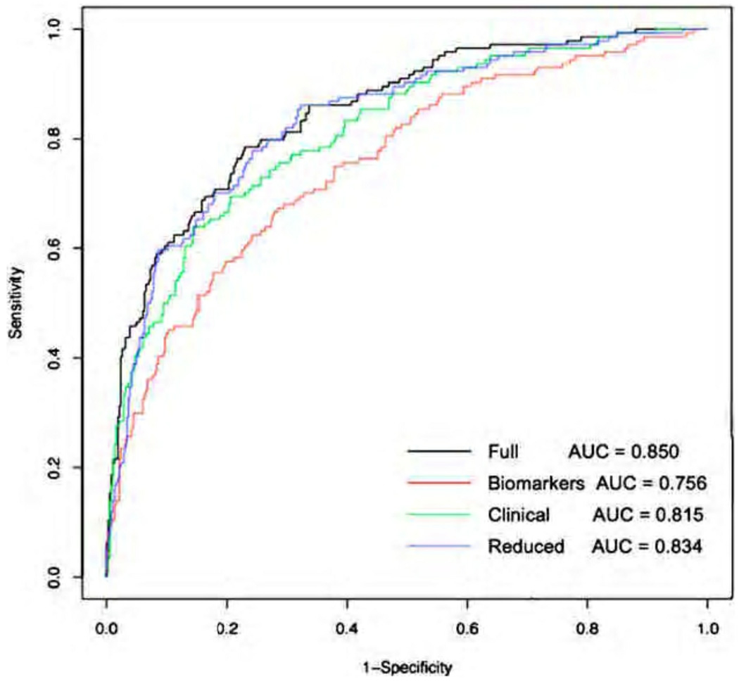
Drawing on the large numbers of biomarkers measured at enrollment in the ARDSNet low tidal volume study, Ware and colleagues tested the ability of a panel of eight biomarkers that had previously been associated with mortality (vWF, SP-D, TNFR1, IL-6, IL-8, ICAM-1 protein C and PAI-1) and six clinical predictors (age, cause of lung injury, APACHE III score, plateau pressure, organ failures and alveolar-arterial difference)12 to discriminate 60-day mortality in patients with ALI/ARDS enrolled in the high PEEP vs. low PEEP trial.95 Using the clinical predictors only, a logistic regression model had an AUC of 0.815. A model combining the eight biomarkers with the six clinical predictors had improved discrimination with an AUC of 0.850 suggesting a modest benefit in terms of adding biomarkers. A reduced model with APACHE III score, age, SP-D and IL-8 had an AUC of 0.834 – Figure 4. In this study, the best performing biomarkers were markers of alveolar epithelial injury (SP-D) and inflammation/neutrophil chemotaxis (IL-8), highlighting the importance of these mechanisms in the pathogenesis of ALI.
Figure 4.
Receiver Operator Characteristic curve for multiple mortality prediction models in patients with acute lung injury. Full model includes six clinical predictors (age, cause of injury, APACHE III, plateau pressure, organ failures, alveolar-arterial difference and eight biomarkers (IL-8, IL-6, TNFR-1, SP-D, Protein C, PAI-1, ICAM-1) and has an AUC 0.850. The reduced model includes APACHE III score, age, surfactant protein D and interleukin 8 with an AUC 0.834. From Ware LB et al. Chest. Feb 2010;137(2):292, with permission.
A similar analysis was undertaken with pre-selected biomarkers of inflammation and coagulation to investigate if these markers were still predictive of clinical outcomes following the widespread institution of low tidal volume ventilation.96 In 50 patients with early ALI the three top markers out of a broad panel were IL-8, ICAM-1 and protein C of which the first two were independently associated with increased mortality in ALI. SP-D was not measured in this cohort. It is notable that IL-8 featured prominently in both data sets that have a combination of biomarkers.12,76
Risk reclassification with multiple biomarkers
Risk reclassification is a relatively new statistical approach that was developed to overcome deficiencies with ROC-based methods, which typically require large odds ratios to demonstrate improvements in the AUC with addition of a novel biomarker to established predictors. Risk reclassification compares the predictive accuracy of two models – a baseline model and a secondary model putatively improved by additional variables such as biomarker data. This comparison generates a net reclassification improvement index based on the proportion of patients newly reclassified to a risk category more closely allied with the outcome examined.97 Working with biomarkers measured in the first two ARDSNet clinical trials Calfee and colleagues98 showed that a panel of five biomarkers (sICAM, vWF, IL-8, SP-D and STNFr-1) significantly improved risk prediction for mortality when compared to a clinical prediction model using APACHE III scores only. This method also was superior in detecting differences in outcome prediction not seen with the ROC-based approach.
BEYOND PROTEIN BIOMARKERS: NOVEL PREDICTORS IN ALI
Stem cells
Adult derived stem cells have been studied as biomarkers in ALI. Circulating endothelial progenitor cells (EPCs) have attracted attention recently as prognostic as well as potential therapeutic targets in ALI.99 A handful of studies have shown a small but consistent effect linking higher circulating levels of EPCs with survival from ALI,100,101 suggesting that mobilization of endothelial progenitor cells in periods of acute stress may be beneficial.
Exhaled breath condensate
The exhaled breath condensate (EBC) is a novel, non-invasive method for analyzing byproducts of metabolism as the lung excretes them. The underlying biologic principal is that injury to the lung leads to differential release of metabolites, which can be recovered in the EBC. Both physical characteristics of the EBC such as pH and metabolites including products of nitric oxide metabolism (nitrosative stress), isoprostanes, hydrogen peroxide and cytokines have been studied. It is not unclear whether these measurements reflect systemic or lung-specific production of metabolites or indeed the anatomic region of the respiratory tract from which they arise. Acidification of the EBC and increases in nitric oxide metabolites have been associated with overdistention from mechanical ventilation.102 A lower EBC pH was also inversely related to Lung Index Severity Score.103 Overall, there is still a lack of data but the use of EBC is appealing because unlike BAL, it samples the lung compartment non-invasively. Coupling measurements of EBC to emerging metabolomic techniques is an interesting avenue of future research.
Genetic approaches
The role of genetic polymorphisms as biomarkers of risk for ALI or poor prognosis is beginning to be explored. The study of genetic markers in ALI encounters many of the same methodological issues as the candidate protein biomarker approach including phenotype definition, power estimation, quality control, population stratification and relevant control identification. These methodological issues may take on higher significance given the relatively modest predicted contribution of single gene polymorphisms to ALI.25
Two approaches are used in genetic studies of ALI: a candidate gene approach and the powerful but labor-intensive genome wide approach to identifying susceptibility loci or genes. The candidate approach is hypothesis-driven and involves choosing candidate genes that may be of likely relevance to the disease process, based on knowledge gained from experimental and clinical studies and testing for an association with ALI. As recently reviewed25 only 31 genetic associations, 21 of which have been replicated, have been shown to have associations with the ALI phenotype. Many of the genes corroborate what is known about the pathophysiology of ALI from individual biomarkers. For example polymorphisms in cytokines both pro (TNFA, IL6, IL8)104,105 and anti (IL10) inflammatory106, epithelial markers (surfactant protein B)107, cell-signaling (mannose-binding lectin108) and the deep internal machinery of the cell (nuclear factor of κ light polypeptide gene enhancer in B cells 1)109 have been demonstrated. The most widely replicated polymorphisms are in the genes for IL6, surfactant protein B and ACE.25 Pathogenic concepts such as dysregulated iron metabolism have been revived by genetic association studies as evidenced in the recently reported ferritin light chain polymorphism.110
Conversely, genome wide association studies (GWAS), are not a priori hypothesis-driven and thus offer a potentially less biased avenue to genetic marker discovery. This approach requires substantial increases in sample size and data analysis, but because of the relative reduction in genotyping costs and development of more standardized approaches to GWAS data analysis, this approach is becoming increasingly popular for discovery and replication studies of complex diseases. The output of GWAS is candidate genes that can be supported by other genome-wide approaches such as expression array and proteomic profiling.
Gene expression studies
Howrylak and colleagues111 explored a gene expression signature for ALI due to sepsis as opposed to sepsis alone that would identify a set of genes uniquely activated in ALI regardless of genetic predisposition. Using an innovative group of classification algorithms they arrived at an eight-gene expression profile in whole blood that was characteristic of ALI. This model had a within-study accuracy of 100% for diagnosis of ALI and when validated still had 89% accuracy albeit with an n = 9. A similar approach was used to study differential gene expression between the early and late phases of ARDS. Peptidase inhibitor 3 (neutrophil elastase inhibitor – PI3 or pre-elafin) gene expression became progressively silenced from acute to recovery stages of ARDS.112 A follow-up validation study examined the clinical significance of this finding in relation to the ratio of neutrophil elastase (HNE) to PI3 in the plasma of ICU patients ‘at risk’ for ARDS. An increase in HNE to PI3 ratio from pre to early ARDS in patients developing the syndrome was observed. In contrast, the ratio fell in an ‘at risk’ patient cohort who remained free of ARDS. Thus, a change in HNE: PI3 balance might be a useful indicator of imminent ARDS.113
In summary, gene expression analysis is a promising methodology for identification of novel biomarkers of ALI. However, it should be noted that the gene expression profile generated depends to a large extent on the cellular mix present in the sample, a factor which is of particular concern in whole blood gene expression studies.
Proteomics and metabolomics
Proteomics is a systems-based methodology for mining the complex changes in protein expression and posttranslational modifications present in biological samples that can occur with disease processes. A variety of approaches have been used including 2-dimensional electrophoresis methods as well as the more powerful approach of liquid chromatography-tandem mass spectrometry. Insulin-growth factor-binding protein-3 (IGFBP-3), a marker of apoptosis, and S100 proteins A8 and A9 proteins, markers of inflammation have been identified in this way.114,115 The next frontier is to move forward from descriptive lists of differentially expressed proteins to mechanistic insights making use of a plethora of cutting edge analytic tools such as principal component analysis, gene-ontology and network analysis to identify nodal points of protein-protein interaction.116,117
The nascent field of metabolomics may also be an avenue for biomarker discovery. Downstream metabolic profiles are analyzed to characterize a physiological or disease-specific state. Critically, metabolomics allows ‘real time’ integration of upstream genomic and proteomic data.118 This approach was explored in a small sample of major trauma patients admitted to the emergency room using nuclear magnetic resonance imaging based metabolomics on whole blood to establish a differential metabolic ‘fingerprint’ between survivors and non-survivors.119 Similarly, Stringer and colleagues120 performed a feasibility study to identify metabolites associated with sepsis-induced ALI. They found quantitative differences in four metabolites: total glutathione, adenosine, phosphatidylserine and sphingomyelin between healthy controls and a small cohort with sepsis-induced ALI. It is premature to assess the potential impact of this methodology but newer more quantitative methodologies should advance the field.
SUMMARY
So are we making progress? In this review we have identified four areas where significant progress has been made over the last 10 years. The first has been the large-scale validation of a number of candidate biomarkers from a range of biologic pathways (IL-8, IL-6, vWF, protein C, PAI-1, SP-D) for prognostication and mortality prediction. The second area is a number of novel predictors, still requiring validation, mediating endothelial permeability (Ang-2), epithelial cell injury (Clara cell protein) and inflammation (decoy receptor 3) that appear to have strong translational potential or to be of particular scientific interest such as endothelial progenitor cells. It is important to note that only 2% of the proteome has been fully characterized, which helps to contextualize the current state of ALI biomarker research. The third area is the advent of genomics and proteomics allied to computationally intensive methods in the field of bioinformatics. These high-dimensional methodologies hold great promise for integrating vast arrays of information, to make predictions about those at risk or those with early disease from genomic or proteomic signatures and to identify novel biomarkers. The final area has been our ability to combine and test all of the above into prognostic indices capable of outperforming individual biomarkers alone.
Although much progress has been made, it should be noted that further progress will be dependent on the availability of large well-phenotyped databases of clinical data and biological samples from patients at risk for and with established ALI. Only with large samples sizes and excellent clinical phenotyping will full operationalization of candidate and novel biomarkers be possible, transforming candidate into clinic-worthy biomarkers, so that our patients may ultimately benefit.
Acknowledgments
This work was supported by HL 103836 and HL 088263 from the National Institutes of Health and an American Heart Association Established Investigator Award.
Footnotes
Publisher's Disclaimer: This is a PDF file of an unedited manuscript that has been accepted for publication. As a service to our customers we are providing this early version of the manuscript. The manuscript will undergo copyediting, typesetting, and review of the resulting proof before it is published in its final citable form. Please note that during the production process errors may be discovered which could affect the content, and all legal disclaimers that apply to the journal pertain.
References
- 1.Hudson LD, Martin TR. Predicting ARDS: problems and prospects. Lancet. 1997 Jun 21;349(9068):1783. doi: 10.1016/S0140-6736(05)61686-8. [DOI] [PubMed] [Google Scholar]
- 2.Ventilation with lower tidal volumes as compared with traditional tidal volumes for acute lung injury and the acute respiratory distress syndrome. The Acute Respiratory Distress Syndrome Network. N Engl J Med. 2000 May 4;342(18):1301–1308. doi: 10.1056/NEJM200005043421801. [DOI] [PubMed] [Google Scholar]
- 3.Eisner MD, Parsons P, Matthay MA, Ware L, Greene K. Plasma surfactant protein levels and clinical outcomes in patients with acute lung injury. Thorax. 2003 Nov;58(11):983–988. doi: 10.1136/thorax.58.11.983. [DOI] [PMC free article] [PubMed] [Google Scholar]
- 4.Biomarkers and surrogate endpoints: preferred definitions and conceptual framework. Clin Pharmacol Ther. 2001 Mar;69(3):89–95. doi: 10.1067/mcp.2001.113989. [DOI] [PubMed] [Google Scholar]
- 5.WHO International Programme on Chemical Safety. Biomarkers in risk assessment: validity and validation. [Accessed 20th October, 2010];2001 http://www.inchem.org/documents/ehc/ech/ech222.htm.
- 6.WHO International Programme on Chemical Safety. Biomarkers in risk assessment: validity and validation. [Accessed 20th October, 2010];1993 http://www.inchem.org/documents/ehc/ech/ech155.htm.
- 7.Strimbu K, Tavel JA. What are biomarkers? Curr Opin HIV AIDS. 2010 Nov;5(6):463–466. doi: 10.1097/COH.0b013e32833ed177. [DOI] [PMC free article] [PubMed] [Google Scholar]
- 8.Spragg RG, Bernard GR, Checkley W, et al. Beyond mortality: future clinical research in acute lung injury. Am J Respir Crit Care Med. 2010 May 15;181(10):1121–1127. doi: 10.1164/rccm.201001-0024WS. [DOI] [PMC free article] [PubMed] [Google Scholar]
- 9.Ware LB, Matthay MA, Parsons PE, Thompson BT, Januzzi JL, Eisner MD. Pathogenetic and prognostic significance of altered coagulation and fibrinolysis in acute lung injury/acute respiratory distress syndrome. Crit Care Med. 2007 Aug;35(8):1821–1828. doi: 10.1097/01.CCM.0000221922.08878.49. [DOI] [PMC free article] [PubMed] [Google Scholar]
- 10.Marshall JC, Reinhart K. Biomarkers of sepsis. Crit Care Med. 2009 Jul;37(7):2290–2298. doi: 10.1097/CCM.0b013e3181a02afc. [DOI] [PubMed] [Google Scholar]
- 11.Willson DF, Thomas NJ, Markovitz BP, et al. Effect of exogenous surfactant (calfactant) in pediatric acute lung injury: a randomized controlled trial. JAMA. 2005 Jan 26;293(4):470–476. doi: 10.1001/jama.293.4.470. [DOI] [PubMed] [Google Scholar]
- 12.Ware LB, Koyama T, Billheimer DD, et al. Prognostic and pathogenetic value of combining clinical and biochemical indices in patients with acute lung injury. Chest. 2010 Feb;137(2):288–296. doi: 10.1378/chest.09-1484. [DOI] [PMC free article] [PubMed] [Google Scholar]
- 13.Tzouvelekis A, Pneumatikos I, Bouros D. Serum biomarkers in acute respiratory distress syndrome an ailing prognosticator. Respir Res. 2005;6:62. doi: 10.1186/1465-9921-6-62. [DOI] [PMC free article] [PubMed] [Google Scholar]
- 14.Olman MA, White KE, Ware LB, et al. Pulmonary edema fluid from patients with early lung injury stimulates fibroblast proliferation through IL-1 beta-induced IL-6 expression. J Immunol. 2004 Feb 15;172(4):2668–2677. doi: 10.4049/jimmunol.172.4.2668. [DOI] [PubMed] [Google Scholar]
- 15.Soreide K. Receiver-operating characteristic curve analysis in diagnostic, prognostic and predictive biomarker research. J Clin Pathol. 2009 Jan;62(1):1–5. doi: 10.1136/jcp.2008.061010. [DOI] [PubMed] [Google Scholar]
- 16.Zhu W, Zeng N, Wang N. Sensitivity, Specificity, Accuracy, Associated Confidence Interval and ROC Analysis with Practical SAS Implementations. [Accessed 14th November, 2010];2010 http://www.nesug.org/Proceedings/nesug10/hl/hl07.pdf. [Google Scholar]
- 17.Shehabi Y, Seppelt I. Pro/Con debate: is procalcitonin useful for guiding antibiotic decision making in critically ill patients? Crit Care. 2008;12(3):211. doi: 10.1186/cc6860. [DOI] [PMC free article] [PubMed] [Google Scholar]
- 18.Kaufman RA. Strategic planning for success : aligning people, performance, and payoffs. [Contributor biographical information. [Google Scholar]
- 19.Ware LB, Matthay MA. The acute respiratory distress syndrome. N Engl J Med. 2000 May 4;342(18):1334–1349. doi: 10.1056/NEJM200005043421806. [DOI] [PubMed] [Google Scholar]
- 20.Levitt JE, Gould MK, Ware LB, Matthay MA. The pathogenetic and prognostic value of biologic markers in acute lung injury. J Intensive Care Med. 2009 May–Jun;24(3):151–167. doi: 10.1177/0885066609332603. [DOI] [PubMed] [Google Scholar]
- 21.Imai Y, Kuba K, Rao S, et al. Angiotensin-converting enzyme 2 protects from severe acute lung failure. Nature. 2005 Jul 7;436(7047):112–116. doi: 10.1038/nature03712. [DOI] [PMC free article] [PubMed] [Google Scholar]
- 22.Tejera P, Wang Z, Zhai R, et al. Genetic polymorphisms of peptidase inhibitor 3 (elafin) are associated with acute respiratory distress syndrome. Am J Respir Cell Mol Biol. 2009 Dec;41(6):696–704. doi: 10.1165/rcmb.2008-0410OC. [DOI] [PMC free article] [PubMed] [Google Scholar]
- 23.Parsons PE, Matthay MA, Ware LB, Eisner MD. Elevated plasma levels of soluble TNF receptors are associated with morbidity and mortality in patients with acute lung injury. Am J Physiol Lung Cell Mol Physiol. 2005 Mar;288(3):L426–L431. doi: 10.1152/ajplung.00302.2004. [DOI] [PubMed] [Google Scholar]
- 24.Okajima K, Harada N, Sakurai G, et al. Rapid assay for plasma soluble E-selectin predicts the development of acute respiratory distress syndrome in patients with systemic inflammatory response syndrome. Transl Res. 2006 Dec;148(6):295–300. doi: 10.1016/j.trsl.2006.07.009. [DOI] [PubMed] [Google Scholar]
- 25.Gao L, Barnes KC. Recent advances in genetic predisposition to clinical acute lung injury. Am J Physiol Lung Cell Mol Physiol. 2009 May;296(5):L713–L725. doi: 10.1152/ajplung.90269.2008. [DOI] [PMC free article] [PubMed] [Google Scholar]
- 26.Matthay MA, Zimmerman GA. Acute lung injury and the acute respiratory distress syndrome: four decades of inquiry into pathogenesis and rational management. Am J Respir Cell Mol Biol. 2005 Oct;33(4):319–327. doi: 10.1165/rcmb.F305. [DOI] [PMC free article] [PubMed] [Google Scholar]
- 27.Bastarache JA, Ware LB, Bernard GR. The role of the coagulation cascade in the continuum of sepsis and acute lung injury and acute respiratory distress syndrome. Semin Respir Crit Care Med. 2006 Aug;27(4):365–376. doi: 10.1055/s-2006-948290. [DOI] [PubMed] [Google Scholar]
- 28.Ware LB, Fremont RD, Bastarache JA, Calfee CS, Matthay MA. Determining the aetiology of pulmonary oedema by the oedema fluid-to-plasma protein ratio. Eur Respir J. 2010 Feb;35(2):331–337. doi: 10.1183/09031936.00098709. [DOI] [PMC free article] [PubMed] [Google Scholar]
- 29.Kitano H. Systems biology: a brief overview. Science. 2002 Mar 1;295(5560):1662–1664. doi: 10.1126/science.1069492. [DOI] [PubMed] [Google Scholar]
- 30.Meduri GU, Headley S, Kohler G, et al. Persistent elevation of inflammatory cytokines predicts a poor outcome in ARDS. Plasma IL-1 beta and IL-6 levels are consistent and efficient predictors of outcome over time. Chest. 1995 Apr;107(4):1062–1073. doi: 10.1378/chest.107.4.1062. [DOI] [PubMed] [Google Scholar]
- 31.Park WY, Goodman RB, Steinberg KP, et al. Cytokine balance in the lungs of patients with acute respiratory distress syndrome. Am J Respir Crit Care Med. 2001 Nov 15;164(10 Pt 1):1896–1903. doi: 10.1164/ajrccm.164.10.2104013. [DOI] [PubMed] [Google Scholar]
- 32.Parsons PE, Moss M, Vannice JL, Moore EE, Moore FA, Repine JE. Circulating IL-1ra and IL-10 levels are increased but do not predict the development of acute respiratory distress syndrome in at-risk patients. Am J Respir Crit Care Med. 1997 Apr;155(4):1469–1473. doi: 10.1164/ajrccm.155.4.9105096. [DOI] [PubMed] [Google Scholar]
- 33.Bouros D, Alexandrakis MG, Antoniou KM, et al. The clinical significance of serum and bronchoalveolar lavage inflammatory cytokines in patients at risk for Acute Respiratory Distress Syndrome. BMC Pulm Med. 2004 Aug 17;4:6. doi: 10.1186/1471-2466-4-6. [DOI] [PMC free article] [PubMed] [Google Scholar]
- 34.Pittet JF, Mackersie RC, Martin TR, Matthay MA. Biological markers of acute lung injury: prognostic and pathogenetic significance. Am J Respir Crit Care Med. 1997 Apr;155(4):1187–1205. doi: 10.1164/ajrccm.155.4.9105054. [DOI] [PubMed] [Google Scholar]
- 35.Suter PM, Suter S, Girardin E, Roux-Lombard P, Grau GE, Dayer JM. High bronchoalveolar levels of tumor necrosis factor and its inhibitors, interleukin-1, interferon, and elastase, in patients with adult respiratory distress syndrome after trauma, shock, or sepsis. Am Rev Respir Dis. 1992 May;145(5):1016–1022. doi: 10.1164/ajrccm/145.5.1016. [DOI] [PubMed] [Google Scholar]
- 36.Takala A, Jousela I, Takkunen O, et al. A prospective study of inflammation markers in patients at risk of indirect acute lung injury. Shock. 2002 Apr;17(4):252–257. doi: 10.1097/00024382-200204000-00002. [DOI] [PubMed] [Google Scholar]
- 37.Donnelly SC, Strieter RM, Kunkel SL, et al. Interleukin-8 and development of adult respiratory distress syndrome in at-risk patient groups. Lancet. 1993 Mar 13;341(8846):643–647. doi: 10.1016/0140-6736(93)90416-e. [DOI] [PubMed] [Google Scholar]
- 38.Cohen MJ, Brohi K, Calfee CS, et al. Early release of high mobility group box nuclear protein 1 after severe trauma in humans: role of injury severity and tissue hypoperfusion. Crit Care. 2009;13(6):R174. doi: 10.1186/cc8152. [DOI] [PMC free article] [PubMed] [Google Scholar]
- 39.Ley K, Laudanna C, Cybulsky MI, Nourshargh S. Getting to the site of inflammation: the leukocyte adhesion cascade updated. Nat Rev Immunol. 2007 Sep;7(9):678–689. doi: 10.1038/nri2156. [DOI] [PubMed] [Google Scholar]
- 40.van Meurs M, Kumpers P, Ligtenberg JJ, Meertens JH, Molema G, Zijlstra JG. Bench-to-bedside review: Angiopoietin signalling in critical illness - a future target? Crit Care. 2009;13(2):207. doi: 10.1186/cc7153. [DOI] [PMC free article] [PubMed] [Google Scholar]
- 41.Fiedler U, Augustin HG. Angiopoietins: a link between angiogenesis and inflammation. Trends Immunol. 2006 Dec;27(12):552–558. doi: 10.1016/j.it.2006.10.004. [DOI] [PubMed] [Google Scholar]
- 42.Jones PF. Not just angiogenesis--wider roles for the angiopoietins. J Pathol. 2003 Dec;201(4):515–527. doi: 10.1002/path.1452. [DOI] [PubMed] [Google Scholar]
- 43.Gallagher DC, Parikh SM, Balonov K, et al. Circulating angiopoietin 2 correlates with mortality in a surgical population with acute lung injury/adult respiratory distress syndrome. Shock. 2008 Jun;29(6):656–661. doi: 10.1097/shk.0b013e31815dd92f. [DOI] [PMC free article] [PubMed] [Google Scholar]
- 44.Christie JD, Wurfel MM, G.E. OK, et al. Genome wide association (gwa) identifies functional susceptibility loci for acute lung injury. Am J Respir Crit Care Med. 2010 [Google Scholar]
- 45.Meyer NJ, Li M, Shah CV, et al. Large scale genotyping in an african american trauma population identifies angiopoeitin-2 variants associated with ALI. Am J Respir Crit Care Med. 2010;179:A3879. [Google Scholar]
- 46.Meyer NJ, Li M, Feng R, et al. ANGPT2 Genetic Variant is Associated with Trauma-Associated Acute Lung Injury and Altered Plasma Angiopoietin-2 Isoform Ratio. Am J Respir Crit Care Med. 2011 Jan 21; doi: 10.1164/rccm.201005-0701OC. [DOI] [PMC free article] [PubMed] [Google Scholar]
- 47.Medford AR, Millar AB. Vascular endothelial growth factor (VEGF) in acute lung injury (ALI) and acute respiratory distress syndrome (ARDS): paradox or paradigm? Thorax. 2006 Jul;61(7):621–626. doi: 10.1136/thx.2005.040204. [DOI] [PMC free article] [PubMed] [Google Scholar]
- 48.Oh H, Takagi H, Suzuma K, Otani A, Matsumura M, Honda Y. Hypoxia and vascular endothelial growth factor selectively up-regulate angiopoietin-2 in bovine microvascular endothelial cells. J Biol Chem. 1999 May 28;274(22):15732–15739. doi: 10.1074/jbc.274.22.15732. [DOI] [PubMed] [Google Scholar]
- 49.Findley CM, Cudmore MJ, Ahmed A, Kontos CD. VEGF induces Tie2 shedding via a phosphoinositide 3-kinase/Akt dependent pathway to modulate Tie2 signaling. Arterioscler Thromb Vasc Biol. 2007 Dec;27(12):2619–2626. doi: 10.1161/ATVBAHA.107.150482. [DOI] [PubMed] [Google Scholar]
- 50.Ware LB, Kaner RJ, Crystal RG, et al. VEGF levels in the alveolar compartment do not distinguish between ARDS and hydrostatic pulmonary oedema. Eur Respir J. 2005 Jul;26(1):101–105. doi: 10.1183/09031936.05.00106604. [DOI] [PMC free article] [PubMed] [Google Scholar]
- 51.van der Heijden M, van Nieuw Amerongen GP, Koolwijk P, van Hinsbergh VW, Groeneveld AB. Angiopoietin-2, permeability oedema, occurrence and severity of ALI/ARDS in septic and non-septic critically ill patients. Thorax. 2008 Oct;63(10):903–909. doi: 10.1136/thx.2007.087387. [DOI] [PubMed] [Google Scholar]
- 52.Franchini M, Lippi G. Von Willebrand factor and thrombosis. Ann Hematol. 2006 Jul;85(7):415–423. doi: 10.1007/s00277-006-0085-5. [DOI] [PubMed] [Google Scholar]
- 53.Rubin DB, Wiener-Kronish JP, Murray JF, et al. Elevated von Willebrand factor antigen is an early plasma predictor of acute lung injury in nonpulmonary sepsis syndrome. J Clin Invest. 1990 Aug;86(2):474–480. doi: 10.1172/JCI114733. [DOI] [PMC free article] [PubMed] [Google Scholar]
- 54.Bajaj MS, Tricomi SM. Plasma levels of the three endothelial-specific proteins von Willebrand factor, tissue factor pathway inhibitor, and thrombomodulin do not predict the development of acute respiratory distress syndrome. Intensive Care Med. 1999 Nov;25(11):1259–1266. doi: 10.1007/s001340051054. [DOI] [PubMed] [Google Scholar]
- 55.Moss M, Ackerson L, Gillespie MK, Moore FA, Moore EE, Parsons PE. von Willebrand factor antigen levels are not predictive for the adult respiratory distress syndrome. Am J Respir Crit Care Med. 1995 Jan;151(1):15–20. doi: 10.1164/ajrccm.151.1.7812545. [DOI] [PubMed] [Google Scholar]
- 56.Langer HF, Chavakis T. Leukocyte-endothelial interactions in inflammation. J Cell Mol Med. 2009 Jul;13(7):1211–1220. doi: 10.1111/j.1582-4934.2009.00811.x. [DOI] [PMC free article] [PubMed] [Google Scholar]
- 57.Newman W, Beall LD, Carson CW, et al. Soluble E-selectin is found in supernatants of activated endothelial cells and is elevated in the serum of patients with septic shock. J Immunol. 1993 Jan 15;150(2):644–654. [PubMed] [Google Scholar]
- 58.Donnelly SC, Haslett C, Dransfield I, et al. Role of selectins in development of adult respiratory distress syndrome. Lancet. 1994 Jul 23;344(8917):215–219. doi: 10.1016/s0140-6736(94)92995-5. [DOI] [PubMed] [Google Scholar]
- 59.Matthay MA, Zemans RL. The Acute Respiratory Distress Syndrome: Pathogenesis and Treatment. Annu Rev Pathol. 2010 Jan 15; doi: 10.1146/annurev-pathol-011110-130158. [DOI] [PMC free article] [PubMed] [Google Scholar]
- 60.Calfee CS, Ware LB, Eisner MD, et al. Plasma receptor for advanced glycation end products and clinical outcomes in acute lung injury. Thorax. 2008 Dec;63(12):1083–1089. doi: 10.1136/thx.2008.095588. [DOI] [PMC free article] [PubMed] [Google Scholar]
- 61.Nakashima T, Yokoyama A, Inata J, et al. Mucins carrying selectin ligands as predictive biomarkers of disseminated intravascular coagulation complication in ARDS. Chest. 2010 Jul 29; doi: 10.1378/chest.09-3082. [DOI] [PubMed] [Google Scholar]
- 62.Pastva AM, Wright JR, Williams KL. Immunomodulatory roles of surfactant proteins A and D: implications in lung disease. Proc Am Thorac Soc. 2007 Jul;4(3):252–257. doi: 10.1513/pats.200701-018AW. [DOI] [PMC free article] [PubMed] [Google Scholar]
- 63.Chroneos ZC, Sever-Chroneos Z, Shepherd VL. Pulmonary surfactant: an immunological perspective. Cell Physiol Biochem. 2010;25(1):13–26. doi: 10.1159/000272047. [DOI] [PMC free article] [PubMed] [Google Scholar]
- 64.Greene KE, Wright JR, Steinberg KP, et al. Serial changes in surfactant-associated proteins in lung and serum before and after onset of ARDS. Am J Respir Crit Care Med. 1999 Dec;160(6):1843–1850. doi: 10.1164/ajrccm.160.6.9901117. [DOI] [PubMed] [Google Scholar]
- 65.Greene KE, Ye S, Mason RJ, Parsons PE. Serum surfactant protein-A levels predict development of ARDS in at-risk patients. Chest. 1999 Jul;116(1 Suppl):90S–91S. [PubMed] [Google Scholar]
- 66.Bersten AD, Hunt T, Nicholas TE, Doyle IR. Elevated plasma surfactant protein-B predicts development of acute respiratory distress syndrome in patients with acute respiratory failure. J Respir Crit Care Med. 2001 Aug 15;164(4):648–652. doi: 10.1164/ajrccm.164.4.2010111. [DOI] [PubMed] [Google Scholar]
- 67.Broeckaert F, Bernard A. Clara cell secretory protein (CC16): characteristics and perspectives as lung peripheral biomarker. Clin Exp Allergy. 2000 Apr;30(4):469–475. doi: 10.1046/j.1365-2222.2000.00760.x. [DOI] [PubMed] [Google Scholar]
- 68.Determann RM, Millo JL, Waddy S, Lutter R, Garrard CS, Schultz MJ. Plasma CC16 levels are associated with development of ALI/ARDS in patients with ventilator-associated pneumonia: a retrospective observational study. BMC Pulm Med. 2009;9:49. doi: 10.1186/1471-2466-9-49. [DOI] [PMC free article] [PubMed] [Google Scholar]
- 69.Kropski JA, Fremont RD, Calfee CS, Ware LB. Clara cell protein (CC16), a marker of lung epithelial injury, is decreased in plasma and pulmonary edema fluid from patients with acute lung injury. Chest. 2009 Jun;135(6):1440–1447. doi: 10.1378/chest.08-2465. [DOI] [PMC free article] [PubMed] [Google Scholar]
- 70.Thygesen K, Alpert JS, White HD, et al. Universal definition of myocardial infarction. Circulation. 2007 Nov 27;116(22):2634–2653. doi: 10.1161/CIRCULATIONAHA.107.187397. [DOI] [PubMed] [Google Scholar]
- 71.Bernard GR, Artigas A, Brigham KL, et al. The American-European Consensus Conference on ARDS. Definitions, mechanisms, relevant outcomes, and clinical trial coordination. Am J Respir Crit Care Med. 1994 Mar;149(3 Pt 1):818–824. doi: 10.1164/ajrccm.149.3.7509706. [DOI] [PubMed] [Google Scholar]
- 72.Phua J, Stewart TE, Ferguson ND. Acute respiratory distress syndrome 40 years later: time to revisit its definition. Crit Care Med. 2008 Oct;36(10):2912–2921. doi: 10.1097/CCM.0b013e31817d20bd. [DOI] [PubMed] [Google Scholar]
- 73.Aman J, van der Heijden M, van Lingen A, et al. Plasma protein levels are markers of pulmonary vascular permeability and degree of lung injury in critically ill patients with or at risk for acute lung injury/acute respiratory distress syndrome. Crit Care Med. 2011 Jan;39(1):89–97. doi: 10.1097/CCM.0b013e3181feb46a. [DOI] [PubMed] [Google Scholar]
- 74.Creagh-Brown BC, Quinlan GJ, Evans TW, Burke-Gaffney A. The RAGE axis in systemic inflammation, acute lung injury and myocardial dysfunction: an important therapeutic target? Intensive Care Med. 2010 Oct;36(10):1644–1656. doi: 10.1007/s00134-010-1952-z. [DOI] [PubMed] [Google Scholar]
- 75.Uchida T, Shirasawa M, Ware LB, et al. Receptor for advanced glycation end-products is a marker of type I cell injury in acute lung injury. Am J Respir Crit Care Med. 2006 May 1;173(9):1008–1015. doi: 10.1164/rccm.200509-1477OC. [DOI] [PMC free article] [PubMed] [Google Scholar]
- 76.Fremont RD, Koyama T, Calfee CS, et al. Acute lung injury in patients with traumatic injuries: utility of a panel of biomarkers for diagnosis and pathogenesis. J Trauma. 2010 May;68(5):1121–1127. doi: 10.1097/TA.0b013e3181c40728. [DOI] [PMC free article] [PubMed] [Google Scholar]
- 77.Tzu J, Marinkovich MP. Bridging structure with function: structural, regulatory, and developmental role of laminins. Int J Biochem Cell Biol. 2008;40(2):199–214. doi: 10.1016/j.biocel.2007.07.015. [DOI] [PMC free article] [PubMed] [Google Scholar]
- 78.Katayama M, Ishizaka A, Sakamoto M, et al. Laminin gamma2 fragments are increased in the circulation of patients with early phase acute lung injury. Intensive Care Med. 2010 Mar;36(3):479–486. doi: 10.1007/s00134-009-1719-6. [DOI] [PMC free article] [PubMed] [Google Scholar]
- 79.Muller WA. Mechanisms of transendothelial migration of leukocytes. Circ Res. 2009 Jul 31;105(3):223–230. doi: 10.1161/CIRCRESAHA.109.200717. [DOI] [PMC free article] [PubMed] [Google Scholar]
- 80.Calfee CS, Eisner MD, Parsons PE, et al. Soluble intercellular adhesion molecule-1 and clinical outcomes in patients with acute lung injury. Intensive Care Med. 2009 Feb;35(2):248–257. doi: 10.1007/s00134-008-1235-0. [DOI] [PMC free article] [PubMed] [Google Scholar]
- 81.Conner ER, Ware LB, Modin G, Matthay MA. Elevated pulmonary edema fluid concentrations of soluble intercellular adhesion molecule-1 in patients with acute lung injury: biological and clinical significance. Chest. 1999 Jul;116(1 Suppl):83S–84S. doi: 10.1378/chest.116.suppl_1.83s. [DOI] [PubMed] [Google Scholar]
- 82.Prabhakaran P, Ware LB, White KE, Cross MT, Matthay MA, Olman MA. Elevated levels of plasminogen activator inhibitor-1 in pulmonary edema fluid are associated with mortality in acute lung injury. Am J Physiol Lung Cell Mol Physiol. 2003 Jul;285(1):L20–L28. doi: 10.1152/ajplung.00312.2002. [DOI] [PubMed] [Google Scholar]
- 83.Pugin J, Verghese G, Widmer MC, Matthay MA. The alveolar space is the site of intense inflammatory and profibrotic reactions in the early phase of acute respiratory distress syndrome. Crit Care Med. 1999 Feb;27(2):304–312. doi: 10.1097/00003246-199902000-00036. [DOI] [PubMed] [Google Scholar]
- 84.Chesnutt AN, Matthay MA, Tibayan FA, Clark JG. Early detection of type III procollagen peptide in acute lung injury. Pathogenetic and prognostic significance. Am J Respir Crit Care Med. 1997 Sep;156(3 Pt 1):840–845. doi: 10.1164/ajrccm.156.3.9701124. [DOI] [PubMed] [Google Scholar]
- 85.Meduri GU, Tolley EA, Chinn A, Stentz F, Postlethwaite A. Procollagen types I and III aminoterminal propeptide levels during acute respiratory distress syndrome and in response to methylprednisolone treatment. Am J Respir Crit Care Med. 1998 Nov;158(5 Pt 1):1432–1441. doi: 10.1164/ajrccm.158.5.9801107. [DOI] [PubMed] [Google Scholar]
- 86.Dentener MA, Vreugdenhil AC, Hoet PH, et al. Production of the acute-phase protein lipopolysaccharide-binding protein by respiratory type II epithelial cells: implications for local defense to bacterial endotoxins. Am J Respir Cell Mol Biol. 2000 Aug;23(2):146–153. doi: 10.1165/ajrcmb.23.2.3855. [DOI] [PubMed] [Google Scholar]
- 87.Villar J, Perez-Mendez L, Espinosa E, et al. Serum lipopolysaccharide binding protein levels predict severity of lung injury and mortality in patients with severe sepsis. PLoS One. 2009;4(8):e6818. doi: 10.1371/journal.pone.0006818. [DOI] [PMC free article] [PubMed] [Google Scholar]
- 88.Ware LB, Eisner MD, Thompson BT, Parsons PE, Matthay MA. Significance of von Willebrand factor in septic and nonseptic patients with acute lung injury. Am J Respir Crit Care Med. 2004 Oct 1;170(7):766–772. doi: 10.1164/rccm.200310-1434OC. [DOI] [PubMed] [Google Scholar]
- 89.Parsons PE, Eisner MD, Thompson BT, et al. Lower tidal volume ventilation and plasma cytokine markers of inflammation in patients with acute lung injury. Crit Care Med. 2005 Jan;33(1):1–6. doi: 10.1097/01.ccm.0000149854.61192.dc. discussion 230–232. [DOI] [PubMed] [Google Scholar]
- 90.Ware CF. Targeting the LIGHT-HVEM pathway. Adv Exp Med Biol. 2009;647:146–155. doi: 10.1007/978-0-387-89520-8_10. [DOI] [PubMed] [Google Scholar]
- 91.Chen CY, Yang KY, Chen MY, et al. Decoy receptor 3 levels in peripheral blood predict outcomes of acute respiratory distress syndrome. Am J Respir Crit Care Med. 2009 Oct 15;180(8):751–760. doi: 10.1164/rccm.200902-0222OC. [DOI] [PubMed] [Google Scholar]
- 92.Bajwa EK, Khan UA, Januzzi JL, Gong MN, Thompson BT, Christiani DC. Plasma C-reactive protein levels are associated with improved outcome in ARDS. Chest. 2009 Aug;136(2):471–480. doi: 10.1378/chest.08-2413. [DOI] [PMC free article] [PubMed] [Google Scholar]
- 93.Zhai R, Sheu CC, Su L, et al. Serum bilirubin levels on ICU admission are associated with ARDS development and mortality in sepsis. Thorax. 2009 Sep;64(9):784–790. doi: 10.1136/thx.2009.113464. [DOI] [PMC free article] [PubMed] [Google Scholar]
- 94.Tseng JS, Chan MC, Hsu JY, Kuo BI, Wu CL. Procalcitonin is a valuable prognostic marker in ARDS caused by community-acquired pneumonia. Respirology. 2008 Jun;13(4):505–509. doi: 10.1111/j.1440-1843.2008.01293.x. [DOI] [PubMed] [Google Scholar]
- 95.Brower RG, Lanken PN, MacIntyre N, et al. Higher versus lower positive end-expiratory pressures in patients with the acute respiratory distress syndrome. N Engl J Med. 2004 Jul 22;351(4):327–336. doi: 10.1056/NEJMoa032193. [DOI] [PubMed] [Google Scholar]
- 96.McClintock D, Zhuo H, Wickersham N, Matthay MA, Ware LB. Biomarkers of inflammation, coagulation and fibrinolysis predict mortality in acute lung injury. Crit Care. 2008;12(2):R41. doi: 10.1186/cc6846. [DOI] [PMC free article] [PubMed] [Google Scholar]
- 97.Pencina MJ, D'Agostino RB, Sr, D'Agostino RB, Jr, Vasan RS. Evaluating the added predictive ability of a new marker: from area under the ROC curve to reclassification and beyond. Stat Med. 2008 Jan 30;27(2):157–172. doi: 10.1002/sim.2929. discussion 207-112. [DOI] [PubMed] [Google Scholar]
- 98.Calfee CS, Ware L, Glidden DV, et al. Use of Risk Reclassification with Multiple Biomarkers Improves Mortality Prediction in Acute Lung Injury. Critical Care Medicine. 2011 doi: 10.1097/CCM.0b013e318207ec3c. in press. [DOI] [PMC free article] [PubMed] [Google Scholar]
- 99.Matthay MA, Thompson BT, Read EJ, et al. Therapeutic potential of mesenchymal stem cells for severe acute lung injury. Chest. 2010 Oct;138(4):965–972. doi: 10.1378/chest.10-0518. [DOI] [PMC free article] [PubMed] [Google Scholar]
- 100.Burnham EL, Mealer M, Gaydos J, Majka S, Moss M. Acute lung injury but not sepsis is associated with increased colony formation by peripheral blood mononuclear cells. Am J Respir Cell Mol Biol. 2010 Sep;43(3):326–333. doi: 10.1165/rcmb.2009-0015OC. [DOI] [PMC free article] [PubMed] [Google Scholar]
- 101.Burnham EL, Taylor WR, Quyyumi AA, Rojas M, Brigham KL, Moss M. Increased circulating endothelial progenitor cells are associated with survival in acute lung injury. Am J Respir Crit Care Med. 2005 Oct 1;172(7):854–860. doi: 10.1164/rccm.200410-1325OC. [DOI] [PubMed] [Google Scholar]
- 102.Owens RL, Stigler WS, Hess DR. Do newer monitors of exhaled gases, mechanics, and esophageal pressure add value? Clin Chest Med. 2008 Jun;29(2):297–312. vi–vii. doi: 10.1016/j.ccm.2008.02.001. [DOI] [PubMed] [Google Scholar]
- 103.Gessner C, Hammerschmidt S, Kuhn H, et al. Exhaled breath condensate acidification in acute lung injury. Respir Med. 2003 Nov;97(11):1188–1194. doi: 10.1016/s0954-6111(03)00225-7. [DOI] [PubMed] [Google Scholar]
- 104.Gong MN, Zhou W, Williams PL, et al. −308GA and TNFB polymorphisms in acute respiratory distress syndrome. Eur Respir J. 2005 Sep;26(3):382–389. doi: 10.1183/09031936.05.00000505. [DOI] [PubMed] [Google Scholar]
- 105.Sutherland AM, Walley KR, Manocha S, Russell JA. The association of interleukin 6 haplotype clades with mortality in critically ill adults. Arch Intern Med. 2005 Jan 10;165(1):75–82. doi: 10.1001/archinte.165.1.75. [DOI] [PubMed] [Google Scholar]
- 106.Gong MN, Thompson BT, Williams PL, et al. Interleukin-10 polymorphism in position −1082 and acute respiratory distress syndrome. Eur Respir J. 2006 Apr 27;(4):674–681. doi: 10.1183/09031936.06.00046405. [DOI] [PMC free article] [PubMed] [Google Scholar]
- 107.Gong MN, Wei Z, Xu LL, Miller DP, Thompson BT, Christiani DC. Polymorphism in the surfactant protein-B gene, gender, and the risk of direct pulmonary injury and ARDS. Chest. 2004 Jan;125(1):203–211. doi: 10.1378/chest.125.1.203. [DOI] [PubMed] [Google Scholar]
- 108.Gong MN, Zhou W, Williams PL, Thompson BT, Pothier L, Christiani DC. Polymorphisms in the mannose binding lectin-2 gene and acute respiratory distress syndrome. Crit Care Med. 2007 Jan;35(1):48–56. doi: 10.1097/01.CCM.0000251132.10689.F3. [DOI] [PMC free article] [PubMed] [Google Scholar]
- 109.Zhai R, Zhou W, Gong MN, et al. Inhibitor kappaB-alpha haplotype GTC is associated with susceptibility to acute respiratory distress syndrome in Caucasians. Crit Care Med. 2007 Mar;35(3):893–898. doi: 10.1097/01.CCM.0000256845.92640.38. [DOI] [PubMed] [Google Scholar]
- 110.Lagan AL, Quinlan GJ, Mumby S, et al. Variation in iron homeostasis genes between patients with ARDS and healthy control subjects. Chest. 2008 Jun;133(6):1302–1311. doi: 10.1378/chest.07-1117. [DOI] [PubMed] [Google Scholar]
- 111.Howrylak JA, Dolinay T, Lucht L, et al. Discovery of the gene signature for acute lung injury in patients with sepsis. Physiol Genomics. 2009 Apr 10;37(2):133–139. doi: 10.1152/physiolgenomics.90275.2008. [DOI] [PMC free article] [PubMed] [Google Scholar]
- 112.Wang Z, Beach D, Su L, Zhai R, Christiani DC. A genome-wide expression analysis in blood identifies pre-elafin as a biomarker in ARDS. Am J Respir Cell Mol Biol. 2008 Jun;38(6):724–732. doi: 10.1165/rcmb.2007-0354OC. [DOI] [PMC free article] [PubMed] [Google Scholar]
- 113.Wang Z, Chen F, Zhai R, et al. Plasma neutrophil elastase and elafin imbalance is associated with acute respiratory distress syndrome (ARDS) development. PLoS One. 2009;4(2):e4380. doi: 10.1371/journal.pone.0004380. [DOI] [PMC free article] [PubMed] [Google Scholar]
- 114.Schnapp LM, Donohoe S, Chen J, et al. Mining the acute respiratory distress syndrome proteome: identification of the insulin-like growth factor (IGF)/IGF-binding protein-3 pathway in acute lung injury. Am J Pathol. 2006 Jul;169(1):86–95. doi: 10.2353/ajpath.2006.050612. [DOI] [PMC free article] [PubMed] [Google Scholar]
- 115.de Torre C, Ying SX, Munson PJ, Meduri GU, Suffredini AF. Proteomic analysis of inflammatory biomarkers in bronchoalveolar lavage. Proteomics. 2006 Jul;6(13):3949–3957. doi: 10.1002/pmic.200500693. [DOI] [PubMed] [Google Scholar]
- 116.Ware LB, Matthay MA. Beyond fishing: the role of discovery proteomics in mechanistic lung research. Am J Physiol Lung Cell Mol Physiol. 2009 Jan;296(1):L12–L13. doi: 10.1152/ajplung.90558.2008. [DOI] [PMC free article] [PubMed] [Google Scholar]
- 117.Gerszten RE, Accurso F, Bernard GR, et al. Challenges in translating plasma proteomics from bench to bedside: update from the NHLBI Clinical Proteomics Programs. Am J Physiol Lung Cell Mol Physiol. 2008 Jul;295(1):L16–L22. doi: 10.1152/ajplung.00044.2008. [DOI] [PMC free article] [PubMed] [Google Scholar]
- 118.Lacy P. Metabolomics of sepsis-induced acute lung injury: a new approach for biomarkers. Am J Physiol Lung Cell Mol Physiol. 2011 Jan;300(1):L1–L3. doi: 10.1152/ajplung.00375.2010. [DOI] [PubMed] [Google Scholar]
- 119.Cohen MJ, Serkova NJ, Wiener-Kronish J, Pittet JF, Niemann CU. 1H-NMR-based metabolic signatures of clinical outcomes in trauma patients--beyond lactate and base deficit. J Trauma. 2010 Jul;69(1):31–40. doi: 10.1097/TA.0b013e3181e043fe. [DOI] [PubMed] [Google Scholar]
- 120.Stringer KA, Serkova NJ, Karnovsky A, Guire K, Paine R, 3rd, Standiford TJ. Metabolic consequences of sepsis-induced acute lung injury revealed by plasma (1)H-nuclear magnetic resonance quantitative metabolomics and computational analysis. Am J Physiol Lung Cell Mol Physiol. 2011 Jan;300(1):L4–L11. doi: 10.1152/ajplung.00231.2010. [DOI] [PMC free article] [PubMed] [Google Scholar]


