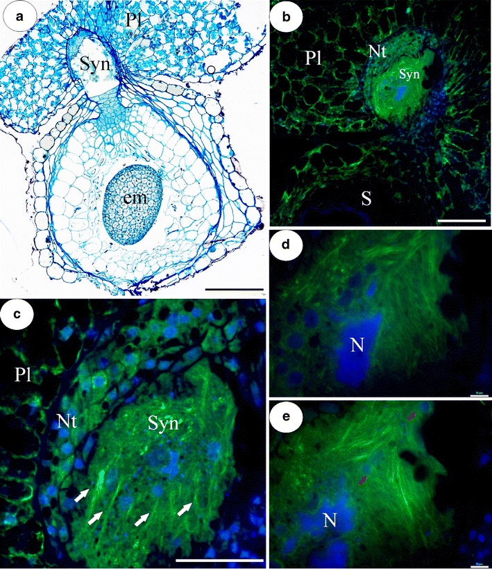
Fig. 1.

a Light micrograph of section through syncytium and seed with embryo of U. intermedia. Syn syncytium, em embryo, Pl placenta; bar = 200 μm. b Section through syncytium and seed, note dense array of actin filaments in syncytium. Syn syncytium, S seed, Nt nutritive tissue, Pl placenta; bar = 200 μm. c F-actin cables (arrows) running parallel to each other, across the syncytium. Syn syncytium, Nt nutritive tissue, Pl placenta; bar = 50 μm. d, e Two optical sections through syncytium showing giant endosperm nucleus (N) and network of numerous F-actin bundles; arrows—nuclei from former nutritive tissue cells; bar = 8 μm
